R E S E A R C H
Open Access
The protective effect of neonatal oral
administration of oleanolic acid against the
subsequent development of
fructose-induced metabolic dysfunction in male and
female rats
Trevor T. Nyakudya
1,3*, Emmanuel Mukwevho
2and Kennedy H. Erlwanger
1Abstract
Background:Consumption of fructose-rich diets has been implicated in the increasing global prevalence of metabolic syndrome (MetS). Interventions during periods of early ontogenic developmental plasticity can cause epigenetic changes which program metabolism for positive or negative health benefits later in life. The
phytochemical oleanolic acid (OA) possesses anti-diabetic and anti-obesity effects. We investigated the potential protective effects of neonatal administration of OA on the subsequent development of high fructose diet-induced metabolic dysfunction in rats.
Method:Male and female (N= 112) suckling rats were randomly assigned to four groups and administered orally: distilled water (DW), oleanolic acid (OA; 60 mg/kg), high-fructose solution (HF; 20%w/v) or OA + HF for 7 days. The rats were weaned onto normal commercial rat chow up to day 55. From day 56, half of the rats in each treatment group were continued on plain water and the rest on a high fructose solution as drinking fluid for 8 weeks. On day 110, the rats were subjected to an oral glucose tolerance test and then euthanased on day 112. Tissue and blood samples were collected to determine the effects of the treatments on visceral fat pad mass, fasting plasma levels of cholesterol, insulin, glucose, triglycerides, insulin resistance (HOMA-IR) and glucose tolerance.
Results:Rats which consumed fructose as neonates and then later as adults (HF + F) and those which consumed fructose only in adulthood (DW + F) had significant increases in terminal body mass (females only), visceral fat mass (males and females), serum triglycerides (females only), epididymal fat (males only), fasting plasma glucose (males and females), impaired glucose metabolism (females only),β-cell dysfunction and insulin resistance (males and females) compared to the other treatment groups (P< 0.05). There were no differences in fasting serum cholesterol levels across all treatment groups in both male and female rats (P> 0.05).
Conclusion:We conclude that neonatal oral administration of OA during the critical window of developmental plasticity protected against the development of health outcomes associated with fructose-induced metabolic disorders in the rats.
* Correspondence:[email protected] 1
School of Physiology, Faculty of Health Sciences, University of the Witwatersrand, 7 York Road, Parktown, Johannesburg 2193, South Africa 3Department of Human Anatomy and Physiology, Faculty of Health Sciences,
University of Johannesburg, Doornfontein, Johannesburg 2028, South Africa Full list of author information is available at the end of the article
Introduction
Metabolic syndrome (MetS), obesity and type 2 dia-betes (T2DM) have increased to epidemic proportions worldwide in the last few decades [1]. Metabolic syn-drome (MetS) is defined as a cluster of physiologic and metabolic disorders that are associated with a marked increase in the risk to develop T2DM and major adverse cardiovascular outcomes such as hyper-tension, atherosclerosis and myocardial infarction [2– 4]. Obesity, on the other hand, is defined by the World Health Organization (WHO) as an increase in the mass of visceral adipose tissue which may or may not adversely affect health [5]. In fact, obesity is regarded as the main causative factor in the develop-ment of MetS and associated disorders such as T2DM [4].
Previously considered a problem in developed Western countries, the explosive increase in the global prevalence of metabolic disorders such as obesity, impaired glucose metabolism and dyslipidaemia now also pose a major burden and challenge to the public health sector in de-veloping countries [6]. Worldwide, there are over 1.5 bil-lion obese adults (over 20 years old) and 43 milbil-lion children (under 5-years old) who are overweight [7]. Presently, it is estimated that over 415 million people are affected by T2DM worldwide, and according to the International Diabetes Federation, this figure is projected to rise to over 642 million by 2040 [8].
Genetic factors, age, ethnicity, urbanisation and life-style choices such as poor dietary habits and a decrease in physical activity have been implicated as the major contributing factors to the rise in the prevalence of health outcomes associated with metabolic disorders [9– 11]. The increase in the consumption of high-energy di-ets, especially those containing fructose as the main in-gredient, has also been linked to the increased incidence of obesity and metabolic dysfunction [12, 13]. A major source of commercial fructose is high fructose corn syrup (HFCS). HFCS is sweet and affordable and as such it is used as the main sweetener in several processed foods such as bread, yoghurts, cookies, breakfast cereals and sugar-sweetened beverages [10,14].
Recent human epidemiological and rodent experi-mental studies have shown the developexperi-mental origins of metabolic disorders and associated diseases by es-tablishing a link between the peri-conceptual, foetal or early infant phases of life and the subsequent de-velopment of adult obesity and the MetS [15–17]. The neonatal period has been identified as a critical window of developmental plasticity during which the nutritional status of the offspring can affect their sub-sequent development and confer epigenetic predispos-ition of the offspring to positive health outcomes or metabolic disorders later in life [18–21]. A growing
number of clinical and experimental studies have shown that nutritional manipulations or stressful events during the neonatal period can influence epigenetic regu-lation of gene expression by changing the timing and dir-ection of DNA methylation, histone modification and transcription of non-coding ribonucleic acid (ncRNA) [22–24]. The epigenetic phenomenon by which nutri-tional, hormonal, pharmacological and other stressful events acting in the critical periods of development, such as gestation and the neonatal period modify the develop-ment of certain physiological functions is known as neo-natal programming [25,26].
The vulnerability to the development of health out-comes associated with metabolic disorders such as T2DM and obesity is determined by exposure to ad-verse early life nutritional environments during gesta-tion and the early postnatal period [16]. Maternal nutritional status during pregnancy and that of the offspring in the early neonatal period are also import-ant in the development of metabolic disorders in the offspring [27]. The diet-induced epigenetic changes triggered in the neonatal period usually only become apparent later in life as either positive health outcomes or manifestations of metabolic disorders following a second hit or challenge [28]. Fructose consumption by the mother and/or offspring during the perinatal period has been as-sociated with the development of metabolic disorders of the offspring later on in adulthood [29–31]. The metabolic developmental plasticity associated with the neonatal period makes it an important window of opportunity that can be targeted by pharmacological or dietary manipula-tions for prophylactic treatments that may result in the programming for positive health outcomes for the rest of the individual’s life.
Medicinal plants and herbs are used to prevent, treat and manage a wide range of diseases including metabolic disorders and their associated complications [33]. The therapeutic properties of medicinal plants can be attributed to the presence of phytochemicals which may act individually, additively or synergistic-ally to improve health [37]. Oleanolic acid (3β -hydro-xyolean-12-en-28-oic acid) is a biologically active pentacyclic triterpenoid phytochemical commonly found in several plant species that belong to the
Oleaceae family such as olives, (Olea europaea) [38,
39]. Oleanolic acid (OA) is also found in virgin olive oil and fruits (apples and dates) [40, 41] and some commonly used medicinal plants (Crataegus pinnafi-tida and Eclipta alba) [42, 43]. Oleanolic acid pos-sesses several potential pharmacological activities that exhibit several therapeutic activities without the ad-verse side effects of the commonly used synthetic pharmacological agents [44]. Oleanolic acid was used for the present study due to these previously de-scribed pharmacological activities which include hepa-toprotection against chemical or fructose-induced liver injury [45, 46], anti-inflammatory properties [47,
48] anti-diabetic action [35, 44, 49, 50], anti-oxidant activities [51–54] and anti-glycosilative effects [55,
56]. Studies in adult animals have shown that OA possesses beneficial effects against the development of diabetes and MetS by preserving β-cell functionality and improving insulin sensitivity [44, 57, 58]. In ro-dent studies, OA has been demonstrated to improve lipid metabolism by downregulating he expression of lipogenic genes [59, 60]. OA also attenuates fructose-induced hyperglycaemia by modulating en-zymes involved in carbohydrate digestion, insulin se-cretion and insulin signalling [58, 61].
In as much as OA has been shown to possess several therapeutic properties in the management of diet-induced metabolic disorders in adulthood, its potential protective effect against the development of health outcomes associ-ated with fructose-induced metabolic syndrome later in life when administered in the neonatal period, a critical window of developmental plasticity needs to be investi-gated. Rats are altricial species consequently, the neonatal period provides a window for ontogenic plasticity and rep-resents a viable interventional phase for neonatal programming.
Methods
The study, conducted in the Central Animal Services (CAS) unit at the Faculty of Health Sciences, University of the Witwatersrand, was done in accordance with the International Standards of Care and Use of Animals in Research and was approved by the Animal Ethics Screening Committee (AESC) of the University of the
Witwatersrand, Johannesburg, South Africa (Ethics clearance number: 2014/47/D).
Housing and animal husbandry
Sprague Dawley (Rattus norvegicus) dams, each with be-tween 8 and 12 pups, were used in this study. The rats were supplied by the CAS, University of the Witwaters-rand, South Africa. Each dam and its respective litter were housed in acrylic cages in which wood shavings were used as bedding. The bedding was changed once a week. The ambient environmental room temperature was maintained at 25 ± 2 °C and a 12-h light and dark cycle followed (with lights on at 07:00 am). Adequate ventilation of the room was maintained at all times. The dams were supplied with standard commercially sourced rat chow (SRC) (Epol®, Johannesburg, South Africa) and water ad libitum throughout the suckling period. The dams were allowed to freely nurse until weaning of the pups on postnatal day (PD) 21. The dams were returned to stock after weaning of their offspring rats. The weaned rats were housed individually as described for the dams above.
Study design and dietary treatments
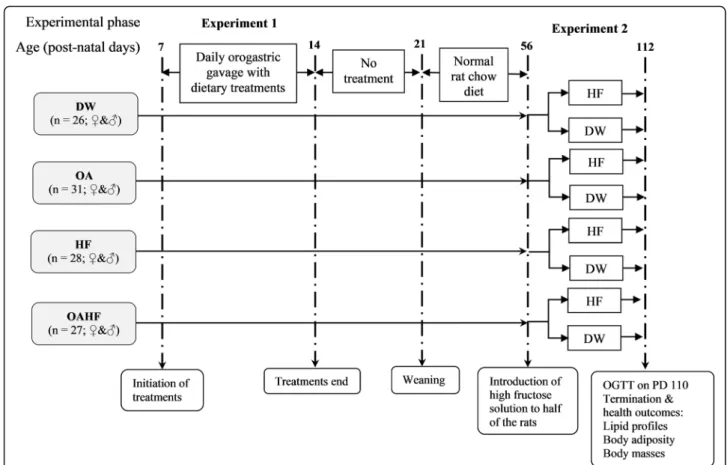
One hundred and twelve male and female pups were used in this interventional study which was conducted in two experimental phases. The timelines and group al-locations for the rats are summarised in Fig. 1. The dams with their pups were received on PD3. All rats were acclimatised for 4 days from PD4 up to the com-mencement of experimental treatment on PD7. During the first experimental phase (PD7-PD14), the first nutri-tional intervention was introduced in order to induce neonatal programming. The pups were weighed daily to monitor growth performance and in order to adjust treatments so as ensure the desired dosage per body mass. The pups were then randomly assigned to one of four dietary treatment groups:
i). Control group (DW)–received distilled water with dimethyl sulphoxide (DMSO) solution (0.5%v/v). The DMSO was used as a vehicle control to dissolve the OA for the other groups (n= 26; 13 males and 13 females).
ii). Oleanolic acid (OA) group–received oleanolic acid (60 mg/kg body mass) reconstituted in DMSO (0.5%v/v) (n= 31; 15 males and 16 females). iii).High fructose solution (HF) group–received 20%
fructose solution (w/v) reconstituted in DMSO (0.5%v/v) (n= 28; 13 males and 15 females). iv).Oleanolic acid and high fructose solution (OAHF)
in DMSO (0.5% v/v) (n= 27; 14 males and 13 females).
All treatments in the first experimental phase (Fig.
1) were administered once a day (between 09:00–
10:00), for seven consecutive days (PD7 to PD13), at a volume of 10 mL/kg body mass via orogastric gav-age using an orogastric tube attached to a 1 mL syr-inge. After treatments in the first experimental phase, pups were allowed to continue nursing with their dams from PD14 to PD20 until they were weaned on PD21. The dams were returned to stock and the pups were housed individually in acrylic cages where they ad libitum access to SRC and plain drinking tap water until PD55.
In the second experimental phase (PD56 up to PD112), the rats in each of the four experimental treatment groups received ad libitum access to SRC, however, half the num-ber of male and female rats (n= 56) in each group re-ceived either plain drinking water whilst the other half received 20% (w/v) fructose solution as drinking fluid.
Fresh fructose solution and water were provided every 2 days. Fructose was given in the second phase, as a sec-ondary dietary insult in order to induce health outcomes associated with MetS [62,63].
In the current study, rats were subjected to either a single or double hit according to the single or double hit hypothesis [64]. The single hit was either an early neo-natal administration of fructose (20% w/v) or the provision of fructose in drinking water (20% w/v) later in adult life. The double hit was characterised by two dietary interventions; a fructose hit in early-life (first-hit) which would predispose the rats to the onset of meta-bolic derangements followed by another dietary fructose intervention in later life (the second-hit).
Oral glucose tolerance test
At the end of the 16 week study period (PD110), an oral glucose tolerance test (OGTT) was performed after an overnight fast. Briefly, fasting blood glucose was deter-mined and then each rat was administered with a dose of 2 g/kg body mass of sterile 50% (w/v) D-glucose Fig. 1Schematic flow diagram showing the experimental groups, stages of development, sequence and timing of interventions and
measurements for the experimental study.DW= gavaged daily with 10 mL/kg body mass of distilled water with 0.5% (v/v) dimethyl sulphoxide in the neonatal phase (n= 26;♀&♂);OA= gavaged daily with 10 mL/kg body mass of oleanolic acid (60 mg/kg) in the neonatal phase (n= 31;
solution (Sigma, Johannesburg, South Africa) via orogas-tric gavage [65]. Thereafter blood glucose concentrations were measured atT= 15, 30, 60, 120 and 180 min using a calibrated glucometer (Contour Plus® glucometer, Bayer (Pty) Ltd., Johannesburg, South Africa). The blood samples were collected following a sterile pin prick of the distal tail vein. The incremental changes in blood glucose after administration of the glucose load were expressed as an area under the curve (AUC) from the time when the fasting blood was drawn (T= 0) until 180 min post-load blood sampling [66].
Terminal procedures
After the OGTT, the rats were placed back onto their re-spective dietary treatments for a further 48 h. Thereafter, the animals were fasted overnight and their fasting blood glucose levels measured using the calibrated glucometer (Contour Plus® glucometer, Bayer (Pty) Ltd., Johannes-burg, South Africa). The rats were then euthanased by an overdose of intra-peritoneally injected sodium pento-barbital (200 mg/kg body mass; Eutha-naze®, Bayer Cor-poration, Johannesburg, South Africa).
Blood and adipose tissue sample collection
Following euthanasia of the rats, blood was collected via cardiac puncture using 21G needles and 10 ml syringes and transferred into heparinised blood collection tubes (BD Vacutainer® Systems, Meylan Cedex, France). The blood samples were centrifuged for 15 min at 5000×g at 20 °C (Sorvall RT® 6000B centrifuge, Rockville, USA). The collected plasma samples were stored at −20 °C for the analysis of insulin and cholesterol.
The abdomen was opened via a midline incision and the visceral (and epipidymal in males) fat pads were dis-sected out and weighed. The left tibia was disdis-sected from the hindlimb of each rat, de-fleshed, dried in an oven (50 °C for 5 days) and the length measured. The length of the tibia, which is less prone to acute variation compared to body mass, was used to calculate the rela-tive masses of visceral and epididymal fat.
Determination of circulating cholesterol and triglyceride concentrations
The plasma concentrations of triglycerides were mea-sured using a calibrated triglyceride metre (Accutrend®, Roche, Mannheim, Germany) in accordance with the manufacturer’s instructions. Plasma cholesterol was measured using a calibrated automatic biochemical analyzer (IDEXX VetTest™ Clinical Chemistry Analyser, IDEXX Laboratories Inc., Westbrook, ME, USA) as per the manufacturer’s instructions.
Measurement of plasma insulin and calculation of the homeostatic model assessment of insulin resistance (HOMA-IR)
Plasma concentrations of insulin were measured using an enzyme-linked immunosorbent assay (ELISA) kit for rats (Elabscience ®, Rat INS ELISA kit, Wuhan, China) following the manufacturer’s instructions. Insulin resist-ance was calculated by means of the homeostatic model assessment index (HOMA-IR) using the relationship be-tween the fasting blood glucose and insulin levels, ac-cording to the following formula:
HOMA-IR = Insulin (μU/mL) × Blood glucose (mM)/ 22.5 [67]. Plasma levels of insulin were converted from ng/g toμU/mL for the calculation of HOMA-IR.
Statistical analyses
Data were expressed as mean ± standard deviation and analysed using GraphPad Prism for Windows Version 7.0 (GraphPad Software Inc., San Diego, USA). The baseline fasting glucose levels were subtracted from all other glu-cose values for each animal and the total area under the curve (AUC) for the OGTT was calculated by the trapez-oidal method to assess glucose tolerance [65]. A two-way repeated measures analysis of variance (ANOVA), with Bonferroni post-hoc test, was used to analyse terminal body mass with day as a within-subjects factor and treat-ment as a between-subjects factor. A one-way ANOVA with Bonferroni post-hoc test was used to compare the means for all parameters measured from different treat-ment groups. The level of significance acceptable was
P≤0.05.
Results
The effect of neonatal oral administration of oleanolic acid on weaning and terminal body masses of fructose-fed male and female rats
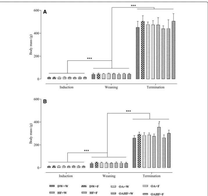
The induction body masses in male rats were similar across all experimental treatment groups (P> 0.05; Fig.2a). There was a significant increase in body masses of male rats in all experimental treatment groups from induction to weaning and from weaning up to termin-ation (P< 0.05; Fig.2a). There were no significant differ-ences in terminal body masses from all experimental treatment groups (P> 0.05; Fig.2a).
Fig. 2The effect of neonatal oral administration of oleanolic acid on weaning and terminal body masses of male (a) and female (b) rats fed a high fructose diet. All data presented as mean ± standard deviation.***Significant increase in body mass from induction to weaning and from weaning to termination (P< 0.005).#Significant increase in terminal body mass for female rats receiving a double hit of fructose (HF + F) compared to all of the other treatment groups (P< 0.005).DW + W= gavaged daily with 10 mL/kg body mass of distilled water with DMSO (0.5% v/v) in the neonatal period followed by ad libitum access to plain drinking water post-weaning and throughout adulthood (male = 7; female = 7);
DW + F= gavaged daily with 10 mL/kg body mass of distilled water with DMSO (0.5%v/v) in the neonatal period followed by ad libitum access to 20% (w/v) fructose solution as drinking fluid in adulthood (male = 6; female = 6);OA + W= gavaged daily with 60 mg/kg body mass oleanolic acid in the neonatal period followed by ad libitum access to plain drinking water post-weaning and throughout adulthood (male = 8; female = 8);
body mass recorded in the rats that received the double hit of fructose (HF + F) (P< 0.05; Fig. 2b). Neonatal ad-ministration of OA therefore prevented the increase in terminal body mass due to a double hit with fructose in female rats.
The effect of neonatal oral administration of oleanolic acid on glycaemic control in fructose-fed male and female rats
Oral glucose tolerance test (OGTT)
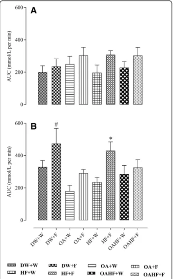
There was no significant difference in the total area under the curve (AUC) of oral glucose tolerance test (OGTT) for male rats across all experimental treatment groups (P> 0.05; Fig.3a).
In female rats, fructose consumption either late in adulthood (DW + F) or as a double hit early in the neo-natal period and late in adulthood (HF + F) resulted in up to 37% increase in the total AUC compared with other experimental treatment groups (P <0.05; Fig. 3b). Neonatal administration of OA prevented the increase
in AUC induced by the late single hit of fructose (OA + F) and a double hit (OAHF+F) fructose effects (P< 0.05; Fig. 3b). There were no significant differences in the total AUC in rats that received neonatal OA and the control group that did not receive fructose (DW + W) (P> 0.05).
Fasting blood glucose, insulin and homeostatic model assessment of insulin resistance (HOMA-IR)
In male rats, fructose consumption either late in adult-hood (DW + F) or a double hit (HF + F) resulted in an increase in fasting levels of glucose and the HOMA-IR compared to other treatment groups (P< 0.05; Table 1). However, there were no significant differences in fasting levels of insulin in male rats across all treatment groups (P> 0.05; Table1). There were no significant differences in the insulin levels in rats that received neonatal OA and the control group that did not receive fructose (DW + W) (P> 0.05). Oral administration of OA in the neonatal period prevented the increase in fasting glucose and HOMA-IR observed as a result of the double hit with fructose (OA + F vs OAHF+F)P> 0.001; Table1).
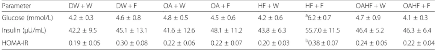
In female rats, a double hit of fructose (early in the neo-natal period and late in adulthood: HF + F) resulted in an increase in fasting levels of glucose and HOMA-IR com-pared with other experimental treatment groups (P< 0.05; Table2). Neonatal oral OA administration with the double hit fructose (OAHF+F) prevented the effects on glucose levels and HOMA-IR observed in the rats that had the double hit of fructose without any other intervention (HF + F) (P< 0.05; Table 2). There were no significant differ-ences in the fasting levels of insulin across all treatment groups in female rats (P> 0.05; Table2).
All data presented as mean ± standard deviation.abc Signi-ficant increase in glucose and HOMA-IR (P< 0.05) for groups receiving fructose late (DW + F) and a double hit (neonatally and in adulthood, HF + F) compared with other treatment groups.DW + W= gavaged daily with 10 mL/kg body mass of distilled water with DMSO (0.5% v/v) in the neonatal period followed by ad libitum access to plain
drinking water post-weaning and throughout adulthood (male = 7; female = 7);DW + F= gavaged daily with 10 mL/ kg body mass of distilled water with DMSO (0.5%v/v) in the neonatal period followed ad libitum access to 20% (w/v) fructose solution as drinking fluid in adulthood (male = 6; female = 6); OA + W= gavaged daily with 60 mg/kg body mass oleanolic acid in the neonatal period followed by ad libitum access to plain drinking water post-weaning and throughout adulthood (male = 8; female = 8); OA + F= gavaged with 60 mg/kg body mass oleanolic acid in the neonatal period followed by ad libitum access to 20% (w/v) fructose as drinking fluid in adulthood (male = 7; female =
8);HF + W= gavaged daily with 10 mL/kg body mass 20%
(w/v) fructose solution in the neonatal period followed by ad libitum access to plain drinking water post-weaning and throughout adulthood (male = 6; female = 7); HF + F= gavaged daily with 10 mL/kg body mass 20% (w/v) fructose solution in the neonatal period followed by ad libitum
Table 1Effect of neonatal oral administration of oleanolic acid and or fructose on fasting blood glucose and plasma insulin concentration and HOMA-IR index in male rats fed a high fructose diet
Parameter DW + W DW + F OA + W OA + F HF + W HF + F OAHF + W OAHF + F
Glucose (mmol/L) 4.9 ± 0.7 5.3 ± 0.3 4.5 ± 0.5 4.7 ± 0.6 4.5 ± 0.4 a6.3 ± 0.7 4.8 ± 0.5 4.5 ± 1.1
Insulin (μU/mL) 45.3 ± 10.5 53.2 ± 10.2 44.6 ± 9.4 51.4 ± 8.7 39.8 ± 5.2 56.9 ± 7.9 41.9 ± 10.2 46.5 ± 16.5
HOMA-IR 0.24 ± 0.06 b0.33 ± 0.06 0.23 ± 0.05 0.27 ± 0.07 0.18 ± 0.03 c0.35 ± 0.12 0.22 ± 0.05 0.22 ± 0.07
All data presented as mean ± standard deviation abc
Significant increase in glucose and abc HOMA-IR (P< 0.05) for groups receiving fructose late (DW + F) and a double hit (neonatally and in adulthood, HF + F) compared with other treatment groups.DW + W= gavaged daily with 10 mL/kg body mass of distilled water with DMSO (0.5% v/v) in the neonatal period followed by ad libitum access to plain drinking water post-weaning and throughout adulthood (male = 7; female = 7);DW + F= gavaged daily with 10 mL/kg body mass of distilled water with DMSO (0.5% v/v) in the neonatal period followed ad libitum access to 20% (w/v) fructose solution as drinking fluid in adulthood (male = 6; female = 6);OA + W= gavaged daily with 60 mg/kg body mass oleanolic acid in the neonatal period followed by ad libitum access to plain drinking water post-weaning and throughout adulthood (male = 8; female = 8);OA + F= gavaged with 60 mg/kg body mass oleanolic acid in the neonatal period followed by ad libitum access to 20% (w/v) fructose as drinking fluid in adulthood (male = 7; female = 8);HF + W= gavaged daily with 10 mL/kg body mass 20% (w/v) fructose solution in the neonatal period followed by ad libitum access to plain drinking water post-weaning and throughout adulthood (male = 6; female = 7);HF + F= gavaged daily with 10 mL/kg body mass 20% (w/v) fructose solution in the neonatal period followed by ad libitum access to 20% (w/v) fructose as drinking fluid in adulthood (male = 7; female = 8);OAHF + W= gavaged daily with 10 mL/kg body mass of a combination of oleanolic acid (60 mg/kg) and 20% (w/v) fructose solution in the neonatal period followed by ad libitum access to plain drinking water post-weaning and throughout adulthood (male = 7; female = 6);OAHF + F= gavaged daily with 10 mL/kg body mass of a combination of oleanolic acid (60 mg/kg) and 20% (w/v) fructose solution in the neonatal period followed by ad libitum access to 20% (w/v) fructose solution as drinking fluid in adulthood (male = 7; female = 7)
Table 2Effect of neonatal oral administration of oleanolic acid and fructose on fasting blood glucose and plasma insulin concentration and HOMA-IR index in female rats fed a high fructose diet
Parameter DW + W DW + F OA + W OA + F HF + W HF + F OAHF + W OAHF + F
Glucose (mmol/L) 4.2 ± 0.3 4.6 ± 0.8 4.8 ± 0.5 4.5 ± 0.6 4.2 ± 0.6 a6.2 ± 0.7 4.7 ± 0.9 4.1 ± 0.3
Insulin (μU/mL) 42.2 ± 9.5 45.1 ± 13.1 41.6 ± 12.6 48.1 ± 11.2 43.8 ± 6.3 55.7.0 ± 11.5 46.4 ± 5.2 46.3 ± 6.4
HOMA-IR 0.19 ± 0.05 0.30 ± 0.08 0.22 ± 0.06 0.22 ± 0.07 0.20 ± 0.03 b0.38 ± 0.07 0.24 ± 0.05 0.22 ± 0.04
All data presented as mean ± standard deviation ab
Significant increase in glucose and HOMA-IR (P< 0.05) for groups receiving a double hit (neonatally and in adulthood, HF + F) compared with other treatment groups.DW + W= gavaged daily with 10 mL/kg body mass of distilled water with DMSO (0.5% v/v) in the neonatal period followed by ad libitum access to plain drinking water post-weaning and throughout adulthood (male = 7; female = 7);DW + F= gavaged daily with 10 mL/kg body mass of distilled water with DMSO (0.5% v/v) in the neonatal period followed ad libitum access to 20% (w/v) fructose solution as drinking fluid in adulthood (male = 6; female = 6);OA + W= gavaged daily with 60 mg/kg body mass oleanolic acid in the neonatal period followed by ad libitum access to plain drinking water post-weaning and
access to 20% (w/v) fructose as drinking fluid in adulthood (male = 7; female = 8); OAHF + W= gavaged daily with 10 mL/kg body mass of a combination of oleanolic acid (60 mg/kg) and 20% (w/v) fructose solution in the neonatal period followed by ad libitum access to plain drinking water post-weaning and throughout adulthood (male = 7; female = 6);OAHF + F= gavaged daily with 10 mL/kg body mass of a combination of oleanolic acid (60 mg/kg) and 20% (w/ v) fructose solution in the neonatal period followed by ad libitum access to 20% (w/v) fructose solution as drinking fluid in adulthood (male = 7; female = 7).
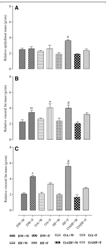
The effect of neonatal oral administration of oleanolic acid on visceral and epididymal fat masses in fructose-fed rats
Male rats that received fructose either late in adulthood, with or without neonatal OA (DW + F and OA + F) or as a double hit (neonatally and late in adulthood, HF + F) had up to 40% increase in the mass of the relative
epididymal fat pads (P< 0.05; Fig.4a). A double hit with fructose (early in the neonatal period and late in adult-hood, HF + F) caused up to 40% increase in relative vis-ceral fat mass (adjusted to relative tibial length) compared to the other experimental treatment groups (P< 0.05; Fig. 4b). Rats which received neonatal oral ad-ministration of OA and the double hit of fructose (OAHF+F) had significantly lower measures of adiposity than the rats which had the double hit of fructose with-out any other treatment intervention (HF + F) (P< 0.05; Fig. 4b), but there was no significant difference when compared to the rats that had the late hit with fructose (OA + F) (P> 0.05; Fig.4b).
In female rats, fructose consumption either late in adulthood (DW + F) or as a double hit (early neonatally and late in adulthood, HF + F) resulted in up to 26% and 65% respectively increase in relative visceral fat masses compared to the other experimental groups (P< 0.05; Fig.4c). Neonatal oral OA administration prevented the late single hit (OA + F group) and the double hit (OAHF +F group) (P< 0.05; Fig. 4c) fructose effects on visceral fat mass. No significant differences were observed be-tween the group that received neonatal OA and the con-trol group which did not receive fructose (DW + W) later in adulthood (P> 0.05; Fig.4c).
The effect of neonatal oral administration of oleanolic acid on the concentration of circulating cholesterol and triglycerides in fructose-fed male and female rats
There were no significant differences in fasting trigly-ceride and cholesterol levels of the male rats across all experimental dietary treatments (P> 0.05; Table3).
The levels of circulating triglycerides in female rats that received only a late fructose hit (DW + F) and those that received the double hit with fructose (in the neo-natal period and in adulthood, HF + F) significantly in-creased by up to 65% than rats from the other experimental treatment groups (P< 0.05; Table4). There were no significant differences in fasting cholesterol
levels of the female rats across all experimental dietary treatments (P> 0.05; Table 4). Oral administration of OA in the neonatal period prevented the increase in the levels of triglycerides observed as a result of either a late single hit or a double hit with fructose (OA + F vs DW + F; OAHF+F vs HF + F respectively;P< 0.05; Table4).
All data presented as mean ± standard deviation.
ab
Significant increase in TG levels in rats that received a late fructose hit (DW + F) and those that received fruc-tose neonatally and as adults (HF + F) compared rats from other experimental treatment groups (P< 0.05).
DW + W= gavaged daily with 10 mL/kg body mass of
distilled water with DMSO (0.5% v/v) in the neonatal period followed by ad libitum access to plain drinking water post-weaning and throughout adulthood (male = 7; female = 7);DW + F= gavaged daily with 10 mL/kg body mass of distilled water with DMSO (0.5% v/v) in the neonatal period followed ad libitum access to 20% (w/v) fructose solution as drinking fluid in adulthood (male = 6; female = 6); OA + W= gavaged daily with 60 mg/kg body mass oleanolic acid in the neonatal period followed by ad libitum access to plain drinking water post-weaning and throughout adulthood (male = 8; fe-male = 8); OA + F= gavaged with 60 mg/kg body mass oleanolic acid in the neonatal period followed by ad libi-tum access to 20% (w/v) fructose as drinking fluid in adulthood (male = 7; female = 8);HF + W= gavaged daily with 10 mL/kg body mass 20% (w/v) fructose solution in the neonatal period followed by ad libitum access to plain drinking water post-weaning and throughout adult-hood (male = 6; female = 7); HF + F= gavaged daily with 10 ml/kg body mass 20% (w/v) fructose solution in the neonatal period followed by ad libitum access to 20% (w/v) fructose as drinking fluid in adulthood (male = 7; female = 8); OAHF + W= gavaged daily with 10 mL/kg body mass of a combination of oleanolic acid (60 mg/kg) and 20% (w/v) fructose solution in the neonatal period followed by ad libitum access to plain drinking water post-weaning and throughout adulthood (male = 7;
Table 3Effect of neonatal oral administration of oleanolic acid and fructose on circulating triglycerides and cholesterol in male rats fed a high fructose diet
Parameter DW + W DW + F OA + W OA + F HF + W HF + F OAHF + W OAHF + F
TG (mmol/L) 1.5 ± 0.2 2.2 ± 0.7 1.5 ± 0.3 1.9 ± 0.4 1.5 ± 0.1 2.1 ± 0.5 1.7 ± 0.7 2.1 ± 0.7
CHOL (mmol/L) 3.7 ± 0.7 3.9 ± 0.4 3.7 ± 0.7 4.1 ± 0.5 3.2 ± 0.7 3.4 ± 0.4 3.8 ± 0.6 3.2 ± 0.9
Table 4Effect of neonatal oral administration of oleanolic acid and fructose on circulating triglycerides and cholesterol in female rats fed a high fructose diet
Parameter DW + W DW + F OA + W OA + F HF + W HF + F OAHF + W OAHF + F
TG (mmol/L) 1.5 ± 0.3 a2.8 ± 0.6 1.5 ± 0.2 1.7 ± 0.3 1.6 ± 0.2 b2.7 ± 0.4 1.4 ± 0.3 1.6 ± 0.4
female = 6); OAHF + F= gavaged daily with 10 mL/kg body mass of a combination of oleanolic acid (60 mg/kg) and 20% (w/v) fructose solution in the neonatal period followed by ad libitum access to 20% (w/v) fructose solu-tion as drinking fluid in adulthood (male = 7; female = 7). TG = triglycerides; CHOL = cholesterol.
Discussion
In this study, we sought to investigate the potential pro-tective effect of neonatal oral administration of oleanolic acid (OA) against the subsequent development of health outcomes associated with metabolic dysfunction induced by the consumption of fructose in adult male and female rats. Fructose caused metabolic derangements in male and female rats, however, this was affected by the timing of the fructose intervention(s). We also noted sex differ-ences in responses to the high fructose diets. We have shown that a double hit of fructose wherein it was ad-ministered in the neonatal period followed by a second-ary dietary insult in adulthood resulted in the development of several negative health outcomes associ-ated with metabolic dysfunction, namely the significant increases in terminal body mass (females only), visceral fat mass (males and females), serum triglycerides (fe-males only), epididymal fat ((fe-males only), fasting plasma glucose (males and females), impaired glucose metabol-ism (females only), β-cell dysfunction and insulin resist-ance (males and females). The single late fructose hit in adulthood resulted in impaired glucose metabolism, in-creased visceral fat pad masses and levels of triglycerides in female but not male rats. Oral administration of OA in the neonatal period successfully attenuated the mani-festation of fructose-induced metabolic disorders in both male and female rats.
The effect of neonatal oral administration of oleanolic acid on terminal body masses in fructose-fed male and female rats
Our findings showed an increase in the body masses of male and female rats across all the experimental treat-ment groups from induction to weaning and from wean-ing to termination. There were no differences in terminal body masses of male rats across all treatment groups. However, the female rats that received a double fructose hit had increased terminal body masses, which was not observed in male rats, and was prevented the administration of OA in the neonatal period. Previous studies have shown that consumption of fructose in-creases energy intake and body weight [68,69]. In older rats, the excess calories are stored as fat while in young animals the energy can be channelled into the metabolic costs of growth. This may explain the difference in our findings regarding the effects of fructose on body mass when compared with previous research findings. The sex
differences in the response to fructose dietary treatments are discussed later. Previous findings in human and ani-mal studies which showed that neonatal consumption of fructose (early single hit) resulted in abnormal body weight gain [70, 71] which is contrary to our findings. The differences could be explained in part by the fruc-tose dosage that was administered and the duration over which the fructose was administered. While increased body mass alone may not represent obesity, the co-existence of other obesogenic variables such as accu-mulation of visceral fat, increased levels of triglycerides (TG) as we have shown, provides confirmation of the obese status especially in the female rats [71].
The oral administration of OA in the neonatal period attenuated the increase in terminal body masses caused by fructose in female rats. This finding suggests that neonatal interventions with OA prevent excessive body mass gain later in adulthood, as such OA may therefore be used in the neonatal period against excessive body mass gain in adulthood. Oleanolic acid (OA) has been reported to decrease food intake [72], and lower plasma glucose levels in murine models of diabetes mellitus even after the dosage has been discontinued [50]. A limi-tation of the current study is that feed and fluid intake were not assessed. Nevertheless, the findings from previ-ous studies support the role of the effect of the OA on food intake and suggest that the effect of OA may be long-lasting. The significantly higher body weights re-corded in female rats that received a double fructose hit and the visceral fat mass in rats that did not receive OA and those that received OA with high-fructose groups suggest that fructose consumption may have played a role in the effect of OA on plasma glucose and triglycer-ide levels. The observed effect of OA on body mass could also be due to the role of neonatal programming. Further investigations using murine models are however required to elucidate the specific molecular mecha-nism(s) through which OA prevents the body mass in-creases induced by fructose consumption.
The effect of neonatal oral administration of oleanolic acid on visceral and epididymal fat masses in fructose-fed adult male and female rats
accumulation in both male and female rats. Due to acute fluctuations in body mass as a result of factors such as the hydration status of animals and gut fill, body mass can be a less reliable index of growth or organ masses, than the linear growth of the long bones which is less likely to fluctuate acutely and thus would have less of a variability on the calculation of relative organ masses [73]. Consequently, the use of tibial length was used as a ‘stable’indicator of linear growth in the calculation of the relative fat masses. While previous studies [71,74] using a late hit with fructose in adults also show similar findings to ours regarding the late hit, it is notable that the impact of the double hit on obesity was much greater than the single hit. The total adipose tissue accumulation plays an important role in the development of metabolic disorders such as insulin resistance (IR), T2DM and CVDs [75]. However, there are some fat depots that are more associ-ated with the development of metabolic risk factors than others [76]. A human study has shown that in men, the excessive accretion of omental and mesenteric adipose tis-sue, both part of visceral adipose tissue (VAT), is strongly associated with the development of CVDs and T2DM [77]. In rodents however, gonadal VAT surrounding testis (epididymal fat) and ovaries is regarded as one of the lar-gest depots that contribute to the development of meta-bolic disorders in rodents on a high-energy cafeteria diet [76]. It is therefore possible that the difference in the epi-didymal fat and the rest of the visceral fat responses ob-served in male rats that received a double hit of fructose could be due to epididymal fat being more vulnerable to accumulate than the other visceral fat depots in response to high-fructose diets. Based on recent neonatal program-ming studies [78,79] and results from this study, we hy-pothesise that neonatal fructose consumption may have programed adipose tissue development during the critical period which resulted in the observed visceral (male and female rats) and epididymal fat (male rats) accumulation later in adulthood. The neonatal programming of visceral fat accumulation and lipid metabolism induced by fruc-tose may have caused the development and manifestation of other MetS-associated outcomes in adulthood upon the introduction of a secondary dietary fructose insult as we have already shown with glucose metabolism in this study. The significantly heavier visceral fat pad masses ob-served in this study in fructose-fed female, but not in male rats, corroborate previous findings which also re-ported an increase in visceral fat deposition and a corresponding increase in body mass following exces-sive consumption of fructose in adulthood [80]. Des-pite the lack of differences in terminal body masses of the male rats that received either a double hit of fructose or a single late hit, as observed in female rats, there were differences in visceral (both males and females) and epididymal (males only) fat mass.
Findings from previous studies also suggest that ex-cessive consumption of fructose increases the levels of plasma triglycerides which may accelerate the ac-cretion of body fat and causing visceral obesity and body mass gain [70, 81], a trend that we have ob-served in the current study, especially in female rats that received a double hit or a single late fructose hit. There are a number of studies in the literature in which fructose solutions, provided along with a chow diet, increase energy intake and body weight [71] glu-cose intolerance, as well as plasma insulin and trigly-ceride levels [68].
Although male and female rats receiving a single late and a double fructose hit exhibited increased visceral fat mass relative to tibial length compared with all other treatment groups, this increase was higher and pro-nounced in female (an average of approximately 26% single hit and an average of approximately 65% double hit) than in male (an average of approximately 40% double hit) rats. This finding suggests that female off-spring were vulnerable to greater fructose-induced adi-posity than their male counterparts, a finding that is in line with the greater body mass increase in fructose-fed females (double hit), also previously reported by Bayol S, Simbi B, Bertrand J and Stickland N [82]. Androgens and female hormones, which control several metabolic pathways [83] and are involved in the pathogenesis of metabolic disorders [84] may have contributed to the observed sexual dimorphic differences in obesity ob-served in this study. Androgens promote cellular glucose uptake and energy utilisation in skeletal muscles and liver [85] thus reducing the tendency to accumulate total body fat in men.
The effect of neonatal oral administration of oleanolic acid on glucose tolerance in fructose-fed adult male and female rats
Both a late single hit and a double hit of 20%w/vfructose solution as drinking fluid caused glucose intolerance (total area under the curve of the OGTT) in the female, but not in male rats. The late single fructose hit resulted in the de-velopment of IR orβ-cell dysfunction (HOMA-IR) in male but not in female rats. However, the double fructose hit caused IR or β-cell dysfunction and hyperglycaemia in both male and female rats. None of the treatments in-duced an increase in the levels of insulin in both male and female rats.
The oral glucose tolerance test (OGTT) is an import-ant clinical tool for the characterisation of metabolic phenoype [88]. It is used to diagnose impaired glucose tolerance and as a standardised test of carbohydrate metabolism by assessing the ability to dispose of an oral glucose load over time [88, 89]. Sustained hypergly-caemia (> 120 min) in plasma glucose constitutes impaired glucose tolerance and can be used together with fasting hyperglycaemia to diagnose patients with T2DM [66].
Our findings on glucose tolerance suggest that a double fructose hit adversely affected glucose metabol-ism by impairing the ability to tolerate a glucose load in female rats and caused IR and possibly pancreaticβ-cell dysfunction in both male and female rats. Insulin resist-ance reported in this study for male and female rats that received a double hit of fructose also corroborates earlier findings by Huynh M, Luiken JJ, Coumans W and Bell RC [80] who also demonstrated the manifestation of IR following a double hit of fructose neonatally and in adulthood. However, results on the levels of insulin in fructose-fed rats from the current study are at variance with the same study by Huynh M, Luiken JJ, Coumans W and Bell RC [80] who reported the development of hyperinsulinaemia in rats that received 10% fructose in the neonatal period and 65% fructose diet in adulthood. The observed variance in levels of insulin can be ex-plained in part by the differences in the quantity and method of fructose administration in adulthood. In the current study, adult rats received 20%w/vfructose solu-tion as a secondary dietary insult, in contrast to Huynh M, Luiken JJ, Coumans W and Bell RC [80] who gave 65%w/w in the feed which possibly provided more calo-ries resulting in hyperinsulinaemia which we did not ob-serve. Although hyperglycaemia alone does not indicate whether there is an insufficiency of insulin secretion [89], it is possible that its development observed in this study following a double hit of fructose in both male and female rats coupled with impaired glucose tolerance and IR suggests that the fructose that was administered in the neonatal period may have been effective in
programming the neonatal rats for the development of hyperglycaemia, impaired glucose tolerance and IR or pancreaticβ-cell dysfunction later in adulthood after ex-posure to a secondary dietary insult.
The development of hyperglycaemia, glucose intoler-ance and IR that was induced by either a single late and double fructose hit was prevented by the neonatal oral administration of OA in male and female rats. These findings expand on previous research that reported the anti-diabetic effects of plant-derived OA in adult rats [49]. Diabetes is characterised by persistent hypergly-caemia, poor glycaemic control and IR [67]. Reactive oxygen species (ROS) have been suspected to play a role in the progression from normal glucose metabolism to impaired glucose tolerance and development of insulin resistance [35]. By upregulating the gene expression of anti-oxidant enzymes, glutathione peroxidase and super-oxide dismutase, OA promotes anti-oxidant cellular de-fences which play a role in its glucose-lowering effects [35]. Previous studies have demonstrated the anti-oxidant and anti-glycative role of OA, as such it is possible that the protective role of OA against the devel-opment of dysregulation of glucose metabolism observed in this study following a late single hit or a double fruc-tose hit, could be due to the ability of OA to scavenge for free radicals and enhancing anti-oxidant cellular de-fence system [58]. The observed improvement of glucose tolerance and IR by OA also suggests that OA may be promoting insulin signal transduction mechanisms and inhibiting poor handling of glucose caused by oxidative stress and IR [35].
OA also protects against oxidative stress-induced IR [35]. Mechanistic studies have shown that triterpenoid compounds such as OA act as hypoglycaemic and anti-obesity agents mainly through (i) reducing the in-testinal absorption of glucose; (ii) decreasing endogen-ous glucose production such as hepatic gluconeogenesis; (iii) increasing insulin sensitivity; (iv) improving lipid metabolism; and (v) promoting body weight loss [90]. In addition to these promising beneficial effects, it is be-lieved that OA and associated triterpenoid protect against diabetes-related comorbidities due to their anti-atherogenic, anti-inflammatory, and anti-oxidant properties [58].
The effect of neonatal administration of oleanolic acid on circulating triglycerides and cholesterol in fructose-fed adult male and female rats
and elevated circulating levels of TGs in rats [71]. The observed hypertriglyceridaemia in female rats following administration of a double fructose hit could be ex-plained in part by the fructose-mediated neonatal pro-gramming of lipogenic genes [91, 92] and the increased vulnerability to develop hypertriglyceridaemia in fructose-fed female rats. Although male rats that re-ceived a late single fructose hit and a double hit of fruc-tose had increased visceral fat accretion, they neither had increased terminal body weights nor TG levels. Re-gional differences in adipose tissue distribution which is affected by sex hormones may have resulted in the ob-served sexual dimorphic differences in circulating TG levels. Fructose is predominantly metabolised in the liver and due to its high lipogenic potential, its excessive con-sumption is likely to increase the metabolic burden on the liver resulting in the development of hepatic steato-sis through de novo lipogenesteato-sis (DNL) [71]. The ob-served increase in the levels of TGs in fructose-fed female animals could also be due to the upregulation of hepatic DNL and secretion of excess hepatic lipids which contributes to the plasma pool of TGs [93].
Fructose consumption as either a late single hit or a double hit did not cause an increase in the levels of chol-esterol levels across all treatment groups in both male and female rats. The manifestation of fructose-induced meta-bolic disorders in adulthood also include hypercholesterol-aemia, especially when the high fructose diet is fortified by fats [94]. In the absence of supplemented fat or very high fructose diets, it is uncommon to induce hypercholesterol-aemia in rats, a finding which is corroborated by our find-ings [95,96].
Conclusion
We have shown that fructose administration had adverse effects on several health outcomes associated with meta-bolic dysfunction. The timing (late or double hit) of the administration of fructose had an effect on the develop-ment of metabolic dysfunction. We also observed sex-specific differences in the metabolic response to dietary fructose, showing the significance of considering sex effects in metabolic studies. We conclude that neo-natal interventional treatment with oleanolic acid during the critical window of developmental plasticity protected against the development of fructose diet-induced health outcomes associated with metabolic dysfunction in male and female Sprague Dawley rats. Following studies in higher animals coupled with molecular analyses to deter-mine its mechanisms, OA should be considered as a nat-ural strategic prophylactic intervention with a lot of potential in the fight against the scourge of metabolic disorders that are impacting significantly on the health systems globally.
Abbreviations
AESC:Animal Ethics Screening Committee; ANOVA: Analysis of variance; AUC: Area under the curve for the oral glucose tolerance test; CAS: Central animal services; CHOL: Cholesterol; CVDs: Cardiovascular diseases; DMSO: Dimethyl sulphoxide; DNL: De novo lipogenesis; DW: Distilled water; ELISA: Enzyme linked immunosorbent assay; HF: High fructose solution (20%
w/v); HFCS: High fructose corn syrup; HOMA-IR: Homeostatic model of insulin resistance; IR: Insulin resistance; MetS: Metabolic syndrome; ncRNA: Non-coding ribonucleic acid; OA: Oleanolic acid; OAHF: Oleanolic acid and high fructose solution (20%w/v) treatment; OGTT: Oral glucose tolerance test; PD: Post-natal day; PGC-1β: Peroxisome proliferator-activated receptor-γ coactivator-1β; ROS: Reactive oxygen species; SRC: Standard rat chow; T2DM: Type 2 diabetes mellitus; TGs: Triglycerides; VAT: Visceral adipose tissue; WHO: World Health Organisation
Acknowledgements
The authors wish to acknowledge the Faculty of Health Sciences Research Committee of the University of the Witwatersrand, Johannesburg, the National Research Foundation of South Africa and, the Department of Higher Education and Training for funding the research. The University of
Johannesburg, Department of Human Anatomy and Physiology is also appreciated for financial support. We also thank the entire staff of the Central Animal Services Unit, University of the Witwatersrand, Johannesburg, for their assistance with the breeding and animal husbandry throughout the experimental period.
Funding
Funding was received from the National Research Foundation grants to KH and EM.
Availability of data and materials
All datasets on which conclusions of the manuscript rely are presented in the main paper are in possession of the corresponding author, Trevor Nyakudya and may be made available in recommended repositories.
Authors’contributions
TN conceptualised the idea of the project collected and analysed the data and wrote the manuscript. EM and KE analysed the data and wrote the manuscript. All authors read and approved the final manuscript.
Ethics approval and consent to participate
The study was done in accordance with the International Standards of Care and Use of Animals in Research and was approved by the Animal Ethics Screening Committee (AESC) of the University of the Witwatersrand, Johannesburg, South Africa (Ethics clearance number: 2014/47/D).
Consent for publication
All authors consented to the publication of the data.
Competing interests
The authors declare that they have no competing interests.
Publisher’s Note
Springer Nature remains neutral with regard to jurisdictional claims in published maps and institutional affiliations.
Author details
1School of Physiology, Faculty of Health Sciences, University of the
Witwatersrand, 7 York Road, Parktown, Johannesburg 2193, South Africa. 2
Department of Biochemistry, Faculty of Natural Sciences & Agriculture, North West University, Mafikeng, Mmabatho 2735, South Africa.3Department of Human Anatomy and Physiology, Faculty of Health Sciences, University of Johannesburg, Doornfontein, Johannesburg 2028, South Africa.
Received: 19 April 2018 Accepted: 22 October 2018
References
2. O'Neill S, O'Driscoll L. Metabolic syndrome: a closer look at the growing epidemic and its associated pathologies. Obes Rev. 2015;16:1–12. 3. Grundy SM. Metabolic syndrome update. Trends Cardiovasc Med. 2016;
26:364–73.
4. Furukawa S, Fujita T, Shimabukuro M, Iwaki M, Yamada Y, Nakajima Y, Nakayama O, Makishima M, Matsuda M, Shimomura I. Increased oxidative stress in obesity and its impact on metabolic syndrome. J Clin Invest. 2017; 114:1752–61.
5. Rodríguez-Ortiz D, Reyes-Pérez A, León P, Sánchez H, Mosti M, Aguilar-Salinas CA, Velázquez-Fernández D, Herrera MF. Assessment of two different diagnostic guidelines criteria (National Cholesterol Education Adult Treatment Panel III [ATP III] and international diabetes federation [IDF]) for the evaluation of metabolic syndrome remission in a longitudinal cohort of patients undergoing roux-en-Y gastric bypass. Surgery. 2016;159:1121–8.
6. Spalding A, Kernan J, Lockette W. The metabolic syndrome: a modern plague spread by modern technology. J Clin Hypertens. 2009;11:755–60. 7. WHO. Obesity and overweight factsheet from the WHO. Health. 2017. 8. RahelićD. Of IDF diabetes atlas--call for immediate action. Lijec Vjesn. 2016;138:57. 9. Armitage JA, Taylor PD, Poston L. Experimental models of developmental
programming: consequences of exposure to an energy rich diet during development. J Physiol. 2005;565:3–8.
10. Bakker K, Apelqvist J, Lipsky B, Van Netten J, Schaper N. The 2015 IWGDF guidance documents on prevention and management of foot problems in diabetes: development of an evidence-based global consensus. Diabetes Metab Res Rev. 2016;32:2–6.
11. WHO. Global Report on diabetes: World Health Organization; 2016. 12. Rippe JM. Fructose, high fructose corn syrup, sucrose and health:
Springer; 2014.
13. Steinmann B, Santer R. Disorders of fructose metabolism. In: Inborn metabolic diseases: Springer; 2016. p. 161–8.
14. Tappy L. Health implications of fructose consumption in humans. Sweeteners. 2017:1–26.
15. Alfaradhi M, Ozanne S. Developmental programming in response to maternal overnutrition. Front Genet. 2011;2:27.
16. Zheng J, Feng Q, Zhang Q, Wang T, Xiao X. Early life fructose exposure and its implications for long-term Cardiometabolic health in offspring. Nutrients. 2016;8:685.
17. Kitsiou-Tzeli S, Tzetis M. Maternal epigenetics and fetal and neonatal growth. Curr Opin Endocrinol Diabetes Obes. 2017;24:43–6. 18. Godfrey KM, Gluckman PD, Hanson MA. Developmental origins of
metabolic disease: life course and intergenerational perspectives. Trends Endocrinol Metab. 2010;21:199–205.
19. Couvreur O, Ferezou J, Gripois D, Serougne C, Crépin D, Aubourg A, Gertler A, Vacher C-M, Taouis M. Unexpected long-term protection of adult offspring born to high-fat fed dams against obesity induced by a sucrose-rich diet. PLoS One. 2011;6:e18043.
20. Gluckman PD, Buklijas T, Hanson MA. The developmental origins of health and disease (DOHaD) concept: past, present, and future. In: The epigenome and developmental origins of health and disease Academic, London; 2015. p. 1–13.
21. Smith CJ, Ryckman KK. Epigenetic and developmental influences on the risk of obesity, diabetes, and metabolic syndrome. Diabetes Metab Syndr Obes. 2015;8:295.
22. Desai N, Roman A, Rochelson B, Gupta M, Xue X, Chatterjee PK, Tam HT, Metz CN. Maternal metformin treatment decreases fetal inflammation in a rat model of obesity and metabolic syndrome. Am J Obstet Gynecol. 2013; 209:136 e131–136. e139.
23. Low FM, Gluckman PD, Godfrey KM. 3 early-life development and epigenetic mechanisms: mediators of metabolic programming and obesity risk. In: Nutrigenomics and Proteomics in Health and Disease: Towards a Systems-level Understanding of Gene-diet Interactions, vol. 42; 2017.
24. Seki Y, Suzuki M, Guo X, Glenn AS, Vuguin PM, Fiallo A, Du Q, Ko Y-A, Yu Y, Susztak K. In Utero Exposure to a High Fat Diet Programs Hepatic Hypermethylation and Gene Dysregulation and Development of Metabolic Syndrome in Male Mice. Endocrinology. 2017;158(9):2860–72.
25. Ozanne SE, Hales CN. Early programming of glucose–insulin metabolism. Trends Endocrinol Metab. 2002;13:368–73.
26. de Moura EG, Cottini MCF. Neonatal programming of body weight regulation and energetic metabolism. Biosci Rep. 2005;25:251–69.
27. Rando OJ, Simmons RA. I’m eating for two: parental dietary effects on offspring metabolism. Cell. 2015;161:93–105.
28. Heindel JJ, Balbus J, Birnbaum L, Brune-Drisse MN, Grandjean P, Gray K, Landrigan PJ, Sly PD, Suk W, Cory Slechta D. Developmental origins of health and disease: integrating environmental influences. Endocrinology. 2015;156:3416–21.
29. Ghezzi AC, Cambri LT, Ribeiro C, Botezelli JD, Mello MA. Impact of early fructose intake on metabolic profile and aerobic capacity of rats. Lipids Health Dis. 2011;10:1.
30. Clayton ZE, Vickers MH, Bernal A, Yap C, Sloboda DM. Early life exposure to fructose alters maternal, fetal and neonatal hepatic gene expression and leads to sex-dependent changes in lipid metabolism in rat offspring. PLoS One. 2015;10:e0141962.
31. Rodríguez L, Panadero MI, Rodrigo S, Roglans N, Otero P, Álvarez-Millán JJ, Laguna JC, Bocos C. Liquid fructose in pregnancy exacerbates fructose-induced dyslipidemia in adult female offspring. J Nutr Biochem. 2016;32:115–22.
32. Roche HM, Phillips C, Gibney MJ. The metabolic syndrome: the crossroads of diet and genetics. Proc Nutr Soc. 2005;64:371–7.
33. Kuete V. African medicinal spices and vegetables and their potential in the Management of Metabolic Syndrome. In: Medicinal Spices and Vegetables from Africa: Therapeutic Potential against Metabolic, Inflammatory, Infectious and Systemic Diseases; 2017. p. 315.
34. Nathan DM, Buse JB, Davidson MB, Ferrannini E, Holman RR, Sherwin R, Zinman B. Medical management of hyperglycemia in type 2 diabetes: a consensus algorithm for the initiation and adjustment of therapy. Diabetes Care. 2009;32:193–203.
35. Wang X, Li YL, Wu H, Liu JZ, Hu JX, Liao N, Peng J, Cao PP, Liang X, Hai CX. Antidiabetic effect of oleanolic acid: a promising use of a traditional pharmacological agent. Phytother Res. 2011;25:1031–40.
36. Hamine S, Gerth-Guyette E, Faulx D, Green BB, Ginsburg AS. Impact of mHealth chronic disease management on treatment adherence and patient outcomes: a systematic review. J Med Internet Res. 2015;17. 37. Gurib-Fakim A. Medicinal plants: traditions of yesterday and drugs of
tomorrow. Mol Asp Med. 2006;27:1–93.
38. Sánchez-Quesada C, López-Biedma A, Gaforio JJ. Oleanolic acid, a compound present in grapes and olives, protects against genotoxicity in human mammary epithelial cells. Molecules. 2015;20:13670–88. 39. Lin C, Wen X, Sun H. Oleanolic acid derivatives for pharmaceutical use: a
patent review. Expert Opin Ther Pat. 2016;26:643–55.
40. Yoshikawa M, Matsuda H. Antidiabetogenic activity of oleanolic acid glycosides from medicinal foodstuffs. Biofactors. 2000;13:231–7. 41. Rodriguez-Rodriguez R. Oleanolic acid and related triterpenoids from
olives on vascular function: molecular mechanisms and therapeutic perspectives. Curr Med Chem. 2015;22:1414–25.
42. Liu J. Oleanolic acid and ursolic acid: research perspectives. J Ethnopharmacol. 2005;100:92–4.
43. Jäger S, Trojan H, Kopp T, Laszczyk MN, Scheffler A. Pentacyclic triterpene distribution in various plants–rich sources for a new group of multi-potent plant extracts. Molecules. 2009;14:2016–31.
44. Castellano JM, Guinda A, Delgado T, Rada M, Cayuela JA. Biochemical basis of the antidiabetic activity of oleanolic acid and related pentacyclic triterpenes. Diabetes. 2013;62:1791–9.
45. Liu J, Liu Y, Klaassen CD. Protective effect of oleanolic acid against chemical-induced acute necrotic liver injury in mice. Zhongguo Yao Li Xue Bao. 1995;16:97–102.
46. Nyakudya TT, Mukwevho E, Nkomozepi P, Swanepoel E, Erlwanger KH. Early postnatal administration of oleanolic acid attenuates the development of non-alcoholic fatty liver disease in fructose fed adult female rats. FASEB J. 2017;31:887–2.
47. Singh G, Singh S, Bani S, Gupta B, Banerjee S. Anti-inflammatory activity of oleanolic acid in rats and mice. J Pharm Pharmacol. 1992;44:456–8.
48. Nkeh-Chungag BN, Oyedeji OO, Oyedeji AO, Ndebia EJ. Anti-inflammatory and membrane-stabilizing properties of two semisynthetic derivatives of oleanolic acid. Inflammation. 2015;38:61–9.
50. Zeng X-Y, Wang Y-P, Cantley J, Iseli TJ, Molero JC, Hegarty BD, Kraegen EW, Ye Y, Ye J-M. Oleanolic acid reduces hyperglycemia beyond treatment period with Akt/FoxO1-induced suppression of hepatic gluconeogenesis in type-2 diabetic mice. PLoS One. 2012;7:e42115.
51. Yin M-C, Chan K-C. Nonenzymatic antioxidative and antiglycative effects of oleanolic acid and ursolic acid. J Agric Food Chem. 2007;55:7177–81. 52. Tsai SJ, Yin MC. Antioxidative and anti-inflammatory protection of oleanolic
acid and ursolic acid in pc12 cells. J Food Sci. 2008;73:H174–8. 53. S-j T, M-c Y. Anti-oxidative, anti-glycative and anti-apoptotic effects of
oleanolic acid in brain of mice treated by D-galactose. Eur J Pharmacol. 2012;689:81–8.
54. S-m T, M-c Y. Antioxidative and antiinflammatory activities of asiatic acid, glycyrrhizic acid, and oleanolic acid in human bronchial epithelial cells. J Agric Food Chem. 2015;63:3196–204.
55. Xi M, Hai C, Tang H, Chen M, Fang K, Liang X. Antioxidant and antiglycation properties of total saponins extracted from traditional Chinese medicine used to treat diabetes mellitus. Phytother Res. 2008;22:228–37.
56. Wang Z-h, Hsu C-C, Huang C-N, Yin M-C. Anti-glycative effects of oleanolic acid and ursolic acid in kidney of diabetic mice. Eur J Pharmacol. 2010;628:255–60. 57. Teodoro T, Zhang L, Alexander T, Yue J, Vranic M, Volchuk A. Oleanolic
acid enhances insulin secretion in pancreaticβ-cells. FEBS Lett. 2008; 582:1375–80.
58. Wang X, Liu R, Zhang W, Zhang X, Liao N, Wang Z, Li W, Qin X, Hai C. Oleanolic acid improves hepatic insulin resistance via antioxidant, hypolipidemic and anti-inflammatory effects. Mol Cell Endocrinol. 2013;376:70–80.
59. de Melo CL, Queiroz MGR, Fonseca SG, Bizerra AM, Lemos TL, Melo TS, Santos FA, Rao VS. Oleanolic acid, a natural triterpenoid improves blood glucose tolerance in normal mice and ameliorates visceral obesity in mice fed a high-fat diet. Chem Biol Interact. 2010;185:59–65.
60. Chen S, Wen X, Zhang W, Wang C, Liu J, Liu C. Hypolipidemic effect of oleanolic acid is mediated by the miR-98-5p/PGC-1βaxis in high-fat diet–induced hyperlipidemic mice. FASEB J. 2017;31:1085–96. 61. Liu C, Li Y, Zuo G, Xu W, Gao H, Yang Y, Yamahara J, Wang J, Li Y.
Oleanolic acid diminishes liquid fructose-induced fatty liver in rats: role of modulation of hepatic sterol regulatory element-binding protein-1c-mediated expression of genes responsible for de novo fatty acid synthesis. Evid Based Complement Alternat Med. 2013;2013. 62. Miller A, Adeli K. Dietary fructose and the metabolic syndrome. Curr Opin
Gastroenterol. 2008;24:204–9.
63. Mock K, Lateef S, Benedito VA, Tou JC. High-fructose corn syrup-55 consumption alters hepatic lipid metabolism and promotes triglyceride accumulation. J Nutr Biochem. 2017;39:32-39.
64. Tamashiro KL, Moran TH. Perinatal environment and its influences on metabolic programming of offspring. Physiol Behav. 2010;100:560–6. 65. Huhn E, Fischer T, Göbl C, Bernasconi MT, Kreft M, Kunze M, Schoetzau
A, Dölzlmüller E, Eppel W, Husslein P. Screening of gestational diabetes mellitus in early pregnancy by oral glucose tolerance test and glycosylated fibronectin: study protocol for an international, prospective, multicentre cohort trial. BMJ Open. 2016;6:e012115.
66. Kwon S, Kim YJ, Kim MK. Effect of fructose or sucrose feeding with different levels on oral glucose tolerance test in normal and type 2 diabetic rats. Nutr Res Pract. 2008;2:252–8.
67. Valle M, St-Pierre P, Pilon G, Anhê FF, Varin T, Marette A. Effects of various natural sweeteners on insulin resistance, inflammation and liver steatosis in a rat model of diet-induced obesity. FASEB J. 2016;30:lb650.
68. Kanarek RB, Orthen-Gambill N. Differential effects of sucrose, fructose and glucose on carbohydrate-induced obesity in rats. J Nutr. 1982;112:1546–54. 69. Elliott SS, Keim NL, Stern JS, Teff K, Havel PJ. Fructose, weight gain, and the
insulin resistance syndrome. Am J Clin Nutr. 2002;76:911–22. 70. Stanhope KL, Havel PJ. Fructose consumption: considerations for future
research on its effects on adipose distribution, lipid metabolism, and insulin sensitivity in humans. J Nutr. 2009;139:1236S–41S.
71. Bocarsly ME, Powell ES, Avena NM, Hoebel BG. High-fructose corn syrup causes characteristics of obesity in rats: increased body weight, body fat and triglyceride levels. Pharmacol Biochem Behav. 2010;97:101–6. 72. Luvuno M, Mbongwa HP, Khathi A. The effects of syzygium
aromaticum-derived triterpenes on gastrointestinal ghrelin expression in streptozotocin-induced diabetic rats. Afr J Trad Complement Altern Med. 2016:13. 73. Yin F, Spurgeon HA, Rakusan K, Weisfeldt ML, Lakatta EG. Use of tibial
length to quantify cardiac hypertrophy: application in the aging rat. Am J Phys Heart Circ Phys. 1982;243:H941–7.
74. Jürgens H, Haass W, Castaneda TR, Schürmann A, Koebnick C, Dombrowski F, Otto B, Nawrocki AR, Scherer PE, Spranger J. Consuming fructose-sweetened beverages increases body adiposity in mice. Obes Res. 2005;13: 1146–56.
75. Wajchenberg BL. Subcutaneous and visceral adipose tissue: their relation to the metabolic syndrome. Endocr Rev. 2000;21:697–738.
76. Bjørndal B, Burri L, Staalesen V, Skorve J, Berge RK. Different adipose depots: their role in the development of metabolic syndrome and mitochondrial response to hypolipidemic agents. J Obes. 2011;2011.
77. Hoffstedt J, Arner P, Hellers G, Lönnqvist F. Variation in adrenergic regulation of lipolysis between omental and subcutaneous adipocytes from obese and non-obese men. J Lipid Res. 1997;38:795–804.
78. Simpson J, Smith AD, Fraser A, Sattar N, Lindsay RS, Ring SM, Tilling K, Davey Smith G, Lawlor DA, Nelson SM. Programming of adiposity in childhood and adolescence: associations with birth weight and cord blood adipokines. J Clin Endocrinol Metab. 2016;102:499–506.
79. Beckford R, Yu J, Das S, Hettich R, Wilson J, Voy B. Mechanisms for programming reduced adiposity through maternal dietary fish oil. FASEB J. 2017;31:–141.145.
80. Huynh M, Luiken JJ, Coumans W, Bell RC. Dietary fructose during the suckling period increases body weight and fatty acid uptake into skeletal muscle in adult rats. Obesity. 2008;16:1755–62.
81. Stanhope KL, Havel PJ. Fructose consumption: potential mechanisms for its effects to increase visceral adiposity and induce dyslipidemia and insulin resistance. Curr Opin Lipidol. 2008;19:16.
82. Bayol S, Simbi B, Bertrand J, Stickland N. Offspring from mothers fed a‘junk food’diet in pregnancy and lactation exhibit exacerbated adiposity that is more pronounced in females. J Physiol. 2008;586:3219–30.
83. Kalyani RR, Dobs AS. Androgen deficiency, diabetes, and the metabolic syndrome in men. Curr Opin Endocrinol Diabetes Obes. 2007;14:226–34. 84. Muller M, Grobbee DE, Den Tonkelaar I, Lamberts SW, Van Der Schouw YT.
Endogenous sex hormones and metabolic syndrome in aging men. J Clin Endocrinol Metab. 2005;90:2618–23.
85. Navarro G, Allard C, Xu W, Mauvais-Jarvis F. The role of androgens in metabolism, obesity, and diabetes in males and females. Obesity. 2015; 23:713–9.
86. Ghanayem BI, Bai R, Kissling GE, Travlos G, Hoffler U. Diet-induced obesity in male mice is associated with reduced fertility and potentiation of acrylamide-induced reproductive toxicity. Biol Reprod. 2010;82:96–104. 87. Cardoso A, Alves M, Mathur P, Oliveira P, Cavaco J, Rato L. Obesogens and
male fertility. Obes Rev. 2017;18:109–25.
88. Ayala JE, Samuel VT, Morton GJ, Obici S, Croniger CM, Shulman GI, Wasserman DH, McGuinness OP. Standard operating procedures for describing and performing metabolic tests of glucose homeostasis in mice. Dis Model Mech. 2010;3:525–34.
89. Ernsberger P, Koletsky RJ. The glucose tolerance test as a laboratory tool with clinical implications. In: Glucose Tolerance: InTech; 2012. 90. Camer D, Yu Y, Szabo A, Huang XF. The molecular mechanisms
underpinning the therapeutic properties of oleanolic acid, its isomer and derivatives for type 2 diabetes and associated complications. Mol Nutr Food Res. 2014;58:1750–9.
91. Herman MA, Samuel VT. The sweet path to metabolic demise: fructose and lipid synthesis. Trends Endocrinol Metab. 2016;27:719–30.
92. Saad AF, Dickerson J, Kechichian TB, Yin H, Gamble P, Salazar A, Patrikeev I, Motamedi M, Saade GR, Costantine MM. High-fructose diet in pregnancy leads to fetal programming of hypertension, insulin resistance, and obesity in adult offspring. Am J Obstet Gynecol. 2016;215:378 e371–378. e376. 93. Basaranoglu M, Basaranoglu G, Bugianesi E. Carbohydrate intake and
nonalcoholic fatty liver disease: fructose as a weapon of mass destruction. Hepatobiliary Surg Nutr. 2014;4:109–16.
94. Okoduwa SIR, Umar IA, James DB, Inuwa HM. Appropriate insulin level in selecting fortified diet-fed, Streptozotocin-treated rat model of type 2 diabetes for anti-diabetic studies. PLoS One. 2017;12:e0170971.
95. Huang BW, Chiang MT, Yao HT, Chiang W. The effect of fat and high-fructose diets on glucose tolerance and plasma lipid and leptin levels in rats. Diabetes Obes Metab. 2004;6:120–6.