CELLULAR & MOLECULAR BIOLOGY LETTERS http://www.cmbl.org.pl
Received: 15 June 2011 Volume 17 (2012) pp 206-216 Final form accepted: 20 January 2012 DOI: 10.2478/s11658-012-0003-x Published online: 28 January 2012 © 2012 by the University of Wrocław, Poland
* Author for correspondence. e-mail: x-wang1@northwestern.edu, tel.: 312-503-0294, fax: 312-503-0296
Abbreviations used: BSP – bisulfite sequencing PCR; FUT4 – fucosyltransferase IV; MSP – methylation-specific PCR; SCC – squamous carcinoma cell; 5-aza-dC – 5-aza-2-deoxycytidine
Short communication
DIFFERENTIAL FUCOSYLTRANSFERASE IV EXPRESSION IN SQUAMOUS CARCINOMA CELLS IS REGULATED
BY PROMOTER METHYLATION
HONGYAN LI1, 2, SHAOMING TONG1, 2, JIWEI LIU3, LI HAN1, XUESONG YANG1, HESHENG HOU2, QIU YAN1 and XIAO-QI WANG4, 1Department of Biochemistry and Molecular Biology, Dalian Medical
University, Dalian, Liaoning, People’s Republic of China, 2College of Life Science, Liaoning Normal University, Dalian, Liaoning, People’s Republic of China, 3Department of Oncology, 1st Affiliated Hospital of Dalian Medical
University, Dalian, Liaoning, People’s Republic of China, 4Departments of Dermatology and Pediatrics, Northwestern University’s Feinberg School
of Medicine, Chicago, Illinois, USA
Abstract: Enhanced fucosyltransferase IV (FUT4) expression correlates with
increased tumor malignancy in many carcinomas. However, little is known about the regulation of FUT4 expression, and whether FUT4 expression is influenced by the methylation status of the FUT4 promoter is unclear. In this study, we demonstrated that FUT4 expression is negatively correlated with the methylation degree of a CpG island in the FUT4 promoter, suggesting that the methylation status of FUT4 promoter regulates the expression of FUT4. The results indicate that manipulating the methylation status of the FUT4 promoter to regulate FUT4 expression may be a novel approach in the treatment of malignant tumors.
Key words: Fucosyltransferase, FUT4 promoter, Methylation, A431 cells,
INTRODUCTION
Fucosyltransferases (FUTs) are the key enzymes that regulate the synthesis of fucosylated oligosaccharides, such as Lewis a (Lea), sialyl Lewis a (sLea), Lewis b (Leb), Lewis X (LeX), sialyl Lewis X (sLeX) and Lewis Y (LeY), which are bound to cell surface glycoproteins or glycolipids [1]. FUTs are classified as -1, 2 FUTs (FUT1 and FUT2), -1, 3/4 FUTs (FUT3, FUT4, FUT5, FUT6, FUT7 and FUT9) and -1, 6 FUT (FUT8) based on their acceptor specificity [2-3]. FUTs play important roles in cancer biology, as increased fucosylation levels of glycoproteins and glycolipids have been reported in a number of cancers [4-6].
FUT4, a key enzyme for the synthesis of 1, 3-fucosylated oligosaccharides such as LeY, catalyzes the transfer of fucose (Fuc) residues from GDP-Fuc to [Fuc1→2Gal1→4GlcNAc1→R] in -1, 3 linkage. Elevated FUT4
expression has been found to correlate with tumor progression [7-9]. Although the impact of FUT4 expression on cell proliferation has been studied [10-11], little information is available on how FUT4 expression is regulated. Accumulating evidence suggests that tumor progression is related to the abnormal hypomethylation of growth regulating genes [12]. These genes, such
as synuclein-γ(SNCG),and paired-box gene 2 (PAX2), are methylated in normal
tissues, but become hypomethylated and highly expressed in cancers [12-13]. Moreover, glycogene expression regulation is involved in promoter methylation status. Incomplete synthesis of carbohydrate determinants, such as sLea and sLeX, occurs through the silencing of glycogenes by DNA methylation in early stage cancers [14]. The promoter region of the human B4GALNT2 gene is heavily hypermethylated in many gastrointestinal cancer cell lines and leads to decreased expression of Sd(a) carbohydrate [15]. However, it is unclear whether
FUT4 expression is directly influenced by the methylation status of its promoter. Using two SCC cell lines, A431 and SCC12 cells, we found that a lower methylation level of FUT4 promoter correlated with a higher FUT4 expression in A431 cells compared with that in SCC12 cells. Furthermore, we demonstrated that treatment with 5-aza-2-deoxycytidine (5-aza-dC), a common methyltransferase inhibitor, significantly decreased the methylation of FUT4 promoter and increased FUT4 expression in SCC12 cells, but did not significantly affect the already low methylation level of FUT4 promoter and high level of FUT4
MATERIALS AND METHODS
Cell culture
The human A431 cell line was obtained from the American Tissue Culture Collection (Manassas, VA), and the SCC12 cell line was provided by Dr. Rheinwald, Harvard University (Boston, MA). Both A431 and SCC12 cells were maintained in DMEM/F12 (1:1, Invitrogen) supplemented with 10% fetal bovine serum (FBS), 100 U/ml penicillin, and 50 μg/ml streptomycin at 37°C under 5% CO2 in humidified air.
Semi-quantitative RT-PCR and real-time PCR
Total RNA was extracted and purified from cells using the TRIzol® reagent (Invitrogen) in the presence of DNase. The first-strand cDNAs were synthesized by RT reaction using oligo-dT primer with M-MLV reverse transcriptase (Takara). Semi-quantitative PCR and real-time PCR were carried out following the conditions listed in the Supplemental Table in Supplementary material at http://dx.doi.org/10.2478/s11658-012-0003-x.
5-aza-dC treatment
After plating for 24 h, cells were treated with either 1-10 M 5-aza-dC (Sigma) or vehicle (2.5-25 l acetic acid/l medium) for 72 h. 5-aza-dC was replenished every 24 h.
Methylation-specific PCR (MSP) and bisulfite sequencing PCR (BSP) assays
Genomic DNA was extracted and purified from cells pretreated with or without 5-aza-dC as indicated elsewhere, then incubated with bisulfite using EZ DNA Methylation-Gold Kit per manufacturer’s instruction (ZYMO REASCHER). The bisulfite-modified DNAs were used as templates for MSP and BSP. MSP and BSP reactions were conducted following the conditions listed in the Supplemental Table. The MSP products were analyzed using 1.5% agarose gel electrophoresis. The BSP products were cloned into pMD18-T vector (Takara) and sequenced.
Western blot and lectin blot analysis
(ECL) detection system (Amersham) was used to determine the expression of
FUT4 and fucosylated proteins.
Flow cytometry assay
Suspended single cells (1x10⁶) prepared as indicated were permeabilized in 0.1% Triton X-100-PBS (4°C, 10 min) before incubating with goat anti-FUT4 antibody (room temperature, 1 h). FITC-conjugated rabbit anti-goat IgG was used to label the FUT4-positive cells followed by FACScan flow cytometer detection.
Immunofluorescence staining
Cells plated on the coverslips were fixed in cold acetone (-20°C, 20 min). 3% BSA was used to block non-specific binding (37°C, 2 h). Cells on the coverslips were incubated with goat FUT4 antibody (1:100) and mouse anti-Golgi marker antibody (AE-6) (1:100, Santa Cruz, CA) overnight at 4°C. TRITC-conjugated rabbit anti-goat secondary antibody (1:50, Santa Cruz, CA) and FITC-conjugated donkey anti-mouse secondary antibody (1:50, Protein Tech Group, Inc) were used to detect the specific antibody binding. Images were captured using an Olympus BX51 microscope (Japan).
Statistical analysis
All data presented were obtained from at least three independent experiments and expressed as means ± standard deviation. Data were analyzed statistically by Student's t test, with p < 0.05 considered to be significant.
RESULTS
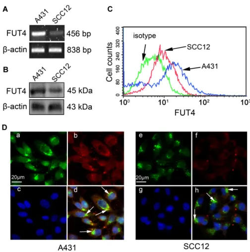
Expression of FUT4 in cells
By semi-quantitative RT-PCR (Fig. 1A), Western blot (Fig. 1B), flow cytometry assay (Fig. 1C) and immunofluorescence staining (Fig. 1D), we found that FUT4
was expressed in both cell lines, but its expression level was greatly enhanced in A431 cells compared to that in SCC12 cells. As shown in Fig. 1D, FUT4 predominantly co-localized with AE-6 in Golgi apparatus.
Methylation status of the CpG island in the FUT4 promoter
primers to amplify the unmethylated fragments yielded more PCR products from A431 cells than from SCC12 cells (Fig. 2B, bottom row). These results suggest that the CpG island in the FUT4 promoter was more methylated in SCC12 cells than in A431 cells. The methylation status of the selected CpG island was also analyzed by BSP assay as described. At least 25 randomly selected clones per cell line were sequenced and the results from 5 representative clones were presented. We found that the 30 CpG sites of the selected CpG island in the
FUT4 promoter were rarely methylated in A431 cells (Fig. 2C), but highly methylated in all of the SCC12 clones tested (Fig. 2D).
Fig. 2. Methylation status of a CpG islandin FUT4 promoter. A – Map of a CpG island in the
FUT4 gene promoter as predicted by MethPrimer software. CpG sites were presented with
vertical bars. The CpG island (-429 to -671 bp) analyzed is highlighted. B – The products of PCR amplified with MSP primers were identified on 1.5% agarose gel. Me: methylation; Un: unmethylation. C, D – The products of PCR amplified with BSP primers were cloned. Five representative sequenced clones from each cell line were presented. Each circle represents one CpG site in the CpG island. ●:methylated CpG site; ○: unmethylated CpG site.
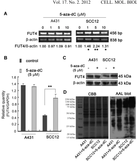
Fig. 4. FUT4 and fucosylated protein expression in 5-aza-dC-treated cells. By (A) semi-quantitative RT-PCR, (B) real-time PCR and (C) Western blot, FUT4 expression was examined after treatment of cells without or with 1-10 M 5-aza-dC. The expression level of -actin or GAPDH was used as an internal control. The band density of PCR products in agarose gel was analyzed using NIH ImageJ program. All data presented are an average of three independent PCR reactions. * p < 0.05; ** p < 0.01. By (D) AAL blot, fucosylated protein expression was detected (right panel), and Coomassie Brilliant Blue (CBB) staining of gels showed comparable amounts of proteins in each lane (left panel).
Effect of 5-aza-dC on the methylation status of the CpG islands in the FUT4
promoter
in the FUT4 promoter in SCC12 cells, but not in A431 cells (not shown). These results imply that 5-aza-dC preferentially demethylates the FUT4 promoter in cells with a hypermethylated FUT4 promoter.
Effect of 5-aza-dC on the expression of FUT4 and fucosylated proteins
By semi-quantitative RT-PCR (Fig. 4A), real-time PCR (Fig. 4B) and Western blot (Fig. 4C), we found that the expression of FUT4 was dramatically increased in SCC12 cells (p < 0.01) when cells were treated with 5 μM 5-aza-dC. In contrast, treatment with different doses of 5-aza-dC (1, 5, or 10 μM) did not further enhance the already high expression level of FUT4 in A431 cells, in which the FUT4 promoter is hypomethylated. These results suggest that FUT4
expression is regulated by its gene promoter methylation status. The effects of 5-aza-dC on the expression of fucosylated proteins in A431 and SCC12 cells were also detected by biotinylated AAL blot (Fig. 4D). The results show that the fucosylation of proteins was significantly increased in 5-aza-dC treated SCC12 cells.
DISCUSSION
In this study, we demonstrated that the methylation of the CpG island in the
FUT4 promoter regulates FUT4 expression. In comparison to the SCC12 cells, the degree of methylation of the CpG island in the FUT4 promoter is significantly lower, and the expression level of FUT4 is dramatically higher, in A431 cells. Treatment of the cells with 5-aza-dC preferentially decreases the methylation of the CpG island in the FUT4 promoter and increases the expression of FUT4 in SCC12 cells, and not in A431 cells. The expression of fucosylated proteins is also significantly elevated in SCC12 cells with 5-aza-dC treatment. Increased FUT4 expression is seen in carcinomas of the lung, stomach, melanoma, and acute myeloid leukemia [1, 7-9]. The mechanism of increased
FUT4 expression in cancers is still largely unknown. Hypomethylation in gene promoter regions is considered as one of the mechanisms for enhanced expression and activation of oncogenes or cancer-promoting genes during carcinogenesis [12]. For example, the expression of the SNCG gene is increased by hypomethylation in breast carcinoma, and elevated SNCG expression stimulates breast cancer proliferation and metastasis [12]. FUT3 overexpression in gastric cells also depends on hypomethylation of its promoter [17]. A similar observation is reported in FUT7 expression [18]. Although increases in the fucosylation level and FUT4 expression were observed in MDA-MB-231 cells treated with a methyltransferase inhibitor, zebularine [19], it is unclear whether
FUT4 expression level is directly correlated with the methylation status of the
adenocarcinoma cell lines [21]. Taken together, these studies indicate that the CpG islands in the FUT4 promoter may play an important role in regulating
FUT4 expression, and this may be dependent on the methylation status of the CpG sites. By comparing the expression level of FUT4 and the methylation status of a CpG island in the FUT4 promoter, we discovered that FUT4
expression level and the degree of methylation of the FUT4 promoter region in A431 and SCC12 cells are negatively correlated.
We have previously found that the proliferation of A431 cells is increased by
FUT4 overexpression, and reduced by knocking down FUT4 expression [10-11]. We have recently found that A431 cells have a relatively high proliferative ability, which correlates with a higher level of FUT4, while SCC12 cells had a relatively low proliferative capability, which correlates with a lower level of FUT4. Consistently, the proliferation of A431 and SCC12 cells is dramatically hindered by knocking down FUT4 expression and significantly increased by
FUT4 overexpression (not shown). This evidence suggested that FUT4
expression affects cancer proliferation. In addition, FUT4 is known to associate with other factors of malignancy such as metastasis [8]. Among FUT4 catalyzed fucosylated oligosaccharide antigens, LeY expression correlates with tumor proliferation, invasion and metastasis in carcinomas of breast, cervix, and ovary [4, 22-24]. Although a few studies have shown that sLeX and sLea act as selectin ligands that can mediate carcinoma metastasis [25-26], there is no direct evidence showing that the binding of LeY and selectins mediates a similar process. These studies indicate that regulation of FUT4 promoter methylation to alter the expression of FUT4 and the fucosylated tumor-associated antigens may be a potential approach to inhibit tumor malignancy.
In conclusion, different methylation levels of the FUT4 promoter play a critical role to regulate FUT4 expression, which in turn may affect the proliferative capabilities in squamous carcinoma cells. The degree of methylation in the
FUT4 promoter may serve as a promising biomarker for squamous cell carcinomas or a target for therapy.
Acknowledgements. This work was supported by National Natural Science
Foundation of China Research Grants No. 30672753 (Liu) and China 973 grant of 2012CB822100 (Yan). This work was also supported by Grant # 204139 from the American Cancer Society, Illinois Division, Inc, the Dermatology Foundation, and the Astellas Pharma US Research Endowment (Wang). We thank Dr. Robert Lavker and Dr. David L.K. Chen for their careful review of this manuscript.
REFERENCES
2. Wang, X., Gu, J., Ihara, H., Miyoshi, E., Honke, K. and Taniguchi, N. Core fucosylation regulates epidermal growth factor receptor-mediated intracellular signaling. J. Biol. Chem. 281 (2006) 2572-2577.
3. Javaud, C., Dupuy, F., Maftah, A., Julien, R. and Petit, J.M. The fucosyltransferase gene family: an amazing summary of the underlying mechanisms of gene evolution. Genetica 118 (2003) 157-170.
4. Madjd, Z., Parsons, T., Watson, N.F., Spendlove, I., Ellis, I. and Durrant, L.G. High expression of Lewis y/b antigens is associated with decreased survival in lymph node negative breast carcinomas. Breast Cancer Res. 7 (2005) R780-787.
5. Miyoshi, E., Moriwaki, K. and Nakagawa, T. Biological function of fucosylation in cancer biology. J. Biochem. 143 (2008) 725-729.
6. Fukushima, K., Satoh, T., Baba, S. and Yamashita, K. alpha1,2-Fucosylated and beta-N-acetylgalactosaminylated prostate-specific antigen as an efficient marker of prostatic cancer. Glycobiology 20 (2010) 452-460.
7. Petretti, T., Schulze, B., Schlag, P.M. and Kemmner, W. Altered mRNA expression of glycosyltransferases in human gastric carcinomas. Biochim.
Biophys. Acta 1428 (1999) 209-218.
8. Ciolczyk-Wierzbicka, D., Bodzioch, M., Gil, D., Zmudzinska, D., Dembinska-Kiec, A. and Laidler, P. Expression of fucosyltransferases contributes to melanoma invasive phenotype. Med. Chem. 3 (2007) 418-424. 9. Stirewalt, D.L., Meshinchi, S., Kopecky, K.J., Fan, W.,
Pogosova-Agadjanyan, E.L., Engel, J.H., Cronk, M.R., Dorcy, K.S., McQuary, A.R., Hockenbery, D., Wood, B., Heimfeld, S. and Radich, J.P. Identification of genes with abnormal expression changes in acute myeloid leukemia. Genes
Chromosomes Cancer 47 (2008) 8-20.
10. Zhang, Z., Sun, P., Liu, J., Fu, L., Yan, J., Liu, Y., Yu, L., Wang, X. and Yan, Q. Suppression of FUT1/FUT4 expression by siRNA inhibits tumor growth. Biochim. Biophys. Acta 1783 (2008) 287-296.
11. Yang, X., Zhang, Z., Jia, S., Liu, Y., Wang, X. and Yan, Q. Overexpression of fucosyltransferase IV in A431 cell line increases cell proliferation. Int. J.
Biochem. Cell Biol. 39 (2007) 1722-1730.
12.Gupta, A., Godwin, A.K., Vanderveer, L., Lu, A. and Liu, J. Hypomethylation of the synuclein gamma gene CpG island promotes its aberrant expression in breast carcinoma and ovarian carcinoma. Cancer Res. 63 (2003) 664-673.
13. Wu, H., Chen, Y., Liang, J., Shi, B., Wu, G., Zhang, Y., Wang, D., Li, R., Yi, X., Zhang, H., Sun, L. and Shang, Y. Hypomethylation-linked activation of PAX2 mediates tamoxifen-stimulated endometrial carcinogenesis.
Nature 438 (2005) 981-987.
14. Kannagi, R., Yin, J., Miyazaki, K. and Izawa, M. Current relevance of incomplete synthesis and neo-synthesis for cancer-associated alteration of carbohydrate determinants--Hakomori's concepts revisited. Biochim.
15. Kawamura, Y.I., Toyota, M., Kawashima, R., Hagiwara, T., Suzuki, H., Imai, K., Shinomura, Y., Tokino, T., Kannagi, R. and Dohi, T. DNA hypermethylation contributes to incomplete synthesis of carbohydrate determinants in gastrointestinal cancer. Gastroenterology 135 (2008) 142-151. 16. Gardiner-Garden, M. and Frommer, M. CpG islands in vertebrate genomes.
J. Mol. Biol. 196 (1987) 261-282.
17. Serpa, J., Mesquita, P., Mendes, N., Oliveira, C., Almeida, R., Santos-Silva, F., Reis, C.A., LePendu, J. and David, L. Expression of Lea in gastric cancer cell lines depends on FUT3 expression regulated by promoter methylation.
Cancer Lett. 242 (2006) 191-197.
18. Syrbe, U., Jennrich, S., Schottelius, A., Richter, A., Radbruch, A. and Hamann, A. Differential regulation of P-selectin ligand expression in naive versus memory CD4+ T cells: evidence for epigenetic regulation of involved glycosyltransferase genes. Blood 104 (2004) 3243-3248.
19. Moriwaki, K., Narisada, M., Imai, T., Shinzaki, S. and Miyoshi, E. The effect of epigenetic regulation of fucosylation on TRAIL-induced apoptosis.
Glycoconj. J. 27 (2010) 649-659.
20. Withers, D.A. and Hakomori, S.I. Human alpha (1,3)-fucosyltransferase IV (FUTIV) gene expression is regulated by elk-1 in the U937 cell line. J. Biol.
Chem. 275 (2000) 40588-40593.
21. Taniguchi, A., Suga, R. and Matsumoto, K. Expression and transcriptional regulation of the human alpha1, 3-fucosyltransferase 4 (FUT4) gene in myeloid and colon adenocarcinoma cell lines. Biochem. Biophys. Res.
Commun. 273 (2000) 370-376.
22. Moro-Rodriguez, E. and Alvarez-Fernandez, E. Losses of expression of the antigens A, Lea and Lex and over-expression of Ley in carcinomas and HG-SIL of the uterine cervix. Diagn. Pathol. 3 (2008) 38.
23. Escrevente, C., Machado, E., Brito, C., Reis, C.A., Stoeck, A., Runz, S., Marme, A., Altevogt, P. and Costa, J. Different expression levels of alpha3/4 fucosyltransferases and Lewis determinants in ovarian carcinoma tissues and cell lines. Int. J. Oncol. 29 (2006) 557-566.
24. Inufusa, H., Adachi, T., Kiyokawa, T., Nakatani, Y., Wakano, T., Nakamura, M., Okuno, K., Shiozaki, H., Yamamoto, S., Suzuki, M., Ando, O., Kurimoto, M., Miyake, M. and Yasutomi, M. Ley glycolipid-recognizing monoclonal antibody inhibits procoagulant activity and metastasis of human adenocarcinoma. Int. J. Oncol. 19 (2001) 941-946.
25. Thurin, M. and Kieber-Emmons, T. SA-Lea and tumor metastasis: the old prediction and recent findings. Genes Chromosomes Cancer 21 (2002) 111-116.
26. Wei, J., Cui, L., Liu, F., Fan, Y., Lang, R., Gu, F., Guo, X., Tang, P. and Fu, L. E-selectin and Sialyl Lewis X expression is associated with lymph node metastasis of invasive micropapillary carcinoma of the breast. Int. J. Surg.