Address for correspondence Dr. Jaspreet Kaur, (Ph.D)
Senior Research Associate, Department of Biochemistry, Shri Ram Murti Smarak Institute of Medical Sciences, Bareilly-243202,
Uttar Pradesh, India. Ph: +91-9458702003
Email: [email protected]
Original Article
Co-supplementation of oral antioxidant to PUVASol
therapy has no effect on oxidative stress index in
patients of unstable vitiligo: A hospital based pilot
study
Introduction
Vitiligo is a common and often heritable,
acquired pigmentation disorder in which
melanocytes in the skin, mucous membranes and
the retina are destroyed.
1Upto 8.8 percent of the
world population is believed to be afflicted with
it.
2,3It is one of the commonest dermatological
problems in India.
2,3It has a special significance
to patients here, because depigmentation is
obvious on dark skin.
4Although the precise etiology of vitiligo is not
known, it has become quite clear that in recent
times complex genetic, immunological, neural
Gaurav Bhardwaj, Jaspreet Kaur
Department of Biochemistry, Shri Ram MurtiSmarak Institute of Medical Sciences, Bareilly-243202, Uttar Pradesh, India.
Abstract
Background In recent years, interest has grown in studying the role of oxidative stress in vitiligo, so measurement of the combined activities of all antioxidants or the Total antioxidant status (TAS) is often used to estimate the overall antioxidant status. Likewise, Total oxidant status (TOS) is measured to determine a patient’s overall oxidation state. Furthermore, the oxidative stress index (OSI), which is calculated as the ratio of TOS to TAS, may be a more accurate index of oxidative stress in the body. This study was done to measure the effect of oral antioxidant supplementation therapy for 1 month on the oxidative stress index in patients of vitiligo.Methods In this pilot study 40 consecutive patients presenting in departmental vitiligo clinic were enrolled and randomly allocated into 2 groups of 20 patients each were prescribed oral prednisolone at a dosage of 1mg/kg for 2 consecutive days/week along with oral 8-methoxypsoralen on 3 alternate days in a week at a dose of 0.6mg/kg body weight. Patients in group B received the following antioxidants orally along with treatment of group A. End point of the study was 1 month of continuous therapy in the respective groups. OSI was calculated before initiating treatment by serum collection.
Results The mean OSI decreased significantly in both the study groups individually (P = 0.037 & 0.040 for Group A and B respectively). However inter-group comparison between the 2 study groups showed no statistically significant difference (P = 0.052).
Conclusion Addition of antioxidants to PUVASol in treatment of unstable vitiligo has no effect on oxidative stress index.
Key words
and self-destructive mechanisms interplay in the
pathogenesis of vitiligo.
5,6According to the auto
cytotoxic hypothesis, oxidative stress has been
suggested to be the initial pathogenic event in
melanocyte
degeneration
with
H
2O
2accumulation in the epidermis of patients with
active disease.
7In recent years, interest has grown in studying
the role played by oxidative stress in vitiligo by
investigating one or more of the antioxidant
markers,
including
Superoxide
Dismutase
(SOD), Catalase (CAT), Glutathione Peroxidase
(GPx), vitamin E, vitamin C.
8-10However, the
measurement of different antioxidant molecules
separately is impractical and has no clinical
significance because the effects of antioxidants
can be additive and measuring individual
antioxidants separately is time consuming and
labor intensive. Hence a measurement of the
combined activities of all antioxidants or the
Total Antioxidant Status (TAS) is often used to
estimate
the
overall antioxidant
status.
11Likewise, Total Oxidant Status (TOS) is
measured to determine a patient’s overall
oxidation state.
12Furthermore, the Oxidative
Stress index (OSI), which is calculated as the
ratio of TOS to TAS, may be a more accurate
index of oxidative stress in the body because it is
a comprehensive measurement of TAS and
TOS.
13Taken together, this study aimed to measure the
effect of oral antioxidant supplementation
therapy for 1 month on the Oxidative Stress
Index in patients of vitiligo.
Methods
Study population
This randomized open clinical trial was
registered in the Clinical Trial Registry-India
(CTRI/2014/11/005223) and conducted in the
Department of Dermatology and Department of
Biochemistry at Shri Ram Murti Smarak
Institute of Medical Sciences, Bareilly (India).
This study was approved by the Institutional
Ethics Committee. The study duration was from
April 2013 to April 2014. In this pilot study 40
consecutive patients presenting in departmental
vitiligo clinic were screened and enrolled based
on inclusion and exclusion criteria. Inclusion
criteria for the study were patients of unstable
vitiligo vulgaris; aged ≥18 years; literate; willing
for investigation, treatment, and regular
follow-up and had not been under any therapeutic
regimen for the previous two months and had
not received drugs containing any antioxidants.
Patients with hepatic or renal impairments,
photodermatoses, past or present history of any
malignancy or immunobullous disorder, any
chronic systemic disorder, patients who took
treatment irregularly, chronic alcoholics and/or
smokers and pregnant or lactating females were
excluded from the study. A washout period of
two months was given for topical and systemic
therapies, respectively, before including the
patients in study.
Written informed consent from all the subjects
was taken before recruitment in this study.
History, Examination, Vitiligo Disease Activity
Score (VIDA)
14, vitiligo European task force
(VETF) for disease activity
15, point counting
method of area estimation16 and other relevant
investigations were recorded in a specially
designed proforma.
after an interval of 2 hours preferably between
10am to 3pm. The sunlight exposure was for 5
minutes initially, and then exposure time was
increased by 5 minutes to a maximum of 30
minutes at every alternate. Topical calcineurin
inhibitor, tacrolimus 0.1% was applied topically
at night for lesion on the face and neck. Patients
in group B received the following antioxidants
orally along with treatment of group A: Vitamin
C 300mg, Vitamin E: 400 IU, Beta Carotene:
30mg, Zinc oxide: 40mg, Sodium selenate:
200µg, Cupric oxide: 2mg, Manganese sulphate:
5mg. End point of the study was 1 month of
continuous therapy in the respective groups. OSI
was calculated in serum samples before
initiating the treatment.
Sample collection
Five ml blood was drawn from median cubital
vein of the patients into plane tubes. To separate
the serum from the plasma sample, it was
centrifuged at 5000 × g for 5 min at room
temperature. All serum samples were stored at -
20°C until time of processing.
Evaluation of total oxidant status (TOS) in all
the study subjects
Oxidant present in the sample oxidizes the
ferrous ion-o-dianisidine complex to ferric ions.
The oxidation reaction is enhanced by glycerol
molecules, which are abundantly present in the
reaction medium. The ferric ions make a colored
complex with Xylenol orange in an acidic
medium. The colour intensity, which can be
measured spectrophotometrically, is related to
the total amount of oxidant molecule present in
the sample. The assay is calibrated with
hydrogen peroxide and results are expressed in
terms of micro molar hydrogen peroxide
equivalent per litre.
12Estimation of total antioxidant status (TAS)
This was measured with help of Quantichrom
TMAntioxidant Assay Kit, CAT# DTAC – 100,
Lot: BD06A17 from BioAssay Systems. 1180
East Ellsworth Road Ann Arbor, Michigan
48108· USA. (www.Bioassaysys.com)
Oxidative stress index (OSI)
It was calculated from a percent ratio of total
peroxide level to the TAS level.
13OSI=[TOS (μmol H2O2 equivalent /L) / TAS
(μmolTrolox equivalent /L) X100.
Clinical response to treatment was assessed by
VIDA score, VETF and point-counting method
of area estimation.
Statistical analysis
SPSS version 20.0 was used for all statistical
analysis. P<0.05 was taken as significant in all
cases. t-test was done to determine the statistical
significance among two groups of data. All data
was represented as mean ±standard deviation.
Results
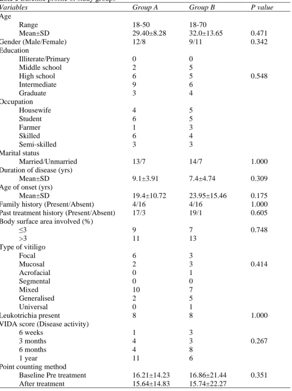
Table 1 Baseline profile of study groups
Variables Group A Group B P value
Age Range Mean±SD 18-50 29.40±8.28 18-70
32.0±13.65 0.471
Gender (Male/Female) 12/8 9/11 0.342
Education Illiterate/Primary Middle school High school Intermediate Graduate 0 2 6 9 3 0 5 5 6 4 0.548 Occupation Housewife Student Farmer Skilled Semi-skilled 4 6 1 6 3 5 5 3 4 3 Marital status
Married/Unmarried 13/7 14/7 1.000
Duration of disease (yrs)
Mean±SD 9.1±3.91 7.4±4.74 0.309
Age of onset (yrs)
Mean±SD 19.4±10.72 23.95±15.46 0.175
Family history (Present/Absent) 4/16 4/16 1.000
Past treatment history (Present/Absent) 17/3 19/1 0.605 Body surface area involved (%)
≤3 >3 9 11 7 13 0.748 Type of vitiligo
Focal Mucosal Acrofacial Segmental Mixed Generalised Universal 6 2 0 0 10 2 0 3 3 1 0 7 5 1 0.414
Leukotrichia present 8 8 1.000
VIDA score (Disease activity) 6 weeks 3 months 6 months 1 year 1 4 4 11 3 3 8 6 0.267
Point counting method Baseline Pre treatment After treatment 16.21±14.23 15.64±14.83 16.86±21.44 15.74±22.27 0.351
history and majority of them had tried both
topical and systemic treatment.
After four weeks of treatment the point counting
method of area estimation showed a decrease in
the mean size of the lesion from 16.21 cm
2to
15.64 cm
2in group A and 16.86 cm
2to 15.74
cm2 in group B but the decrease was statistically
not significant (P = 0.351)
(Table 1)
.
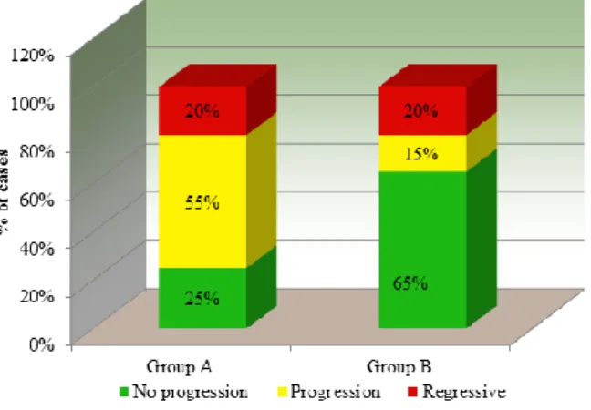
VETF scoring system
(Figure 1)
showed no
Figure 1 Comparison of VFTF System to assess spreading after treatment between group A and group B
among cases and controls after four weeks of
respective therapies (P = 0.017).
As per
Table 2 & 3
, after four weeks of
treatment, mean TOS decreased insignificantly
in both the study groups (P = 0.263 & 0.398 for
Group A and B respectively). However this
difference between the 2 study groups was not
statistically significant (P = 0.076). Mean TAS
also showed a statistically insignificant drop in
group A & B from baseline (P = 0.911 & 0.575
respectively).
Noteworthy, the mean oxidative stress index
(OSI) decreased significantly in both the study
groups individually (P = 0.037 & 0.040 for
Group A and B respectively). However
inter-group comparison between the 2 study inter-groups
showed no statistically significant difference (P
= 0.052).
None of the patients observed any side effects
warranting withdrawal of any patient in either of
the groups during study duration.
Discussion
The
balance
between
oxidation
and
antioxidation is believed to be critical in
maintaining healthy biological systems. Under
physiological
conditions,
the
human
antioxidative defense system including, e.g.-
SOD, CAT, GPx, glutathione (GSH) and others,
allow the elimination of excess ROS such as
superoxide anions (O
2.-), hydroxyl radicals
(OH
.), alkoxyl radicals (RO
.) and peroxyradicals
(ROO
.).
8However, our endogenous antioxidant
defense
systems
is
incomplete
without
exogenous originating reducing compounds such
as vitamin C, vitamin E carotenoids and
Table 2 Baseline stress indices in study groups
Group A (n=20) Group B (n=20)
P Value Mean ± SD Min - Max Mean ± SD Min - Max
Total oxidative stress
before treatment 91.85± 47.50 13.50- 198.20 107.13± 38.22 48.20- 180.30 0.250 Total antioxidant stress
before treatment 528.82± 340.24 237 - 1692 525.40± 226.81 258 - 1078 0.552 Oxidative stress index
before treatment 19.34± 9.15 4.80 - 36.32 22.46 ± 8.81 9.65 - 40.70 0.267 Table 3 Stress indices in study groups after treatment
Group A Group B
P Value
Mean± SD Min - Max Mean ± SD Min - Max
Total Oxidative stress
after treatment 75.85± 63.97 13.50- 282.20 95.79± 43.79 29.10- 201.60 0.076 Total antioxidant stress
after treatment 510.18± 384.56 220 - 1910 513.46± 209.68 313 - 1068 0.974 Oxidative stress index
polyphenols that play an essential role in many
antioxidant mechanisms in living organisms.
Therefore, there is continuous demand for
exogenous antioxidants in order to prevent
oxidative stress, representing a disequilibrium
redox state in favor of oxidation.
8Increased oxidative stress is observed in the
active
vitiligo
patient.
17According
to
autocytotoxic hypothesis in active disease,
oxidative stress is the initial pathogenic event in
melanocyte
degeneration
with
H2O2
accumulation in the epidermis of patients with
active disease.
18The oxidative stress can either be from increased
generation of free radicals or decreased
destruction of these. It is not a localized
phenomenon but a more generalized process.
This may be one of the explanations for
developing newer lesions in vitiligo patients in
the course of the disease.
19In recent years, interest has grown in studying
the role played by oxidative stress in vitiligo by
investigating one or more of the antioxidant
markers, including SOD, CAT, GPx, vitamin E,
vitamin C.
8-10However there are conflicting
evidence for the same. Some researchers report
increased total antioxidant levels, others report
no change or even decreased levels of these
markers like SOD, GPx, malondialdehyde
(MDA), Nitric Oxide (NO), and CAT.
9,10,20,21But these studies didn’t measure the TOS and
TAS. Because the effects of antioxidants and
oxidants could be additive and measuring
individual oxidants and antioxidants separately
is time consuming and labor intensive, a
measurement of the combined activities of all
oxidants and antioxidants is often used to
estimate the overall antioxidant status.
22In the
present study after 4 weeks of treatment a non
significant total oxidative stress and total
antioxidant stress decreased was observed in
both the groups. Noteworthy the difference
between the two was statistically not significant
even after addition of exogenous antioxidants to
the study patients in group B.
Furthermore, the OSI, which is calculated as the
ratio of TOS to TAS, is a more accurate index of
oxidative stress in the body because it is a
comprehensive measurement of TAS and TOS.
22Significant decrease in the OSI was observed
within group A & B individually. But when this
decrease was compared between the two study
groups this came out to be statistically not
significant. This shows that addition of
antioxidants had no added advantage. These
results are similar to the finding observed by
Jayanth et al.
who observed no distinct
advantage of adding antioxidants to the standard
regimen of photochemotherapy for treating
patients of vitiligo.
23The VETF proposed a system that combines
analysis of extent, stage of disease (staging), and
disease progression (spreading).
15In the present
study majority of the patients had complete
depigmentation and all of them were spreading.
After 4 weeks of treatment the BSA and the type
of lesion remained unchanged however addition
of antioxidant had no effect on spreading of
active vitiligo.
Limitations of the study
The present study should be performed with
more patients and treatment needs to be
continued for a longer duration along with a
longer follow-up.
Acknowledgements
The authors thank SRMS IMS Trust, Bareilly
for providing the platform for this research
work.
References
1. Allam M, Riad H. Concise review of recent studies in vitiligo. Qatar Med J 2013; 2013(2): 1–19.
2. Alikhan A, Felsten LM, Daly M, Petronic-Rosic V. Vitiligo: A comprehensive overview part I. Introduction, epidemiology, quality of life, diagnosis, differential diagnosis, associations, histopathology, etiology, and work-up. J Am Acad Dermatol 2011; 65: 473–91.
3. Kruger C, Schallreuter KU. A review of the worldwide prevalence of vitiligo in children/adolescents and adults. Int J Dermatol 2012; 51: 1206–12.
4. Pahwa P, Mehta M, Khaitan BK, Sharma VK, Ramam M. The psychosocial impact of vitiligo in Indian patients. Indian J Dermatol Venereol Leprol 2013; 79: 679-85.
5. Ortonne JP, Bahodoran P, Fitzpatrick TB, Mosher DB, Hori Y. Hypomelanoses and hypermelanoses. In: Freedberg IM, Eisen AZ, Wolff K, Austen KF, Goldsmith LA, Katz SI, editors. Fitzpatrick's Dermatology in General Medicine. 6 th ed. New York: McGraw Hill; 2003. p. 839-47.
6. Kovacs SO. Vitiligo. J Am Acad Dermatol 1998; 38: 647-66.
7. Maresca V, Roccella M, Roccella F, Camera E, Del Porto G, Passi S, et al . Increased sensivity to peroxidative agents as a possible pathogenic factor of melanocyte damage in vitiligo. J Invest Dermatol 1997; 109: 310-3. 8. Bouayed J, Bohn T. Exogenous Antioxidants-Double-Edged Swords in Cellular Redox State: Health Beneficial Effects at Physiologic Doses versus
Deleterious Effects at High Doses. Oxid Med Cell Longev 2010; 3(4): 228-37. 9. Jain A, Mal J, Mehndiratta V, Chander R,
Patra SK. Study of oxidative stress in vitiligo. Indian J Clin Biochem 2011; 26(1): 78-81.
10. Deo SS, Bhagat AR, Shah RN. Study of oxidative stress in peripheral blood of Indian vitiligo patients. Indian Dermatol Online J 2013; 4(4): 279-82.
11. Erel O. A novel automated direct measurement method for total antioxidant capacity using a new generation, more stable ABTS radical cation. Clin Biochem 2004; 37(4): 277-85.
12. Erel OA. New automated colorimetric method for measuring total oxidant status. Clin Biochem 2005; 38 (12): 1103-11. 13. Wu R, Feng J, Yang Y, Dai C, Lu A, Li J, et
al. Significance of Serum Total Oxidant/ Antioxidant Status in Patients with Colorectal Cancer. PLoS ONE 2017; 12(1): e0170003.
14. Njoo MD, Das PK, Bos JD, Westerhof W. Association of the koebner phenomenon with disease activity and the therapeutic responsiveness in vitiligo vulgaris. Arch Dermatol 1999; 135(4):407-13.
15. Taieb A, Bordeaux CHU. Vitiligo. Pigment Cell Res 2007; 20(5):418.
16. Alghamdi KM, Kumar A, Taieb A, Ezzedine K. Assessment methods for the evaluation of vitiligo. J Eur Acad Dermatol Venereol 2012; 26(12): 1463-71.
17. Ines D, Sonia B, Riadh BM, Amel EG, Slaheddine M, Hamida T et al. A comparative study of oxidant-antioxidant status in stable and active vitiligo patients. Arch Dermatol Res 2006; 298(4): 147-52. 18. Maresca V, Roccella M, Roccella F, Camera
E, Del Porto G, Passi S, et al. Increased sensitivity to peroxidative agents as a possible pathogenic factor of melanocyte damage in vitiligo. J Invest Dermatol 1997; 109: 310-3.
19. Sravani PV, Babu NK, Gopal KVT, Rao GRR, Rao AR, Moorthy B, et al. Determination of oxidative stress in vitiligo by measuring superoxide dismutase and catalase levels in vitiliginous and non-vitiliginous skin. Indian J Dermatol Venereol Leprol 2009; 75: 268-71.
Population. Kathmandu Univ Med J 2014; 46(2): 132-6.
21. Singh D, Malhotra SK, Gujral U. Role of oxidative stress in autoimmune pathogenesis of vitiligo. Pigment Int 2016; 3: 90-5 22. Akoglu G, Emre S, Metin A, Akbas A,
Isikoglu S, Sener S, Kilinc F. Evaluation of total oxidant and antioxidant status in
localized and generalized vitiligo. Clin Exp Dermatol. 2013; 38(7): 701-6.