383
© 2017 by the Serbian Biological Society
Glycoxidated ferritin induces the release of microparticles positive for Toll-like receptors
derived from peripheral blood CD14+ cells
López-Soto Luis Fernando1, Galván-Moroyoqui José Manuel1, Martínez-Soto Juan Manuel2,
Almada-Balderrama Martín3, Rosales-Ruiz Ashley Patricia4, Álvarez-Hernández Gerardo1, Camacho Villa Alma
Yolanda5, Bolado Martínez Enrique4, Soto-Guzmán Adriana1 and Candia-Plata Maria del Carmen1,*
1 Departamento de Medicina y Ciencias de la Salud, Universidad de Sonora, Hermosillo Sonora, México 2 Programa de Doctorado en Ciencias de la Salud, Universidad de Sonora, Hermosillo Sonora, México 3 Programa de Maestría en Ciencias de la Salud, Universidad de Sonora, Hermosillo Sonora, México 4 Departamento de Ciencias Químico-Biológicas, Universidad de Sonora, Hermosillo Sonora, México 5Departamento de Medicina, Universidad Durango Santander, Campus Hermosillo, Sonora, México
*Corresponding author: [email protected]
Received: June 14, 2016; Revised: August 20, 2016; Accepted: August 22, 2016; Published online: October 31, 2016
Abstract: Both increased serum ferritin levels and Toll-like receptor (TLR) activation show independent association with the inflammatory processes. During inflammation, cell activation and apoptosis are accompanied by the release of membrane-derived microparticles (MPs), which are considered to be mediators of intercellular communication as they induce specific responses in target cells. The aim of this study was to determine whether glycated and glycoxidated ferritin induce in vitro
release TLR microparticles from CD14+ peripheral blood mononuclear cells. Peripheral blood mononuclear cells were stimulated with glycated, glycoxidated and native ferritin. The release of microparticles from CD14+ cells, the presence of TLR2+ and TLR4+ on the microparticles surface and the presence of interleukins-6 and -8 (IL-6 and IL-8) inside the microparticles after stimulation were determined by flow cytometry. The role of nuclear factor κB (NF-κB) was evaluated by pretreatment of the cells with the Bay 11-7085 inhibitor. Glycated and glycoxidated ferritin induced the release of mic-roparticles from CD14+ cells, the majority of which expressed TLR2+ and TLR4+ on their surface and contained IL-6 and IL-8. These effects were dependent on NF-κB activation. Our findings show that glycated and glycoxidated ferritin might be involved in the release of microparticles and stimulation of inflammatory responses.
Key words: microparticles; Toll-like receptor (TLR); ferritin; glycation; inflammation
How to cite this article: López-Soto LF, Galván-Moroyoqui JM, Martínez-Soto JM, Almada-Balderrama M, Rosales-Ruiz AP, Álvarez-Hernández G, Camacho Villa AY, Bolado Martínez E, Soto-Guzmán JA, Candia-Plata MDC. Glycoxidated ferritin induces the release of microparticles positive for toll-like receptors derived from peripheral blood CD14+ cells. Arch Biol Sci. 2017;69(3):383-90.
INTRODUCTION
Increased levels of serum ferritin and inflammatory markers such as tumor necrosis factor alpha (TNFα) and C-reactive protein (CRP) are frequently related to systemic inflammatory diseases [1-4]. Chronic subclinical inflammation is also mediated by p38 mitogen-activated protein kinase (p38 MAPK), reac-tive oxygen species (ROS) [5], protein kinase C (PKC) and nuclear factor-κB (NF-κB) among other factors [6]. In patients with diabetes mellitus this leads to an increased risk of atherosclerosis, a risk factor for vascular complications in diabetes [7]. Previous work has shown that ferritin genes are susceptible to induc-tion in the course of plaque formainduc-tion [8], however the potential mechanisms by which ferritin could be
involved in these inflammatory events are currently unknown.
plaques and murine models of atherosclerosis [6]. In particular, oxidized low-density lipoprotein (oxLDL) and AGE-products of LDL (AGE-LDL) trigger TLR4-dependent signaling pathways and induce production of the proinflammatory cytokine IL-6 in human and mouse macrophages [6]. Recently, it was shown that glycated ferritin induces the in vitro activation and expression of TLR2 and TLR4 in CD14+ blood pe-ripheral macrophages [9].
During inflammatory processes, cell activation and apoptosis are accompanied by the release of membrane-derived microparticles (MPs) [10]. MPs are submicron vesicles shed from a variety of cells that contain surface molecules and cytoplasmic con-stituents of their parent cells and are considered as biomarkers for several cardiovascular and inflamma-tory disorders. MPs are also mediators of intercellular communication as they induce specific responses in target cells [11]. Furthermore, plasma cell-derived MPs and urine cell-derived MPs appear to be posi-tively associated with adverse clinical events [12, 13]. The relationship between TLRs, glycoxidated fer-ritin and elevated ferfer-ritin serum levels has not been completely elucidated. Therefore, in this study we ex-amined the in vitro effects of glycated and glycoxidat-ed ferritin on the composition of the surface and inter-nal content of MPs released from circulating venous CD14+ monocytes from healthy adult volunteers.
MATERIALS AND METHODS
Experiments were performed using a pool of fasting venous blood samples drawn from five healthy adult volunteers. The donors of blood samples signed an informed consent form and were clinically assessed to ensure that they did not have any inflammatory disorder and had not undergone any steroid and/or antiinflammatory therapy. Blood samples were drawn into heparinized tubes for cell stimulation and flow cytometry. All procedures were conducted in accord-ance with international ethical standards. The study was approved by the Bioethics and Research Com-mittee of the Department of Medicine and Health Sciences, University of Sonora.
Materials
PerCP/Cy5.5 anti-human CD14 (clone HCD14), FITC human CD282 (TLR2) (clone TL2.1), PE anti-human TLR4 (clone HTA125), FITC anti-anti-human IL-6 (clone MQ2-13A5) and PE anti-human IL-8 (clone E8N1) were obtained from BioLegend (San Diego, CA). Fixation buffer (Cat. 420801), permeabilization wash buffer 10x (Cat. 421002) and cell staining buff-er (cat. 420201) wbuff-ere also obtained from BioLegend (San Diego, CA). Calibration 1.0 and 0.1 μM particle size latex fluorescent beads (Cat. L2778 and L9904, respectively), ferritin type I, from horse spleen (Cat. F4503), zymosan A from Saccharomyces cerevisiae
(Cat. Z4250), Bay 11-7085 [14] (Cat. B5681) and D-(+)-glucose (Cat. 5767) were obtained from Sigma-Aldrich® (Saint Louis, MO). All other reagents used were of the highest grade available.
Native ferritin modification by glycation and glycoxidation
A total of 1 mg native ferritin was dissolved in 1 mL of 0.1 mmol/L phosphate buffer solution (PBS), pH 7.4, containing 1 mmol/L EDTA, 0.1 mg/mL chloram-phenicol and 3 mmol/L NaN3.The protein sample was then incubated with 0.4 M glucose at 37°C for 1 week under a nitrogen atmosphere. Glycated ferritin was then reduced with NaBH4 for 1 h at 4°C and dialyzed against 0.1 M PBS containing 0.1 mmol/L EDTA for 24 h [15]. For oxidation, glycated ferritin (1 mg/mL) was dialyzed against 5 mmol/L CuSO4 in PBS for 24 h at 37°C in the dark. Next, the reaction was stopped by incubation with PBS containing 200 μmol/L EDTA and 40 μmol/L 3,5-Di-tert-4-butylhydroxytoluene for 24 h. The protein glycation process was monitored by SDS/ PAGE (data not shown) [9, 15]. Finally, the glycated, glycoxidated and non-modified ferritin concentrations were measured using the Bradford technique [16].
Cell stimulation
A total of 1x10⁶ mononuclear cells from the heparin
mL native ferritin were incubated at room temperature for 2 h. Stimulation assays under the same conditions using 50 ng/mL zymosan A were performed as posi-tive controls for TLR2 and TLR4 expression [14]. To test the role of NF-κB, cells were also pretreated with 15 μmol/L Bay 11-7085 for 30 min [17]. Subsequently, the supernatants were collected and processed for flow cytometry. All assays were performed in triplicate.
Fluorescence-activated cell sorting (FACS) analysis of microparticles
MPs contained in supernatants from the stimulated and non-stimulated cell suspensions were incubated with 20 µL of anti-CD14-PerCP/Cy5.5, anti-TLR2-FITC and anti-TLR4-PE [18, 19] for 30 min at room temperature in the dark. FACS analysis was performed using 1 and 0.1 μM particle size latex fluorescent beads as a control for microparticle size. A total of 100000 MP events per assay were evaluated using a FACScalibur flow cytom-eter (Becton Dickinson, San Diego, CA) and Summit Software Informer®. To determine the content of IL-6 and IL-8 in the CD14+ MPs, MP surface staining was performed for anti-CD14-PerCP/Cy5.5. The MPs were fixed in 0.5 mL/tube fixation buffer (BioLegend Cat. No. 420801) in the dark for 20 min at room
tempera-ture. To permeabilize the MPs, permeabilization wash buffer (Cat. No. 421002; 1X in DI water) was added to the samples. Finally, the fixed and permeabilized MPs were incubated with the anti-IL-6-FITC and anti-IL-8-PE [18, 19] for 30 min at room temperature in the dark.
Statistical analysis
All statistical analyses were performed using the SPSS 20 software (Armonk, NY). Data are presented as the mean +/- standard deviation (SD), unless otherwise indicated. The Student’s t-test was used to compare mean expression of basal control and the average ex-pression produced by the stimulation of cells. A value of p<0.05 was considered to be statistically significant.
RESULTS
Glycated and glycoxidated ferritin increases the release of MPs from CD14+ cells
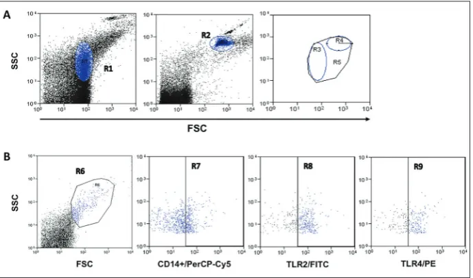
The specific MPs were identified by their forward scatter and side scatter using 0.1 μm and 1.0 μm la-tex fluorescent beads to determine the region of MPs that they represent (Fig. 1A). The percentage of MPs
derived from CD14+ was significantly increased from the basal control when cells were stimulated with 50 ng/mL Gly-ferritin or 50 ng/mL GlyOx-ferritin. Also, the positive control for activation zymosan A pro-duced a significant increase in the MPs from CD14+. The observed effect of non-modified ferritin was com-parable to the baseline (Fig. 2).
Glycated and glycoxidated ferritin increases the expression of TLR2 and TLR4 in CD14+ microparticles
Stimulation of peripheral blood cells with Gly-ferri-tin and GlyOx-ferriGly-ferri-tin induced a significant increase in the percent expression of TLR2 on the surface of CD14+ microparticles (Fig. 3, lower panel). Inten-sity of the TLR2 expression was also estimated by the mean intensity fluorescence (MIF), which signifi-cantly increased (Fig. 3, upper panel). Stimulation of CD14+ with Gly-ferritin and GlyOx-ferritin produced
Fig. 2. CD14+ microparticles.The blood mononuclear cells were stimulated with 50 ng/mL of ferritin, Gly-ferritin, GlyOx-ferritin or zymosan A, and were pre-treated for 30 min with 15 μM BAY to inhibit the NF-κB pathway. The graph represents the mean±SD of three independent experiments and is expressed as the percentage of CD14+ MPs from the total MP value. (*) denotes the significant difference versus the basal control; the inverted commas (“) denote significant difference between the stimulus and BAY pretreatment, with P≤0.05 bythe Student’s t-test.
Fig. 3. TLR2 expression in CD14+ microparticles.The blood mononuclear cells were stimulated with 50 ng/mL of ferritin, Gly-ferritin, GlyOx-ferritin or zymosan A, and pre-treated for 30 min with 15 μM BAY to inhibit the NF-κB pathway. The graphs represent the mean±SD of three independent experiments and are expressed as the medium fluorescence intensity of TLR2+ in CD14+ MPs (upper panel), and as the percentage of TLR2+ MPs from CD14+ total MPs (lower panel). (*) denotes comparisons versus basal; inverted commas (“) are comparisons between the stimulus and BAY pretreatments, with P≤0.05 bythe Student’s
t-test.
a discrete but significant rise in TLR4 expression on the surface of total CD14+ microparticles (Fig. 4, lower panel). However, no differences in MIF were observed upon stimulation with ferritin and Gly-Ox-Ferritin, and only the positive control zymosan A significantly increased the TLR4-FITC MIF on the CD14+ microparticles (Fig. 4, upper panel).
Role of NF-κB in TLR2 and TLR4 expression in CD14+ microparticles
To explore the role of nuclear factor-κB (NF-κB) in the production of MPs, the CD14+ cells were first incubated with the NF-κB inhibitor, Bay 11-7085 [14]. The BAY inhibitor abolished the release of MPs from CD14+ cells and the percentage of CD14+ cells was comparable to the basal control (Fig. 2). Pretreatment with BAY significantly reduced the percent expression and MIF of TLR2 in CD14+ MPs previously induced by Gly-ferritin and GlyOx-ferritin (Fig. 3). We also
observed that pretreatment with the BAY inhibitor af-fected the percent expression of TLR4 in CD14+ MPs stimulated with Gly-ferritin, GlyOx-ferritin and the zymosan A control (Fig. 4, lower panel). Furthermore, the MIF of TLR4 in CD14+ MPs was significantly induced by zymosan A (Fig. 4, upper panel).
Glycated and glycoxidated ferritin increases IL-6 and IL-8 expression in CD14+ MPs
To evaluate the activation of TLR signaling pathways, we measured the content of IL-6 and IL-8 in CD14+ MPs. The cells were pretreated with native ferritin, Gly-ferritin, GlyOx-ferritin or the positive control zy-mosan A. To determine the role of NF-κB in the pro-duction of cytokines, the cells were pretreated with the NF-κB inhibitor BAY after stimulation as described above [14]. Stimulation of peripheral blood cells with Gly-ferritin and GlyOx-ferritin induced a significant increase in the percentage of IL-6 and IL-8 in CD14+ MPs (Fig. 5, upper and lower panel, respectively). Also, the positive control zymosan A significantly increased the percentage of IL-6 and IL-8 in CD14+ MPs (Fig. 5, upper and lower panel, respectively). Pretreatment with BAY significantly reduced the ex-pression of IL-6 and IL-8 in CD14+ MPs, previously induced by Gly-ferritin and GlyOx-ferritin (Fig. 5, upper and lower panel, respectively). We observed the same effect of pretreatment with BAY on the propor-tional expression of cytokines in CD14+ MPs stimu-lated with zymosan A (Fig. 5, p<0.05).
DISCUSSION
Studies have shown that ferritin can act as an acute phase reactant; also, ferritin has been linked to cer-tain metabolic diseases and their components [2, 3, 20]. The latter assumption has been derived only from statistical correlations, with the mechanisms through which ferritin participates in such disorders remain-ing unclear [3, 21]. On the other hand, it has been shown that ferritin can undergo oxidative damage by carbonyl compounds, such as methylglyoxal or ROS, which have been related to the increase in the iron content of cells that can be deleterious to cells [21, 22].
Advanced glycation end-products (AGEs) are a diverse group of highly oxidant compounds with pathogenic significance in diabetes as well as in sev-eral other chronic diseases. AGEs are created through a nonenzymatic reaction between reducing sugars and free amino groups of proteins, lipids or nucleic acids [7]. AGEs of low density lipoprotein are capable of activating an inflammatory-type response in different target cells, activating the MyD88-dependent signaling NF-κB pathway when recognized by TLRs, primarily TLR2 and TLR4 [6, 23]. Recent evidence suggests that
in vitro glycated ferritin is capable of activating TLR2
and TLR4 expression and inducing the release of the proinflammatory cytokines IL-6 and IL-8 in human peripheral blood macrophages [9].
AGEs may also stimulate the release of MPs from vascular endothelial cells [24]. Depending on their content, the MPs could represent markers for treat-ment and prognosis of chronic and inflammatory dis-eases [5]. This could be because the MPs are released from activated cells by outward blebbing and vesicula-tion of the plasma membrane following breakdown of the cytoskeleton of the parental cells. MPs carry cy-toplasmic and/or nuclear components of the parental cell that can alter the activity of recipient cells through the transfer of their cargo [5, 25, 26]. For these reasons it is important to determine the expression of specific molecules on the surface and within the MPs.
In this study, we looked at the effects of glycated and glycoxidated ferritin on the percentage of MPs derived from monocytes and the expression of TLR2 and TLR4 on their surface. The internal IL-6 and IL-8 contents of MPs released from circulating CD14+ monocytes from healthy volunteers were also stud-ied under the described experimental conditions. To this end, the protein was modified in vitro using the method described by Rivandi et al. [15]. We stimu-lated human peripheral blood monocytes (PBMCs) with glycated and glycoxidated ferritin for 2 h and an increase in the number of monocyte-derived MPs was observed, whereas stimulations with the native protein did not alter the number of MPs released from PB-MCs. This suggests that monocytes respond to modi-fied ferritin by secreting MPs. This response could be similar to that induced by ox-LDL in endothelial cells as shown in a previous report [24]. However, based on the design of the present study it has to be stated
that the influence of glycated ferritin on CD14+ cells might be not only direct but also indirect. Namely, as peripheral blood mononuclear cells, and not purified CD14+ cells, were used in the experiments there was the possibility that some of the observed effects were mediated through CD14- cells that were responsive to the influence of glycated ferritin.
We found that glycated and glycoxidated ferritin increased the number of MPs with TLR2 and TLR4 on their surface. The expression of TLR2 and TLR4 was decreased in the presence of the BAY inhibitor, suggesting that ferritin, modified by glycation and gly-coxidation, activates the NF-κB signaling pathway of monocytic cells. These data are consistent with previ-ous reports, indicating that the release of MPs from macrophages is stimulated by activation of the NF-κB signaling pathway with TLR ligands [27, 28].
MPs are considered an information vector for other target cells because they can transfer pro- and antiinflammatory cytokines and also transfer chem-okine and cytchem-okine receptors [29-31]. We ascertained the presence of IL-6 and IL-8 in monocyte-derived MPs and found that glycated and glycoxidated ferritin increases the number of MPs released by monocytes containing IL-6 and IL-8. Furthermore, this stimu-lation increases the concentration of IL-6 and IL-8 within these MPs in a manner that is dependent on the NF-κB signaling pathway.
Our results showed that the MPs released by CD14+ monocytes after stimulation with glycated and glycoxidated ferritin express TLR2 and TLR4 on their surface and carry out inflammatory messages based on their IL-6 and IL-8 contents. In diverse metabolic diseases, increased levels of cytokines in MPs may be important because of their potential effects on target cells [25, 26, 30, 31].
CONCLUSIONS
an inflammatory response that could contribute to the inflammation observed in diseases where ferritin levels and inflammation markers are increased, such as diabetes mellitus [1, 3, 4, 20]. A limitation of our in
vitro system is the fact that we observed phenomena
outside of the organism. However, with this approach, we now have better insight into the potential role of ferritin in inflammatory diseases. Further studies are necessary to confirm this hypothesis.
Acknowledgements: We are grateful to Miriam Garcia, Cecil-ia Soto and Sandra Amarillas for their technical support. This work was financially supported by a grant from CONACyT (No. 149110). Martinez-Soto and Almada-Balderrama are supported by a CONACyT training grant.
Authors’ contribution: López-Soto LF and Galván-Moroyoqui JM contributed equally to this work. López-Soto LF, Galván-Mo-royoqui JM and Soto-Guzmán A provided the research direction and protocols and performed the experimental work, performed the statistical analysis and provided the research report. Martín-ez-Soto JM and Rosales-Ruíz AP performed the experimental work Almada-Balderrama M and Camacho Villa AY provided the protocols and performed the experimental work. Álvarez-Hernández G and Bolado Martínez E validated and analyzed the data. Candia-Plata MC designed the study, oversaw the study and is the guarantor of the manuscript.
Conflict of interest disclosure: The authors have no conflicts of interest, neither financial nor academic.
REFERENCES
1. Ashourpour M, Djalali M, Djazayery A, Eshraghian MR, Taghdir M, Saedisomeolia A. Relationship between serum ferritin and inflammatory biomarkers with insulin resistance in a Persian population with type 2 diabetes and healthy peo-ple. Int J Food Sci Nutr. 2010;61(3):316-23.
2. Aso Y, Takebayashi K, Wakabayashi S, Momobayashi A, Suga-wara N, Terasawa T, Naruse R, Hara K, Suetsugu M, Morita K, Inukai T. Relation between serum high molecular weight adi-ponectin and serum ferritin or prohepcidin in patients with type 2 diabetes. Diabetes Res Clin Pract. 2010;90(3):250-5. 3. Li J, Wang R, Luo D, Li S, Xiao C. Association between serum
ferritin levels and risk of the metabolic syndrome in Chinese adults: a population study. PLoS One. 2013;8(9):e74168. 4. Williams MJ, Poulton R, Williams S. Relationship of serum
ferritin with cardiovascular risk factors and inflammation in young men and women. Atherosclerosis. 2002;165(1):179-84. 5. Roseblade A, Luk F, Rawling T, Ung A, Grau GE, Bebawy M.
Cell-derived microparticles: new targets in the therapeutic management of disease. J Pharm Pharm Sci. 2013;16(2):238-53.
6. Hodgkinson CP, Laxton RC, Patel K, Ye S. Advanced glycation end-product of low density lipoprotein activates the toll-like
4 receptor pathway implications for diabetic atherosclerosis. Arterioscler Thromb Vasc Biol. 2008;28(12):2275-81. 7. Hu H, Jiang H, Ren H, Hu X, Wang X, Han C. AGEs and
chronic subclinical inflammation in diabetes: disorders of immune system. Diabetes Metab Res Rev. 2015;31(2):127-37. 8. Pang JH, Jiang MJ, Chen YL, Wang FW, Wang DL, Chu SH, Chau LY. Increased ferritin gene expression in atherosclerotic lesions. J Clin Invest. 1996;97(10):2204-12.
9. Galván-Moroyoqui JM C-PM, Martínez-Soto JM, Soto-Guzmán JA, Bolado Martínez E, Camacho-Villa AY, López-Soto LF. Glycated ferritin induces activation and expression of tlr2 and tlr4 in human peripheral blood macrophages. Pharma Innov J. 2015;3(12):44-8.
10. Hargett LA, Bauer NN. On the origin of microparticles: From “platelet dust” to mediators of intercellular communication. Pulm Circ. 2013;3(2):329-40.
11. Hoyer FF, Giesen MK, Nunes Franca C, Lutjohann D, Nick-enig G, Werner N. Monocytic microparticles promote athero-genesis by modulating inflammatory cells in mice. J Cell Mol Med. 2012;16(11):2777-88.
12. Burger D, Thibodeau JF, Holterman CE, Burns KD, Touyz RM, Kennedy CR. Urinary podocyte microparticles iden-tify prealbuminuric diabetic glomerular injury. J Am Soc Nephrol. 2014;25(7):1401-7.
13. Giannopoulos G, Oudatzis G, Paterakis G, Synetos A, Tampaki E, Bouras G, Hahalis G, Alexopoulos D, Tousou-lis D, Cleman MW, Stefanadis C, Deftereos S. Red blood cell and platelet microparticles in myocardial infarction patients treated with primary angioplasty. Int J Cardiol. 2014;176(1):145-50.
14. Ikeda Y, Adachi Y, Ishibashi K, Miura N, Ohno N. Activation of toll-like receptor-mediated NF-kappa beta by zymosan-derived water-soluble fraction: possible contribution of endo-toxin-like substances. Immunopharmacol Immunotoxicol. 2005;27(2):285-98.
15. Ravandi A, Kuksis A, Shaikh NA. Glucosylated glycerophos-phoethanolamines are the major LDL glycation products and increase LDL susceptibility to oxidation: evidence of their presence in atherosclerotic lesions. Arterioscler Thromb Vasc Biol. 2000;20(2):467-77.
16. Bradford MM. A rapid and sensitive method for the quantita-tion of microgram quantities of protein utilizing the principle of protein-dye binding. Anal Biochem. 1976;72:248-54. 17. Pierce JW, Schoenleber R, Jesmok G, Best J, Moore SA,
Col-lins T, Gerritsen ME. Novel inhibitors of cytokine-induced IkappaBalpha phosphorylation and endothelial cell adhesion molecule expression show anti-inflammatory effects in vivo. J Biol Chem. 1997;272(34):21096-103.
18. Bessich JL, Nymon AB, Moulton LA, Dorman D, Ashare A. Low levels of insulin-like growth factor-1 contribute to alve-olar macrophage dysfunction in cystic fibrosis. J Immunol. 2013;191(1):378-85.
20. Jehn M, Clark JM, Guallar E. Serum ferritin and risk of the metabolic syndrome in U.S. adults. Diabetes Care. 2004;27(10):2422-8.
21. An SH, Lee MS, Kang JH. Oxidative modification of ferritin induced by methylglyoxal. BMB Rep. 2012;45(3):147-52. 22. Yoon JH, An SH, Kyeong IG, Lee MS, Kwon SC, Kang JH.
Oxidative modification of ferritin induced by hydrogen per-oxide. BMB Rep. 2011;44(3):165-9.
23. Miller YI, Chang MK, Binder CJ, Shaw PX, Witztum JL. Oxi-dized low density lipoprotein and innate immune receptors. Curr Opin Lipidol. 2003;14(5):437-45.
24. Nomura S, Shouzu A, Omoto S, Nishikawa M, Iwasaka T, Fukuhara S. Activated platelet and oxidized LDL induce endothelial membrane vesiculation: clinical significance of endothelial cell-derived microparticles in patients with type 2 diabetes. Clin Appl Thromb Hemost. 2004;10(3):205-15. 25. Angelillo-Scherrer A. Leukocyte-derived microparticles in
vascular homeostasis. Circ Res. 2012;110(2):356-69. 26. Montoro-Garcia S, Shantsila E, Marin F, Blann A, Lip GY.
Circulating microparticles: new insights into the biochemical
basis of microparticle release and activity. Basic Res Cardiol. 2011;106(6):911-23.
27. Gauley J, Pisetsky DS. The release of microparticles by RAW 264.7 macrophage cells stimulated with TLR ligands. J Leukoc Biol. 2010;87(6):1115-23.
28. Spencer DM, Gauley J, Pisetsky DS. The properties of micropar-ticles from RAW 264.7 macrophage cells undergoing in vitro activation or apoptosis. Innate Immun. 2014;20(3):239-48. 29. Angelot F, Seilles E, Biichle S, Berda Y, Gaugler B, Plumas J,
Chaperot L, Dignat-George F, Tiberghien P, Saas P, Garnache-Ottou F. Endothelial cell-derived microparticles induce plas-macytoid dendritic cell maturation: potential implications in inflammatory diseases. Haematologica. 2009;94(11):1502-12. 30. Distler JH, Huber LC, Gay S, Distler O, Pisetsky DS. Mic-roparticles as mediators of cellular cross-talk in inflammatory disease. Autoimmunity. 2006;39(8):683-90.