651
HIPPO SIGNALING PROTEIN MST1 REGULATES OSTEOCLAST DIFFERENTIATION
BY INTERACTING WITH INTEGRIN LINKED KINASE (ILK) AND MODULATING
ACTIN STRUCTURES
Xiao-Han Huang1, Pan Su2 and Wu-Yin Li3,*
1Department of Bone and Joint Diseases, Luoyang Orthopedic-Traumatological Hospital, Luoyang, Henan 471002, P.R. China
2Department of Section Ankle Injury, Luoyang Orthopedic-Traumatological Hospital, Luoyang, Henan 471002, P.R. China
3Department of Orthopedics, Luoyang Orthopedic-Traumatological Hospital, Luoyang, Henan 471002, P.R. China
*Corresponding author: [email protected]
Received: September 29, 2015; Revised: November 15, 2015; Accepted: November 25, 2015; Published online: June 03, 2016
Abstract: Hippo signaling is implicated in balancing cell proliferation, differentiation and death in multiple organs. How-ever, its role in specific bone cell types such as osteoclasts, and its significance in maintaining overall bone tissue homeostasis remain largely unknown. In this study, we investigated the role of the Hippo pathway in osteoclast differentiation. Human primary monocyte cells were treated with receptor activator nuclear factor kappaB ligand (RANKL) and evaluated for osteoclast differentiation by marker protein analysis, tartrate-resistant acid phosphate (TRAP) and resorption assays. Our results showed that Ste20-like kinase 1 (MST1) underwent the maximum change after RANKL treatments and is negatively associated with osteoclast differentiation. Furthermore, proteomic approaches involving co-immunoprecipitation and mass spectrometry identified MST1 interaction with integrin-linked kinase (ILK) which is lost during RANKL induced differentiation. Finally, using RNAi and ectopic expression experiments we observed that MST1-ILK interaction negatively inhibits osteoclast differentiation at the level of actin ring structure formation, which is facilitated by ILK. Together, our data highlight a role for the Hippo pathway protein, MST1, in negatively regulating osteoclast differentiation through its interaction with integrin signaling. Given that integrin signaling is progressively implicated in pathological osteolysis, augmenting this pathway could have therapeutic implications.
Key words:osteoclast; Hippo signaling; Ste20-like kinase 1 (MST1); integrin linked kinase (ILK); integrin signaling; actin ring structures.
INTRODUCTION
Organ functionality in complex multicellular organisms is maintained by balancing proliferation, differentiation and cell death. Recent research studies have identified a tumor suppressor pathway called the Hippo signal-ing pathway that primarily regulates cell proliferation, death and differentiation [1]. Research over the last de-cade into Hippo signaling has used Drosophila genet-ics extensively to identify pathway components, and in recent times their mammalian homologs are also being identified and intensively investigated [2,3].
Although several tissue-specific variations are implicated, the core to Hippo signaling is a kinase
cascade, wherein mammalian serine/threonine kinase 20 (STE20)-like proteins (MST1/2) phosphorylate large tumor suppressor (LATS1/2) kinase proteins, which in turn phosphorylate the major Hippo path-way effector proteins, Yes-associated protein (YAP) and transcriptional coactivators with PDZ-binding motif proteins (TAZ) to cause its cytoplasmic seques-tration. Upon inductive signals, YAP/TAZ proteins are dephosphorylated and translocate to the nucleus, interacting with the transcription enhancer factors − TEA domain family (TEAD 1-4), to induce gene expression and facilitate cell proliferation [4-7].
func-tion are regulated by a combinatorial acfunc-tion of two cell types: osteoclast and osteoblast [8,9]. Osteoblasts are derived from mesenchymal cells and are primarily responsible for new bone formation, while osteoclasts are derived from monocyte/macrophage cells and are responsible for bone resorption [8,9]. Defects in the underlying signaling cascades regulating the osteo-clast/osteoblast balance lead to bone disease [8-15]. In particular, increased osteoclast mass and activity is associated with diseases such as osteoporosis [8-15]. Furthermore, the inflammatory secretions present in disease conditions are also progressively implicated in osteoclast differentiation and activity, but the intramo-lecular events leading to the normal differentiation or pathological state are largely unknown.
Multiple studies have underscored the importance of the tumor necrosis factor (TNF) receptor (TNFR)/ TNF-like proteins (i) osteoprotegerin (OPG), (ii) re-ceptor activator of nuclear factor (NF)-κB (RANK) and (iii) RANK ligand (RANKL) in regulating osteo-clast differentiation and function. More importantly, from these studies it is now known that RANKL or TNF-α combined with macrophage colony-stimulat-ing factor (MCSF) alone is sufficient to induce osteo-clastogenesis from bone marrow macrophage cells in vitro [10-15]. However, deeper understanding of how diverse physiological and pathological signals modu-late the RANKL or pathway and osteoclast functions would allow intervening therapeutics to regulate bone erosion. Given that the Hippo pathway is associated with cell differentiation in most tissues, it is intriguing to hypothesize a similar role in these processes.
In this study, we evaluated the changes in Hippo pathway genes associated with osteoclast differentia-tion, and identified their involvement in regulating actin-ring structures associated with osteoclast dif-ferentiation. We also identified a link between the Hippo signaling pathway and integrin signaling dur-ing osteoclast differentiation.
MATERIALS AND METHODS
Osteoclast differentiation
Human primary osteoclast precursor cells (primary monocyte cells) were maintained in DMEM supple-mented with bullet kit (Lonza, USA). For differentia-tion, cells were plated at a density of 15000 cells/cm2 and supplemented with RANKL (100 ng/mL) com-bined with M-CSF (100 ng/mL) (Sigma-Aldrich, USA) for 7 days, with media changes after every two days.
RNAi and overexpression
siRNA sequences targeting MST1, YAP, TAZ, TEAD1, LATS2 or ILK (Santacruz Biotechnology, USA) were transfected and evaluated after 48 h. For overexpres-sion, cells were either transiently transfected with an empty vector or as full-length constructs of MST1 or ILK (Origene, USA). RANKL treatments were induced 48 h post siRNA treatment or expression of transfected plamid in all of the experiments. For combined siRNA and expression plasmid, both were combined as a single transfection 48 h after RANKL treatments were induced.
Tartrate-resistant acid phosphate (TRAP) staining
RANKL-treated culture systems were fixed in 10% buffered formalin and stained for TRAP (Sigma-Al-drich, USA). The presence of three or more nuclei was used as a criterion to identify osteoclasts.
Osteoclastic resorption (pit formation assay)
Osteoclast differentiation was induced in OsteoAssay plates (Corning, USA) for 1 week and the cells were removed with 10% sodium hypochlorite, washed with water and air-dried. Resorption pits were measured using PixeLink Capture SE software.
RT-PCR
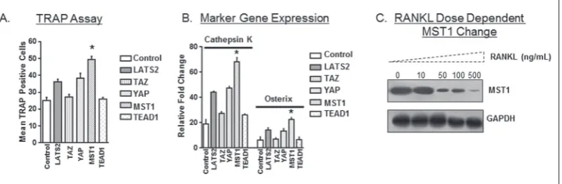
previ-ously [16]. As our cells were derived from primary cul-tures, we evaluated osteoclast differentiation through marker proteins that represent mature osteoclasts, while also evaluating for proteins that are known to inhibit osteoblast differentiation. By this method, if we noticed a consistent trend due to a gene knockdown, it clearly indicated a role in osteoclast differentiation. Based on this rationale, a specific marker protein in-dicative of osteoclast differentiation (Cathepsin K) was selected to identify osteoclasts [17] while also en-suring the trend to inhibit osteoblast differentiation (Sp7) [18]. PCR was performed with a 7500 Applied Biosystems instrument using TaqMan probes with the universal PCR Master Mix (Life Technologies, USA).The following TaqMan probes were used to evaluate gene expression analysis: Ctsk (Cathepsin K): Mm00484039_m1; Sp7 (Osterix): Mm00504574_m1; Gapdh (Glyceraldehyde 3-phosphate dehydrogenase): Mm99999915_g1 (Applied Biosystems, USA). Un-treated samples were used as reference to determine the changes in gene expression.
Co-immunoprecipitation (Co-IP) and immunoblotting
Cell lysates with and without RANKL treatments (48 h) were immunoprecipitated (2µg of antibody per 0.5mg of lysate protein) for MST1 and ILK proteins (Cell Signaling Technology, USA) and analyzed for interacting protein by mass spectrometry or immu-noblotted for specific proteins as described previously [19]. Briefly, the cells were lysed using a RIPA buffer (50 mM Tris-HCl, pH 7.4, 150 mM NaCl, 1% Nonidet P-40, 1 mM EDTA, 10 mg/ml aprotinin, 1 mg/ml leu-peptin, 0.1 M phenylmethylsulfonyl fluoride, 0.5 M sodium fluoride and 1 mM sodium orthovanadate). Equal amounts of proteins were separated by 4-20% gel under reducing conditions, transferred to a nitrocel-lulose membrane (Bio-Rad, USA), and blotted using specific antibodies for proteins. (Cell Signaling Tech-nology, USA). The membrane was then incubated with respective secondary antibodies and developed using Super Signal West Dura (Pierce Biotechnology, USA).
Mass spectrometry
The Co-IP proteins were in-gel digested and then injected for LC-MS/MS (MALDI/TOF–TOF, 4700 Proteomics Analyzer, Applied Biosystems) analy-sis. The protein identifications were made using the Trans-Proteomic Pipeline software running on a sor-cerer platform, and Mascot (version 2.3.02) was used as the search database. The identified proteins were then analyzed using ProHits software, and peptide coverage of 2% was used as the threshold to identify interacting proteins.
Actin staining assay
Cells were fixed with 4% paraformaldehyde for 20 min at room temperature, phosphate-buffer washed and blocked with 3% BSA for 15 min. Cells were then incubated with rhodamine phalloidin (Life Technolo-gies, USA) for 45 min. Cells were then counterstained for nuclei and images collected.
Statistical analysis
All statistical computing and graphical presentations were prepared using GraphPad Prism software. The data from three independent experiments (n=3), each conducted in triplicate, were analyzed and presented as the means±standard deviations (SD). Differences are assessed by ANOVA and accepted as statistically significant at P<0.05.
RESULTS
Hippo pathway is involved in osteoclast differentiation
analysis after RANKL treatments (Fig. 1A and B). These results suggest a role for the Hippo pathway in osteoclast differentiation. To further understand the Hippo pathway-mediated regulation in osteoclast dif-ferentiation, we evaluated for changes in Hippo path-way proteins after RANKL treatments. Surprisingly, no change in most of the tested genes was observed, while a dose-dependent change in the levels of MST1 (Fig. 1C) and a marginal change in MST2 levels were observed (data not shown). These results suggest a critical role of Hippo pathway proteins MST1/2 in osteoclast differentiation. Similarly, previous obser-vation has also shown that MST2 KD could increase osteoclast differentiation [20]. However, as MST1 showed a more prominent change than MST2 in our system, we evaluated the Hippo pathway with osteo-clast differentiation through MST1.
MST1 inhibits osteoclast differentiation
To further understand the relationship between the MST1 protein and osteoclast differentiation, we over-expressed the MST1 protein, and examined for osteo-clast differentiation. In contrast to MST1 knockdown, we observed a decrease in osteoclast differentiation after overexpression of full-length MST1 (Fig. 2A, B and Fig. 1A). These observations were further evalu-ated for functional significance through pit forma-tion assay. In accordance with the osteoclast formaforma-tion
trends, MST1 KD cells showed increased pit forma-tion, while overexpressed cells showed reduced pit formation (Fig. 2C) Together these data confirm that MST1 is involved in negatively regulating osteoclast differentiation.
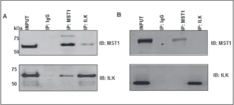
MST1 interacts with ILK
To understand how MST1 is involved in regulating osteoclast differentiation, we identified interacting proteins by Co-IP, followed by mass spectrometry. We compared the interacting proteins in both un-treated and RANKL-un-treated conditions, and selected the proteins that are exclusively present in untreated, and reduced or lost in RANKL treatments. Peptide Table 1. List of MST1 interacting protein differences identified through mass spectrometry.
Protein Name Peptides Identified by MS/MS and MASCOT Sequence coverage Sequence Ion score
ILK
G[45-66]R R[161-176]T I[335-346]Q A[393-406]A
45/0.0021 38/0.008 42/0.014 45/0.0025
11%
14-3-3 ζ/δ V[61-74]RS[28-41]R 72/3.5e-0650/0.0011 11%
Alpha-actinin L[302-311]RV735-746]R 91/1.7e-0745/0.0057 2%
Minimum sequence coverage threshold for identifying interactors-2%; MS/MS – tandem mass spectrometry; MASCOT – search database
Fig. 1. Hippo pathway involvement in osteoclast differentiation. (A-B) Osteoclast differentiation upon knock-down of Hippo pathway genes, LATS2, TAZ, YAP, MST1 and TEAD1. Non-silenced group was used as control.
coverage of 2% was used as the threshold and the list of proteins identified through such selection is pro-vided in Table1. Of them, integrin-linked kinase (ILK) was identified as a prominent interacting protein with ~11% protein coverage, and related to osteoclast dif-ferentiation [21]. To further confirm this MST1-ILK interaction, independent Co-IP experiments were per-formed (Fig. 3A and B). Overall, our results indicated that MST1 interacts with ILK and is lost in osteoclast differentiation after RANKL treatments.Given that MST1 levels decreased with RANKL treatment (Fig. 1), it is as question if the MST1-ILK interaction loss is either a direct loss or an effect acquired due to MST1 protein reduction after RANKL treatments. Although
speculative, the complete loss of ILK as identified through both mass spectrometry and Co-IP experi-ments, suggests that it could be an additive effect due to both direct interaction loss and reduction in MST1 protein levels. Future experiments on MST1 degrada-tion and expression analysis after RANKL treatment could help in understand the mode of loss of inter-action loss. Our data indicate the loss of MST1-ILK interaction after RANKL treatments.
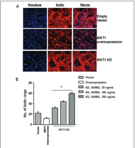
MST1-ILK interaction inhibits actin ring structure formation
Given that ILK is known to mediate integrin signal-ing and is usually found at actin-integrin connection sites, we inferred that actin cytoskeleton rearrange-ments associated with osteoclast were modulated due to MST1 and ILK interaction. After RANKL treat-ments, the control vector-infected cells differentiated normally, exhibiting osteoclastic actin ring structures, while MST1-overexpressing cells showed reduced dif-ferentiation and actin ring structure formation (Fig. 4A and B), suggesting negative regulation by MST1. Accordingly, MST1 KD cells showed a RANKL-dose-dependent increase in both actin ring structures and overall osteoclast differentiation (Fig. 4A and B). These results suggest that MST1 inhibits osteoclast Fig. 2. MST1 negatively regulates osteoclast differentiation.Overexpression and knockdown of MST1 were
per-formed and evaluated for changes in osteoclast differentiation by TRAP and pit formation assays. (A-B) TRAP assay. A – overexpression of MST1 showed a reduction in TRAP positive cells, while knockdown increased it (Fig. 1A). B – TRAP staining changes upon overexpression and knockdown of MST1. C – pit formation assay. Vec-tor – empty vecVec-tor of overexpression plasmid, MST1 – overexpression, MST1 KD – knockdown group. Statistical significance was seen between control group and KD or overexpression group (n=3, p<0.05).
differentiation at the level of actin ring structure for-mation, which is important for the progression of osteoclast differentiation and maturation.
MST1-ILK interaction negatively inhibits ILK activity
Given that actin rearrangements are mediated by ILK [22], and with our observation of the negative influence of MST1, we wondered whether MST1-ILK interaction negatively inhibits ILK-mediated actin re-arrangements. To that end, we overexpressed both the wild-type (WT) and kinase-deficient ILK mutant in MST1 KD cells and checked for changes in osteoclast differentiation. Our results showed a reduction in os-teoclast differentiation in ILK mutant cells. Similar results were obtained in double KD of ILK and MST1 cells (data not shown). In contrast, the cells overex-pressing WT ILK showed an increase in osteoclast Fig. 4. MST1 inhibits actin ring structure formation. Overexpres-sion and knockdown of MST1 were performed and evaluated for changes in actin ring structure by phalloidin staining. A – actin rings observed after expression and knockdown of MST1 (RANKL 100 ng/mL). B – quantification of actin ring structures after over-expression and knockdown of MST1 (n=3; p<0.05).
Fig. 5. MST1-ILK interaction negatively inhibits the ILK. Over-expression of wild-type (WT) or mutant (MUT) form of ILK per-formed along with MST1 knockdown and evaluated for changes in osteoclast differentiation. A – osteoclast number evaluated by the TRAP assay. B – actin ring structures evaluated by phalloidin staining (n=3; p<0.05).
differentiation (Fig. 5A). This change in osteoclast dif-ferentiation is also associated with actin ring structure formation (Fig. 5B). Together, these results suggest that ILK activity is required for osteoclast differentia-tion, and this activity is inhibited by MST1.
DISCUSSION
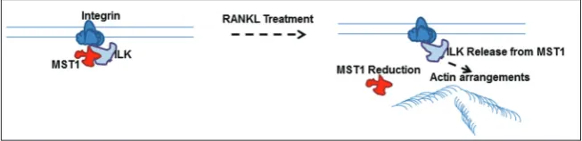
RANKL-based in vitro osteoclast differentiation sys-tems have been widely used to dissect the molecular mechanisms associated with osteoclasts differentiation and function [23-25]. In this study, using human pre-cursor cells and RANKL treatments, we have provided evidence that the Hippo signaling pathway protein, MST1, negatively regulates osteoclast differentiation. Furthermore, we have identified MST1-ILK interac-tion and its significance in regulating osteoclast actin structures, based on which we have proposed a model for MST1-ILK mediated osteoclast differentiation.
differentia-Fig. 6. A model for MST1-ILK-mediated osteoclast differentiation. Hippo pathway protein MST1 interacts with ILK and inhibits its activity to maintain undifferentiated conditions. After exposure to signals that induce differentiation, MST1 interaction with ILK is lost, caus-ing ILK to become active, which in turn mediates the formation of the actin rcaus-ing structure and osteoclast differentiation.
tion. Similar results were also obtained from MST2-based studies [20], suggesting that Hippo signaling in general might act in negatively regulating osteoclast differentiation, and could be exerted by the action of MST1/2.
Multiple studies have highlighted the significance of integrin signaling in promoting osteoclast differen-tiation [26-28]. However, molecular crosstalk between signaling pathways makes it difficult to segregate the components associated with osteoclast differentiation. Our results using a combination of Co-IP and mass spectrometry have identified an interaction between the Hippo pathway protein MST1 and the integrin signaling pathway protein ILK. Previous studies on ILK have also shown it to function as a cytoplasmic effector for integrin receptors and to modulate mul-tiple cellular functions [29]. More importantly, osteo-clast-specific ablation of ILK in mice showed reduced bone resorption function [21]. Consistent with this observation, our results also show that ILK inhibi-tion as mediated by MST1 shows reduced osteoclast differentiation and maturation, which would directly impact its overall functions.
Specific actin arrangements such as podosome- and actin-ring structures are known to be associated with the state and function of osteoclasts, and are effectors in ILK and integrin signaling [30-32]. Our results on ILK inhibition and MST1 overexpression showed defects in these structure formations, sug-gesting that Hippo pathway negatively regulates this structure formation at the level of its formation.
The results presented herein are summarized in a model for MST1-ILK-mediated osteoclast differentia-tion (Fig. 6). Our data indicate that MST1 interacts with ILK in undifferentiating conditions, and upon exposure to stimuli that induce differentiation, such as RANKL, when the MST1-ILK interaction is lost, causing ILK to be active, which in turn mediates actin ring structure formation and osteoclast differentia-tion. Nevertheless, it is also possible that there could be additional regulators that respond to stimuli and add to the complexity of the pathway.
CONCLUSION
Our study highlights a previously unidentified link between the Hippo and integrin signaling pathways. Although it is unclear whether this is a direct inter-action, given the role of integrin signaling in osteo-clast differentiation, the interaction implicated here is significant and could have a regulatory function. We report that this interaction regulates downstream actin organization. However, it is worth pointing out that the model is partly derived from overexpression and knockdown experiments, and further studies are required to confirm its relevance in vivo systems.
Authors’ contribution: Xiao-han Huang and Pan Su contrib-uted equally to this work. Xiao-han Huang, Pan Su, Wu-Yin Li conceived and devised the experimental plans, Xiao-han Huang, Pan Su performed the experiments and Xiao-han Huang, Pan Su, Wu-Yin Li wrote the manuscript.
REFERENCES
1. Yu FX, Guan KL. The Hippo pathway: regulators and regula-tions. Genes Dev. 2013;27(4):355-71.
2. Staley BK, Irvine KD. Hippo signaling in Drosophila: recent advances and insights. Dev Dyn. 2012;241(1):3-15.
3. Harvey KF, Zhang X, Thomas DM. The Hippo pathway and human cancer. Nat Rev Cancer. 2013;13:246-57.
4. Hong W, Guan KL. The YAP and TAZ transcription co-acti-vators: key downstream effectors of the mammalian Hippo pathway. Semin Cell Dev Biol. 2012;23(7):785-93.
5. Hansen CG, Moroishi T, Guan KL. YAP and TAZ: a nexus for Hippo signaling and beyond. Trends Cell Biol. 2015;25(9):499-513.
6. Hergovich A. Regulation and functions of mammalian LATS/ NDR kinases: looking beyond canonical Hippo signalling. Cell Biosci. 2013;3(1):32.
7. Hergovich A, Stegert MR, Schmitz D, Hemmings BA. NDR kinases regulate essential cell processes from yeast to humans. Nat Rev Mol Cell Biol. 2006;7(4):253-64.
8. Boyle WJ, Simonet WS, Lacey DL. Osteoclast differentiation and activation. Nature. 2003;423(6937):337-42.
9. Teitelbaum SL. Bone resorption by osteoclasts. Science. 2000;289(5484):1504-8.
10. Teitelbaum SL Osteoclasts, integrins, and osteoporosis. J Bone Miner Metab. 2000;18(6):344-9.
11. Ikeda K, Takeshita S. Factors and mechanisms involved in the coupling from bone resorption to formation: how osteoclasts talk to osteoblasts. J Bone Metab. 2014;21(3):163-7.
12. Lee SK, Lorenzo J. Cytokines regulating osteoclast formation and function. Curr Opin Rheumatol. 2006;18(4):411-8. 13. Mabey T, Honsawek S. Cytokines as biochemical markers for
knee osteoarthritis. World J Orthop. 2015;6(1):95-105. 14. Marks SC Jr. Osteoclast biology: lessons from mammalian
mutations. Am. J. Med. Genet. 1989;34(1):43-53.
15. McLean W, Olsen BR. Mouse models of abnormal skel-etal development and homeostasis. Trends Genet. 2001;17(10):S38-43.
16. Hayden RS, Fortin JP, Harwood B, Subramanian B, Quinn KP, Georgakoudi I, Kopin AS, Kaplan DL. Cell-tethered ligands modulate bone remodeling by osteoblasts and osteoclasts. Adv Funct Mater. 2014;24(4):472-9.
17. Sørensen MG, Henriksen K, Schaller S, Henriksen DB, Nielsen FC, Dziegiel MH, Karsdal MA. Characterization of osteoclasts derived from CD14+ monocytes isolated from peripheral blood. J Bone Miner Metab. 2007;25:36-45. 18. Yoshida CA, Komori H, Maruyama Z, Miyazaki T, Kawasaki
K, Furuichi T, Fukuyama R, Mori M, Yamana K, Nakamura K, Liu W, Toyosawa S, Moriishi T, Kawaguchi H, Takada K,
Komori T. SP7 inhibits osteoblast differentiation at a late stage in mice. PLoS One. 2012;7(3):e32364.
19. Subramanian B, Ko WC, Yadav V, DesRochers TM, Perrone RD, Zhou J, Kaplan DL. The regulation of cystogenesis in a tissue engineered kidney disease system by abnormal matrix interactions. Biomaterials. 2012;33(33):8383-94.
20. Lee J, Youn BU, Kim K, Kim JH, Lee DH, Seong S, Kim I, Han SH, Che X, Choi JY, Park YW, Kook H, Kim KK, Lim DS, Kim N. Mst2 Controls Bone Homeostasis by Regulating Osteoclast and Osteoblast Differentiation. J Bone Miner Res. 2015;30(9):1597-607.
21. Dossa T, Arabian A, Windle JJ, Dedhar S, Teitelbaum SL, Ross FP, Roodman GD, St-Arnaud R. Osteoclast-specific inactiva-tion of the integrin-linked kinase (ILK) inhibits bone resorp-tion. J Cell Biochem. 2010;110(4):960-7.
22. Qian Y, Zhong X, Flynn DC, Zheng JZ, Qiao M, Wu C, Ded-har S, Shi X, Jiang BH. ILK mediates actin filament rearrange-ments and cell migration and invasion through PI3K/Akt/ Rac1 signaling. Oncogene. 2005;24(19):3154-65.
23. Takahashi N, Udagawa N, Suda T. A new member of tumor necrosis factor ligand family, ODF/OPGL/TRANCE/RANKL, regulates osteoclast differentiation and function. Biochem Biophys Res Commun. 1999;256(3):449-55.
24. Takeshita S, Kaji K, Kudo A. Identification and characteriza-tion of the new osteoclast progenitor with macrophage phe-notypes being able to differentiate into mature osteoclasts. J Bone Miner Res. 2000;15(8):1477-88.
25. Roux S, Orcel P. Bone loss. Factors that regulate osteoclast differentiation: an update. Arthritis Res. 2000;2(6):451-6. 26. Duong LT, Lakkakorpi P, Nakamura I, Rodan GA.
Integrins and signaling in osteoclast function. Matrix Biol. 2000;19(2):97-105.
27. Zou W, Teitelbaum SL. Integrins, growth factors, and the osteoclast cytoskeleton. Ann N Y Acad Sci. 2010;1192:27–31. 28. Nesbitt S, Nesbit A, Helfrich M, Horton M. Biochemical characterization of human osteoclast integrins. Osteoclasts express alpha v beta 3, alpha 2 beta 1, and alpha v beta 1 integrins. J Biol Chem. 1993;268(22):16737-45.
29. Dedhar S, Williams B, Hannigan G. Integrin-linked kinase (ILK): a regulator of integrin and growth-factor signalling. Trends Cell Biol. 1999;9(8):319-23.
30. Teitelbaum SL. The osteoclast and its unique cytoskeleton. Ann N Y Acad Sci. 2011;1240:14-7.
31. Matsubara T, Myoui A, Ikeda F, Hata K, Yoshikawa H, Nishimura R, Yoneda T. Critical role of cortactin in actin ring formation and osteoclastic bone resorption. J Bone Miner Metab. 2006; 24(5):368-72.