Microorganisms surviving in the root canal system despite the chemomechnical preparation are the main reason for reinfection and endodon-tic treatment failure [1]. Among them,
Enterococ-cus faecalis is the most commonly isolated bacte-rium from teeth with periapical periodontitis with or without periodontal lesions [2–4]. Because of its ability to form intra- and extraradicular biofilms,
ORIGINAL PAPERS
Joanna Barbara Grącka-Mańkowska
1, A–D, Beata Zarzycka
2, A–C, F,
Halina Pawlicka
1, C, E, FBactericidal Efficacy of Different Diode Laser
Operation Modes Against
Enterococcus faecalis
Compared with Sodium Hypochlorite Irrigation
Skuteczność bakteriobójcza promieniowania lasera diodowego względem
Enterococcus faecalis
w porównaniu z irygacją podchlorynem sodu
1 Department of Endodontics, Medical University of Lodz, Łódź, Poland
2 Department of Microbiology and Laboratory Medical Immunology, Medical University of Lodz, Łódź, Poland
A – research concept and design; B – collection and/or assembly of data; C – data analysis and interpretation; D – writing the article; E – critical revision of the article; F – final approval of article
Abstract
Background. Microorganisms surviving in the root canal system despite the chemomechanical preparation are the main reason for reinfection and endodontic treatment failure. The introduction of laser-assisted endodontics resulted in higher success rates and less recurrent infections.
Objectives. To compare the antibacterial action of diode laser irradiation (CV – 1500 W, IMP – 3000 W), photo-activated disinfection (PAD), and 5.25% sodium hypochlorite (NaOCl) on Enterococcus faecalis in an infected tooth model.
Material and Methods. Fifty freshly extracted bovine teeth with single canals were prepared to apical size 45° ISO, autoclaved and contaminated with Enterococcus faecalis. After a 48-hour incubation the specimens were randomly divided into four treatment groups and one control group (n = 10): Group 1 was irradiated with 1500 mW (con-tinuous wave diode laser irradiation); Group 2 was irradiated with 3000 mW (impulse diode laser irradiation, 300 ms – pulse, 300 ms – pause); Group 3 was subjected to photo-activated disinfection and Group 4 was irrigated with a 5.25% NaOCl solution for 15 min. Group 5 was rinsed with saline solution (control). After treatment, the dentin samples were collected and the number of surviving bacteria was determined by classical quantitative microbiologic evaluation.
Results. The differences in the mean number of the colony forming units (CFU/mg) between all the groups ana-lyzed were statistically significant (p < 0.001). The highest number of microorganisms was observed in the control group (52.7 x 10³ CFU/mg). Both diode laser applications resulted in a significant reduction in the number of
E. faecalis cells: G1 – 1.4 x 10³ CFU/mg, G2 – 4.2 x 10³ CFU/mg, whereas in the group subjected to photo-activated disinfection the extent of reduction was significantly less (G3 – 42.8 x 10³ CFU/mg).
Conclusions. The present study demonstrates that CW and pulse diode laser applications are both suitable for root canal disinfection, but do not eradicate all bacteria (Dent. Med. Probl. 2016, 53, 2, 216–221).
Key words: photodynamic therapy, disinfection, root canal treatment. Słowa kluczowe: terapia fotodynamiczna, dezynfekcja, leczenie kanałowe.
The research project funded by a grant from the Medical University of Lodz, grant number: 502-03/2-044-02/502-24-025.
Dent. Med. Probl. 2016, 53, 2, 216–221
the proton-pump mechanism and many differ-ent virulence factors (i.e. collagen binding protein – Ace, lipoteichoic acid – LTA, enterococcus aggre-gation substance – EAS), E. faecalis penetrates the tubular network deeper than other microorganisms and is more resistant to calcium hydroxide as an in-tracanal medication between appointments [5–8]. Once in a biofilm, pathogens are more resistant to antibiotics or antiseptic agents, and are able to withstand host-defense measures, such as anti-body-mediated phagocytic killing, unlike those in planktonic forms [9, 10]. There are numerous stud-ies proving that different combinations of antisep-tic agents in various solutions (chlorhexidine, calci-um hydroxide, sodicalci-um hypochlorite) are not able to eradicate Enterococcus faecalis completely. Micro-organisms colonize dentinal tubules as deeply as 1100 µm from the canal lumen, while the irrigation solutions used in the standard chemomechanical treatment penetrate only up to 130 – 300 µm deep in the dentinal tubules [11, 12]. Additional consid-erations in this regard are anatomical features like anastomoses, lateral canals, isthmuses and apical deltas, which are difficult to reach with endodontic instruments. Thus, some regions of the root canal system may remain untouched after conventional chemomechanical preparation [13].
The introduction of laser-assisted endodontics resulted in higher success rates and less recurrent infections. Laser light penetrates deeply into den-tinal tissues (> 1100 µm), and can also be redirect-ed in multiple directions by dentinal tubules act-ing as fiber-optic channels. Both factors improve the disinfecting capabilities of laser light [12].
The bactericidal effects of laser light are based on dose-dependent generation of heat. To avoid the risk of charring the dentin or possibly causing thermal injury to the periodontal tissues, the pulse operation module of the diode laser or the pho-to-activated disinfection procedure is suggested in some studies [14]. Low power laser activates a pho-tosensitizer (a dye such as toluidine blue ortho), which has a bactericidal effect on various species, even within oral biofilms [15].
Taking into consideration these notions, the present in vitro study was performed to compare the efficacy of three different diode laser operation modes, (CW 1500 mW, pulse 3000 mW, photo-acti-vated disinfection (PAD) with a standard irrigation solution (5.25% NaOCl) in root canals contaminat-ed with E. faecalis. A comparison of the effective-ness of these laser-based procedures in eliminating such a resistant pathogen has not been conducted as a research project and has not been published either. The following study exceeds the existing publica-tions in the laser-assisted endodontics domain, be-cause it provides an analysis of different laser-based
bacterial eradication methods compared with stan-dard antiseptic methods (sodium hypochlorite irri-gation). Single-rooted bovine teeth were chosen as a substitute to human teeth to allow the simultane-ous laser/NaOCl applications in all the groups, as bovine dentin is a proved satisfactory replacement for human dentin in microbiological studies [16].
Material and Methods
Preparation of Teeth
Fifty freshly extracted single-rooted bovine teeth were decoronated with a diamond flame-shaped bur. Calculus and periodontal soft tissues were removed from the external root surface with curettes, and the root canals were prepared with Largo®Peeso Ream-ers 2-3-4 (Dentsply, Maillefer, Ballaigues, Switzer-land) to an apical size equal to #45 ISO to standard-ize the specimens. The smear layer was removed by sequential irrigations of 17% EDTA and 5.25% NaOCl, for 5 min each. The canals were then rinsed with saline solution and dried with paper points. All the samples were sterilized by autoclaving (121°C, 30 min) to remove all preexisting bacteria.
Bacteria and Culture Conditions
A pure bacterial culture of Enterococcus faeca-lis (ATCC 11420) was obtained from the Culture Collection (Polish Academy of Sciences, Wrocław, Poland) and transferred onto a microbank system (MVIM VIA Bank, UK). Before inoculation, the bacterial sample was incubated for 24 h in a broth culture medium (Brain Heart Infusion, supple-mented with 7% sheep blood; Oxoid Ltd., Basing-toke, UK) at 37°C under aerobic conditions. After the incubation period, the turbidity of the broth was measured. The concentration of the inoculum was adjusted to a degree of turbidity of 0.5 accord-ing to the McFarland scale, usaccord-ing a CrystalSpec™ nephelometer (Becton Dickinson, Warsaw, Po-land), which corresponds to a bacterial concentra-tion of 1.5 x 10⁸ CFU/ml. All the samples were then
immersed in the prepared broth and incubated at 37°C for 24 h under aerobic conditions to allow the propagation of the bacteria into the dentinal tu-bules. After incubation, the teeth were allocated randomly to the experimental or control groups.
Laser Irradiation
Group 1 (n = 10) was treated with a high-pow-er diode lasa high-pow-er (DiodeLX Mini, Lasotronix, Warsaw, Poland) emitting at a wavelength of 980 nm and set at a power of 1500 mW, operated in CW mode.
Group 2 (n = 10) was treated with the same laser device set at a power of 3000 mW, operated in pulsed mode, using a pulse duration of 300 ms and a pulse interval of 300 ms. Irradiation fol-lowed the oscillatory technique developed by Gut-knecht et al. [17]. A fiber tip with a diameter of 200 µm was inserted into each root canal at a dis-tance of 1 mm from the apical foramen and was withdrawn coronally with helicoidal movements at a speed of approximately 2 mm/sec. The la-ser was adjusted for an effective average output power of 1500 mW for Group 1 and 3000 mW for Group 2, measured directly on the fiber tip using a laser power meter (Gentec UNO UP19K-15S-H5 – 15 J, Gentec Electro-Optics, Quebec City, Can-ada) before each irradiation cycle. This procedure ensures standardized irradiation schemes for each specimen.
Group 3 (n = 10) was subjected to photo-ac-tivated disinfection. Toluidine Blue Ortho (TBO) (Lasotronix, Warsaw, Poland) as a photosensi-tizing agent, was introduced into each canal for 60 s. After this pre-irradiation time, irradiation was performed with a diode laser (Diode LX Mini, Lasotronix, Warsaw, Poland) emitting at a wave-length of 635 nm and set at a power of 100 mW. Laser light was applied through a single-use end-odontic diffusor, which was gently moved up and down the canal during the irradiation time of 30 s.
Group 4 (n = 10) was irrigated with 5.25% NaOCl solution for 15 min with a side-vented end-odontic needle, size 30 ga (CanalPro™, Coltène/ Whaledent Inc., Cuyahoga Falls, OH, USA).
Group 5 (n = 10) was rinsed with saline so-lution using side-vented endodontic needles, size 30 ga, and served as a negative control.
Root Canal Sampling
The dentin chips were collected with Largo® Peeso Reamers 5-6 (Dentsply, Maillefer, Ballai-gues, Switzerland) and placed onto sterile plates, weighed before and after dentin collection on an electronic balance (AS, Radwag, Poland). The por-tions were then transferred to Eppendorf test tubes
containing 2 ml of sterile physiological saline so-lution and sonificated for 30 s (Ultrasonic Disin-tegrator, MSE, UK). For each sample, three seri-al 10-fold dilutions were prepared: 10–1, 10–2, 10–3,
providing 150 portions. One hundred microliters of each dilution were applied to a selective cul-ture medium (Enterococcosel Agar, Graso, Po-land) and incubated at 37°C for 24 h under aero-bic conditions. All procedures were conducted in a laminar flow chamber, using sterile instruments to avoid contamination.
Plate Count/Bacteriological
Evaluation
After the incubation period, the number of CFU/mg of dentin was counted for each plate.
Statistical Analysis
The mean value and the standard deviation of CFU values were calculated. As the data did not have a normal distribution, a nonparametric test was warranted. For significant differences be-tween the CFU, values were subjected to the Krus-kal-Wallis test. For group comparisons the Mann-Whitney U-test was used.
Results
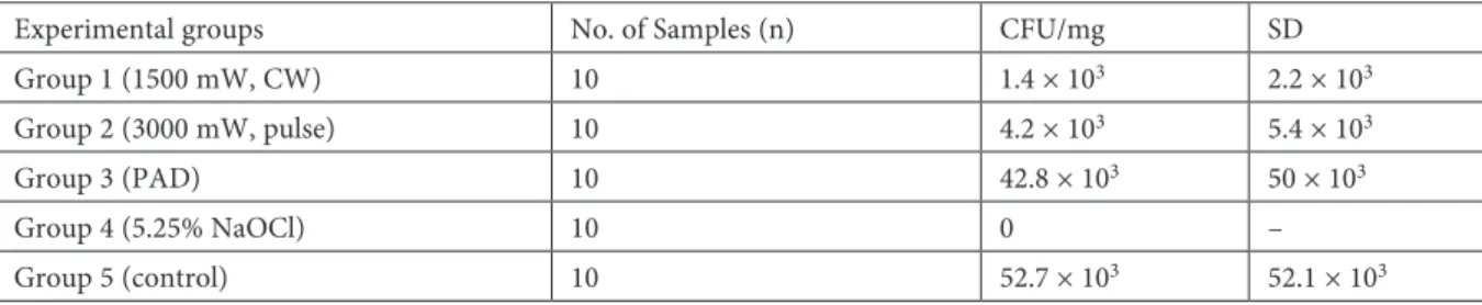
The results are given in Figure 1. The highest number of CFU/mg was observed in the negative control group (Group 5, rinsed with saline solution only). The complete eradication of E. faecalis was achieved in the 5.25% NaOCl group (Group 4, pos-itive control), as no bacterial cells were detected on the Enterococcosel Agar plates in any of the sam-ples. All the groups’ CFU/mg results were com-pared with Group 4 and Group 5 results and the groups subjected to laser-based eradication meth-ods results (CFU/mg) were compared separately (in pairs). A statistically important reduction of CFU/mg was observed in Group 1 (1500 mW, CW) and Group 4 (5.25% NaOCl) – positive control, Table 1. Efficacy of the diode laser, photo-activated disinfection (PAD) and 5.25% NaOCl irrigation in eliminating
Enterococcus faecalis (p < 0.001)
Experimental groups No. of Samples (n) CFU/mg SD
Group 1 (1500 mW, CW) 10 1.4 × 10³ 2.2 × 10³
Group 2 (3000 mW, pulse) 10 4.2 × 10³ 5.4 × 10³
Group 3 (PAD) 10 42.8 × 10³ 50 × 103
Group 4 (5.25% NaOCl) 10 0 –
in comparison with Group 5 (negative control), p < 0.001. Among the groups of samples treated with laser-based bacterial eradication methods (Group 1, 2 and 3), only the 1500 mW applied in the continuous wave mode was effective (Group 1), statistically important reduction of E. faecalis.
Discussion
Root canal treatment failure, resulting in peri-apical lesion formation may be caused by an in-fection with Enterococcus faecalis [1, 3, 18, 19]. The high resistance of the microorganism to com-monly applied disinfectants or inter-appointment medications [8] contributes to its high preva-lence in primary and secondary endodontic infec-tions [9], and has prompted a search for effective elimination methods [20, 21].
Different laser devices are used currently in root canal treatment [4, 22, 23]. The high power diode laser, emitting at a wavelength of 980 nm, was first introduced for use in endodontics in 1997 [22]. Its light is better absorbed by water than dental tissues, which results in greater dentin pen-etration and less laser light-dentin interactions, especially compared with the Nd-YAG laser [4]. Enamel prisms and dentin tubules acting as op-tical fibers permit laser light propagation as far as 1100 µm, and possibly more, from the canal lu-men [24]. Microorganisms are capable of invading the tubular 3D network up to a depth of 1100 µm. However, the conventionally used irrigation so-lutions penetrate periluminal dentin to no more than 130 µm deep [12]. Three different mecha-nisms of laser light bactericidal action have been
reported: A rise in the temperature of the bacte-rial environment above lethal values, a temper-ature rise inside the bacteria (due to laser light-sensitive chromophores), and light-induced mod-ulation of enzymatic activity [23]. The high power diode laser (810 nm) was previously used to dis-infect root canal systems in the pulsed operation mode with a repetition rate of 1.5 Hz and an out-put power of 4 W [22] or 3 W [4]. In the present study the bactericidal effect of the high power di-ode laser operated in the pulse mdi-ode (Group 2) was not as significant, possibly because only one irradiation cycle was performed instead of sever-al cycles in the previous studies [4, 22]. The aim of our study was to compare the bactericidal ef-fect of a single application of different procedures that are used in current practice. Single irradia-tion with a 980-nm diode laser operated at a pow-er output of 1500 mW in the continuous wave (CW) mode (Group 1) resulted in a significant re-duction of E. faecalis CFU/mg in comparison with the control group (Group 5). Complete E. fae-calis eradication was demonstrated in the 5.25% NaOCl-irrigated group (Group 4), which is in ac-cordance with other studies [23, 25]. This effect re-sults from the antimicrobial action of hypochlo-rous acid and active chlorine [26]. The irrigation time in the present study corresponds to the che-momechanical preparation time of root canals of average difficulty [27]. The method of sampling applied in the present study did not entail con-tinuing use of NaOCl on the agar plates, which is why no neutralizing agent was necessary [27]. Sev-eral methods of sample collection were applied in previous studies: Use of paper points, rinsing [28], and immersion in culture broth or in physiolog-ical saline solution [29, 30]. In the present study the dentin shaving method was applied to guar-antee precise quantification of the recovered vol-umes. The literature largely presents satisfactory disinfection rates achieved by several irradiation cycles with the high power diode laser operated in pulse mode [22, 31]. In the present study a single irradiation cycle of each laser-treated group was performed to standardize the procedure. Repeti-tive laser pulse irradiation could probably improve the bactericidal effect. High E. faecalis resistance to photo-activated disinfection is commonly asso-ciated with its Gram-positive cell wall structure, and/or the natural resistance of starved bacterial cells to adverse conditions [32]. Despite dentin tu-bules acting as optical fibers, some bacteria may invade anatomical root canal system complexi-ties deeper than others and form biofilms, thereby hindering complete eradication [13].
Further investigation of laser-assisted endo-therapies should be conducted to establish
ap-Fig. 1. Effect of CW and pulse mode diode laser irradiation, photo-activated disinfection and sodium hypochlorite irrigation on the survival of Enterococcus faecalis in the infected tooth model. Vertical bars rep-resent the mean number of recovered cells
1.4×10³ 4.2×10 3
42.8×10³
0
52.7×10³
0 10 000 20 000 30 000 40 000 50 000 60 000
1500 mW 3000 mW
propriate laser parameters and eliminate the risk of recurrent periapical infections. According to the authors, research that mimics in vivo condi-tions, in which different microorganisms interact among themselves, would be of invaluable clinical significance.
The present study demonstrates that the most effective laser-based method in E. faecalis elimina-tion is CW laser light applicaelimina-tion (1500 mW). Both pulse diode laser application and photo-activated disinfection can be suitable for root canal disinfec-tion, but do not eradicate all bacteria.
References
[1] Sundqvist G., Figdor D., Persson S., Sjögren U.: Microbiologic analysis of teeth with failed endodontic treat-ment and the outcome of conservative re-treattreat-ment. Oral Surg. Oral Med. Oral Pathol. Oral Radiol. Endod. 1998, 85, 86–93.
[2] Hubble T.S., Hatton J.F., Nallapareddy S.R., Murray B.E., Gillespie M.J.: Influence of Enterococcus faecalis
proteases and the collagen-binding protein, ace, on adhesion to dentin. Oral Microbiol. Immunol. 2003, 18, 121–126. [3] Kaufman B., Spångberg L., Barry J., Fouad A.F.: Enterococcus spp. in endodontically treated teeth with and
without periradicular lesions. J. Endod. 2005, 31, 851–856.
[4] De Souza E.B., Cai S., Simionato M.R.L., Lage-Marques J.L.: High-power diode laser in the disinfection in depth of the root canal dentin. Oral Surg. Oral Med. Oral Pathol. Oral Radiol. Endod. 2008, 106, e68–72.
[5] Spratt D.A., Pratten J., Wilson M., Gulabivala K.: An in vitro evaluation of the antimicrobial efficacy of ir-rigants on biofilms of root canal isolates. Int. Endod. J. 2001, 34, 300–307.
[6] Distel J.W., Hatton J.F., Gillespie M.J.: Biofilm formation in medicated root canals. J. Endod. 2002, 28, 689–693. [7] Stuart C.H., Schwartz S.A., Beeson T.J., Owatz C.B.: Enterococcus faecalis: its role in root canal treatment
fail-ure and current concepts in retreatment. J. Endod. 2006, 32, 93–98.
[8] Evans M., Davies J.K., Sundqvist G., Figdor D.: Mechanisms involved in the resistance of Enterococcus faecalis
to calcium hydroxide. Int. Endod. J. 2002, 35, 221–228.
[9] Lins R.X., de Oliveira Andrade A., Hirata Junior R., Wilson M.J., Lewis M.A.O., Williams D.W.: Antimi-crobial resistance and virulence traits of Enterococcus faecalis from primary endodontic infections. J. Dent. 2013, 41, 779–786.
[10] Stewart P.S., Costerton J.W.: Antibiotic resistance of bacteria in biofilms. Lancet, 2001, 358, 135–138.
[11] Klinke T., Klimm W., Gutknecht N.: Antibacterial effects of Nd:YAG laser irradiation within root canal dentin. J. Clin. Laser Med. Surg. J. Clin. Laser Med. Surg. 1997, 15, 29–31.
[12] Berutti E., Marini R., Angeretti A.: Penetration ability of different irrigants into dentinal tubules. J. Endod. 1997, 23, 725–727.
[13] Diallo B., Diatta M.: Evaluation de la hauteur du delta apical des dents antérieures maxillaires. Intérêt chirurgi-cal. Odontostomatol. Trop. 2002, 25, 33–36.
[14] Asnaashari M., Safavi N.: Disinfection of contaminated canals by different laser wavelengths, while performing root canal T\therapy. J. Lasers Med. Sci. 2013, 4, 8–16.
[15] Dobson J., Wilson M.: Sensitization of oral bacteria in biofilms to killing by light from a low-power laser. Arch. Oral Biol. 1992, 37, 883–887.
[16] Camargo C.H.R., Siviero M., Camargo S.E.A., de Oliveira S.H.G., Carvalho C.A.T., Valera M.C.: Top-ographical, diametral, and quantitative analysis of dentin tubules in the root canals of human and bovine teeth. J. Endod. 2007, 33, 422–426.
[17] Gutknecht N., Moritz A., Conrads G., Sievert T., Lampert F.: Bactericidal effect of the Nd:YAG laser in
in vitro root canals. J. Clin. Laser Med. Surg. 1996, 14, 77–80.
[18] Siqueira J.F.: Aetiology of root canal treatment failure: why well-treated teeth can fail. Int. Endod. J. 2001, 34, 1–10. [19] Love R.M.: Enterococcus faecalis – a mechanism for its role in endodontic failure. Int. Endod. J. 2001, 34, 399–405. [20] Haapasalo M., Endal U., Zandi H., Coil J.M.: Eradication of endodontic infection by instrumentation and
ir-rigation solutions. Endod. Top. 2005, 10, 77–102.
[21] Hems R.S., Gulabivala K., Ng Y-L., Ready D., Spratt D.A.: An in vitro evaluation of the ability of ozone to kill a strain of Enterococcus faecalis. Int. Endod. J. 2005, 38, 22–29.
[22] Moritz A., Gutknecht N., Goharkhay K., Schoop U., Wernish J., Sperr W.: In vitro irradiation of infected root canals with a diode laser: Results of microbiologic, infrared spectrometric, and stain penetration examina-tions. Quintessence Int. 1997, 28, 205–210.
[23] Meire M.A., De Prijck K., Coenye T., Nelis H.J., De Moor R.J.G.: Effectiveness of different laser systems to kill
Enterococcus faecalis in aqueous suspension and in an infected tooth model. Int. Endod. J. 2009, 42, 351–359. [24] Vaarkamp J., ten Bosch J., Verdonschot E.: Propagation of light through human dental enamel and dentine.
Caries Res. 1995, 29, 8–13.
[25] Radcliffe C.E., Potouridou L., Qureshi R., Habahbeh N., Qualtrough A., Worthington H.: Antimicro-bial activity of varying concentrations of sodium hypochlorite on the endodontic microorganisms Actinomyces is-raelii, A. naeslundii, Candida albicans and Enterococcus faecalis. Int. Endod. J. 2004, 37, 438–446.
[26] Zehnder M.: Root canal irrigants. J. Endod. 2006, 32, 389–398.
[27] Eldeniz A.U., Ozer F., Hadimli H.H., Erganis O.: Bactericidal efficacy of Er,Cr:YSGG laser irradiation against
[28] Schoop U., Moritz A., Kluger W., Patruta S., Goharkhay K., Sperr W.: The Er:YAG laser in endodontics: results of an in vitro study. Lasers Surg. Med. 2002, 30, 360–364.
[29] Schoop U., Kluger W., Moritz A., Nedjelik N., Georgopoulos A., Sperr W.: Bactericidal effect of different laser systems in the deep layers of dentin. Lasers Surg. Med. 2004, 35, 111–116.
[30] Moritz A., Jakolitsch S., Goharkhay K., Schoop U., Kluger W., Mallinger R.: Morphologic changes cor-relating to different sensitivities of Escherichia coli and Enterococcus faecalis to Nd:YAG laser irradiation through dentin. Lasers Surg. Med. 2000, 26, 250–261.
[31] Orstavik D., Haapasalo M.: Disinfection by endodontic irrigants and dressings of experimentally infected den-tinal tubules. Endod. Dent. Traumatol. 1990, 6, 142–149.
[32] Portenier I., Waltimo T., Ørstavik D., Haapasalo M.: The susceptibility of starved, stationary phase, and growing cells of Enterococcus faecalis to endodontic medicaments. J. Endod. 2005, 31, 380–386.
Address for correspondence:
Joanna Barbara Grącka-Mańkowska Department of Endodontics Medical University of Lodz Pomorska 251
92-213 Lodz, Poland
E-mail: [email protected] Conflict of Interest: None declared
Received: 25.02.2016 Revised: 13.03.2016 Accepted: 16.03.2016