Overlapping IgG4 responses to self and environmental antigens
in endemic pemphigus foliaceus
Ye Qian*, Joseph S. Jeong*, Jian Ye†, Bim Dang*, Maha Abdeladhim‡, Valeria Aoki§, Gunter Hans-Filhio¶, Evandro A. Rivitti§, Jesus G. Valenzuela‡, and Luis A. Diaz*
*Department of Dermatology, University of North Carolina at Chapel Hill, Chapel Hill, NC 27599,
USA
†National Center for Biotechnology Information, National Library of Medicine, National Institutes of
Health, Bethesda, MD 20894
‡Vector Molecular Biology Section, LMVR, National Institute of Allergy and Infectious Diseases,
NIH, Rockville, MD
§Departamento de Dermatologia, Universidade de Sao Paulo, Brazil
¶Departamento de Dermatologia, Universidade Federal de Mato Grosso do Sul, Brazil
Abstract
The etiology of human autoimmune diseases in general remains largely unknown, although the genetic and environmental interplay may be relevant. This applies to the autoimmune diseases of the skin such as the pemphigus phenotypes and others. In this group, there is an endemic form of pemphigus foliaceus [also known as Fogo Selvagem (FS)] where the pathogenic IgG4
autoantibody response to the self-antigen, Desmoglein 1 (Dsg1) cross-react with the LJM11 sand fly salivary gland antigen. In this investigation we dissected the IgG4 autoantibody repertoires utilized by FS patients in response to endogenous self Dsg1 and exogenous LJM11 sand fly antigen. Based on analyses of the genetic clonal signatures of these antibodies, our results indicate that there is a significant overlap between these two responses as all identified IgG4 monoclonal antibodies cross-react to both Dsg1 and LJM11 antigens. Germline H and L chain V gene antibodies generated according to mutated cross-reactive monoclonal antibodies preserved their reactivity to both antigens. Our findings suggest that both Dsg1 autoantigen and LJM11
environmental antigen could be the initial antigenic stimulants for the IgG4 autoimmune responses in FS. These results support our hypothesis that LJM11 antigen plays a substantial role in
triggering the IgG4 autoantibody development in FS, and provide new insights on how non-infectious environmental antigen(s) may drive the generation of autoantibodies in IgG4-related autoimmune diseases.
HHS Public Access
Author manuscript
J Immunol. Author manuscript; available in PMC 2017 March 01.
Published in final edited form as:
J Immunol. 2016 March 1; 196(5): 2041–2050. doi:10.4049/jimmunol.1502233.
Author Manuscript
Author Manuscript
Author Manuscript
Introduction
Fogo Selvagem (FS) is an endemic form of pemphigus foliaceus (PF) found in certain states of Brazil (1, 2). The hallmark of this disease is the presence of intraepidermal vesicles due to epidermal cell detachment (acantholysis) (3) induced by pathogenic IgG4 anti-desmoglein 1 (Dsg 1) autoantibodies (autoAbs) (4–8). FS shows similar clinical, histological and
immunological features to those observed in non-endemic PF (9, 10). Epidemiologic and immunogenetic studies suggest that both genetic and environmental factors contribute to the development of FS (1, 11, 12). Previous studies suggest that exposure to hematophagous insect bites in genetically predisposed individuals may be a risk factor for FS (12). To strengthen this hypothesis we have shown that IgG4 anti-Dsg1 autoAbs cross-react with LJM11 sand fly salivary gland antigen (13), which suggests that the development of IgG4 Abs may be linked to immune responses to environmental antigens. Compared to
investigations on the pathogenesis and genetic predisposition of autoimmune diseases, etiological studies regarding environmental triggers of these diseases are lacking due to low prevalence and the clinical heterogeneity of the diseases (14–19). Similarly, the random nature of autoimmune skin diseases in North America makes it difficult to assess their etiological commonality and further dissect their causes. In this regard the endemic nature of FS provides an invaluable model and rare opportunity to study the environmental factors within the FS endemic region and their contribution to the development of FS.
IgG4 Abs are known to be elevated in patients with FS (20–22), other bullous dermatoses (23), as well as autoimmune pancreatitis (24), Mikulicz's disease (primary Sjogren's syndrome) (25), and other diseases (26). Recently, the terms “IgG4-related disease” and ‘IgG4-related skin disease” have been proposed (26–28). Among some autoimmune diseases, increased serum levels of total IgG4 are often observed and certain specific histopathological features, such as IgG4 plasma cell infiltration in effected tissues or organs, are present. (26–28). On the other hand, increased circulating anti-Dsg1 IgG4 autoAbs are characteristic of FS/PF as these anti-epidermal autoAbs are pathogenic and are detected bound to the surface of detached keratinocytes in lesional and perilesional epidermis of these patients (4). In FS/PF the lesional skin does not show IgG4 B cell or plasma cell infiltrates. In 1989 Rock et al demonstrated that the IgG4 response in FS is pathogenic (20). Later studies confirmed that the bulk of pathogenic anti-Dsg1 autoAbs in FS are predominantly IgG4 (21). IgG4 anti-Dsg 1 Abs from FS patients are pathogenic in mice (8, 29); similar to those in PF (30) using an IgG passive transfer mouse model. One study showed that progression from the preclinical to the clinical stage of the disease is associated with a dramatic rise in IgG4 anti-Dsg1 autoAbs (21) and that the level of anti-Dsg1 IgG4 Abs can be used as a predictor of FS (22). Our recent finding that IgG4 autoAbs in FS cross-react with an LJM11 sand fly antigen (13) suggests that the development of IgG4 autoAbs in patients may be linked to exposure to an environmental antigen. The studies of the association of environmental factors, such as infectious agents, with the development of autoimmune diseases have a long history (15, 17–19, 31–34). However, the studies of non-infectious antigens and their association with the development of autoimmune diseases are scarce. Since sand flies are prevalent in FS endemic areas, it is possible that individuals living in these endemic regions, especially among those FS susceptible individuals, may
Author Manuscript
Author Manuscript
Author Manuscript
generate an immune response to antigens from sand fly saliva (i.e. LJM11). The LJM11 antigen, a member of the “yellow family” of salivary proteins is one of the most
immunogenic component among the sand fly saliva proteins and is also a marker for human exposure of sand fly bites (35). Given that anti-LJM11 Abs cross-react to autoantigen Dsg1 (13, 36) strongly suggests the existence of a “cross-reactive” epitope(s) on both LJM11 and Dsg1. Since exposure to exogenous antigens may induce the autoimmune response
to ”cross-reactive” autoantigens in autoimmune diseases (33, 34, 37–39), we hypothesize that the immune response directed against this “cross-reactive” epitope(s) subsequently initiates the generation of IgG4 autoAbs in FS susceptible individuals.
The aim of this investigation was to determine if the IgG4 autoAb responses to Dsg1 and LJM11 sand fly antigens overlap at the Ab V gene level on FS patients from endemic areas. Our study indicates that there is a striking overlap between these two IgG4 responses and LJM11 could be an inciting antigen for autoAb development, strongly suggesting that LJM11 sand fly antigen plays an important role in the etiology of FS.
Materials and Methods
Immortalized B cell samples from FS patients
Immortalized B cell samples from FS patients in this investigation have been described before (40) and kept at the University of North Carolina Dermatology Research Laboratories. These studies are approved by the institutional review boards from the University of North Carolina at Chapel Hill.
IgG4 Ab phage display library construction
To amplify the VH and VL genes for Ab phage display (APD) library construction, mRNA from the immortalized B cells of three FS patients (13) were isolated and their cDNA were synthesized. The L chain V genes were amplified as originally described (41). To obtain IgG4 only H chain genes, we used the 5’ primers described from the original method (41), while the 3’ primers for VH genes from the original method (41) were replaced by an IgG4 specific primer based on an IgG4 Ab constant region specific sequence designed by
Efremov et al (42). A 209 bp DNA segment from the heavy chain constant region (including 36bp from the IgG4 hinge region) was amplified along with the H chain V genes transcripts. The resulting IgG4 scFv (single-chain fragment variable) comprises L and H chain V segments and partial constant region of IgG4 H chain (Supplemental Figure 1). The subsequent APD library construction was according to the original method described (41). To introduce FLAG-tag at the N-terminal of the resulting scFv for more convenient detection and purification, the original pComb3xSS vector was modified. The primer sequences used for PCR are:
5’ATAGGCCAGGCCGGCCAGCACCATCACCATCACCATTAGGAGGGTGGTGGCT CTG3’ (for removing HA at C-terminal) and
5’ATAGGCCGCCTGGGCCAACTTGTCATCGTCATCCTTGTAATCCGCAGCTTGGG CCACGGTAGCGAA3’ (for adding FLAG at N-Term). PCR cycle: 94°C-4 min for one cycle; 94°C-30 sec/55°C-30 sec/72°C-5 min for 30 cycles; 72°C-10 min for one cycle. This
Author Manuscript
Author Manuscript
Author Manuscript
PCR product was SfiI digested and cloned to modified pComb3xSS vector (with FLAG-tag at the N-terminal).
Isolation of anti-Dsg1, anti-LJM11, and cross-reactive mAbs
The constructed IgG4 APD libraries were panned as described (41) against either Dsg1 or LJM11 for three rounds, or the alternation of Dsg1 and LJM11 for total of four rounds (Supplemental Table I). These selected clones were amplified and their H and L chain sequenced. Selected clones were subjected to scFv protein expression and production. Produced scFv mAbs were purified by either Nickel column (Qiagen, CA) or anti-FLAG magnetic beads (Sigma, MO) according to manufacturers’ instruction for further testing. Human Dsg1 and sand fly LJM11 protein were purified as described (13, 35, 36, 40, 43).
ELISA and other mAbs
The specificity of those purified scFv mAbs were tested by ELISA as previous described (13, 36, 40, 44). Negative control scFv (TT1) was kindly provided by Drs. Don Siegel and Stephen Kacir from University of Pennsylvania and was subcloned in our modified
pComb3xSS vector with an N-terminal FLAG-tag. Anti-FLAG-HRP (Sigma, MO) was used as secondary Ab for detection of all purified scFv mAbs.
Revertant scFv mAbs
The germline sequences of the H and L chain of two mAbs (4E4 and 2D11) isolated previously (40) were identified using The National Center for Biotechnology Information (IgBlast) (45). The H and L chain V genes of the germline formats of these two mAbs were synthesized (Genscript, NJ) and subcloned into our modified pComb3xSS vector. The revertant scFv mAbs (4E4-R and 2D11-R) and their mutated counterpart scFv mAbs (4E4 and 2D11) were expressed and purified as described (41).
VH gene analysis
The identified scFv mAb H chain genes were analyzed using Ab gene database at The National Center for Biotechnology Information (IgBlast) (45). Vector NTI (Invitrogen, MA) is used for multiple alignments of the VH genes.
Statistics
The significant difference between the ratio of replacement and silent mutations (R/S) for CDRs and FWRs is assessed using Chi-square test.
Results
Construction of IgG4 Ab Phage Display Libraries from FS B cells
Since the main pathogenic autoAbs are of the IgG4 subclass in FS (20, 21), we sought to focus on the characterization of IgG4 Abs from FS patients. The APD library constructed using the method originally described by Barbas et al (41) includes the Ab repertoire from all 4 IgG subclasses in a constructed IgG phage display library and the original IgG subclass identity could not be determined from resulting scFv Abs from the library. However,
Author Manuscript
Author Manuscript
Author Manuscript
because of the predominant presence of IgG4 Abs in autoimmune skin diseases (4–8) and other IgG4-related diseases (26), the identification and isolation of IgG4 mAbs from these patients became increasingly necessary to understand the mechanism of IgG4 Ab
development in these patients. Thus, we have modified the method for our particular purpose to obtain only IgG4 mAbs (Supplemental Figure 1). The advantages of the resulting mAbs are that: (a) the scFv Abs in these libraries derived from FS B cells comprise IgG4 Abs only, and (b) the scFv contain partial constant and hinge regions of IgG4 immediately following VH domain, and their IgG4 subclass identity can be further confirmed. Employing this strategy, we have constructed 3 IgG4 phage display libraries using mRNA isolated from 3 FS patients’ EBV immortalized B cells.
Isolation of anti-Dsg1 and anti-LJM11 IgG4 mAbs by panning against Dsg1 and LJM11
We have shown that FS patients have significantly higher IgG4 anti-Dsg1 and LJM11 Abs compared to normal controls (HC) from both FS (Limao Verde in Brazil, LV) and non-FS (US) endemic region (36), suggesting that there are IgG4 immune responses against both the autoantigen and environmental antigen. Additionally, that two anti-Dsg1 IgG4 mAbs isolated from FS patients previously using hybridoma method cross-react to both Dsg1 and LJM11 (13) suggests the presence of cross-reactive epitope(s). Thus, it is possible that IgG4 Abs are present in FS patients reacting to both Dsg1 and LJM11.
To determine whether there is an overlap between these anti-Dsg1 and anti-LJM11 IgG4 responses, we identified the anti-Dsg1 and anti-LJM11 IgG4 Abs from the IgG4 APD libraries constructed from B cells of 3 FS patients to study the IgG4 Ab repertoires by panning against either Dsg1 and/or LJM11 (Supplemental Table I) (41). Colonies were randomly selected from Dsg1 and LJM11 panning of these three IgG4 APD libraries. Their plasmids were purified and subject to sequencing using Ompseq and HRML-F primers as described (41). The Ab H and L chain gene sequencing results of selected Ab clones from IgG4 APD libraries were analyzed using Ab gene database at The National Center for Biotechnology Information (IgBlast) (45), and their four framework regions (FWRs) and three complementary determining regions (CDRs) determined. Totals of 39 (GCDS), 58 (FGS), 39 (JLDO) clones from Dsg1 and LJM11 panning, were successfully sequenced and their VH and VL gene usages were determined. The additional H chain constant region sequences of these mAbs were also analyzed and confirmed to be of IgG4 subclass, indicating that only IgG4 mAbs were identified from our constructed IgG4 APD libraries.
Same parental B cell origins by IgG4 mAb clones isolated from panning of IgG4 APD libraries against either Dsg1 autoantigen or LJM11 environmental antigen
The H chain V regions of mAbs from pemphigus are known to contribute most of the Dsg binding. For example, Di Zenzo et al (46) demonstrated that H chain V gene somatic mutations are required for mAbs to maintain its binding to Dsg3 while L chain V gene mutations are not required for their binding to Dsg3. Studies from Stanley group indicated that the H chain of an anti-Dsg mAb could pair with L chains encoded by different VL genes without losing its Dsg specificity (47–50). Consistent with all these previous findings, we also found that most of independent mAbs identified from either Dsg1 or LJM11 panning of FS IgG4 APD libraries have different L chains paired with the same H chains (data not
Author Manuscript
Author Manuscript
Author Manuscript
shown). Therefore, all our genetic analyses of these mAbs in this investigation are centered on the H chain V gene segments. Among all three CDRs, the CDR3 region is the most diverse part of the antibody V regions and a unique CDR3 sequence represents the genetic clonal signature for a group of Abs derived from the same parental B cell. Based on their H chain V gene usage and the clonal signature of clonally related Abs (51), these clones can be grouped to 4, 6, and 4 independent clones from 3 FS patients, respectively (Table I). It should be noted that the VH genes of two IgG4 mAbs (GCDS-2D11 and FGS-4E4) that were isolated using hybridoma method from our previous study (40) are also identified from these clones isolated in our current study. The H chain of GCDS-2D11 is encoded by IGHV2-5 while that of FGS-4E4 is encoded by IGHV3-48. Both clusters of mAbs encoded by these two VH genes and their respective CDR3s are identified from their corresponding IgG4 APD libraries (both VH genes are in bold in Table I). It indicates that antigen-specific mAbs can be identified using both hybridoma and APD libraries, further validating our newly developed method for construction and panning of IgG4 APD libraries to identify only IgG4 mAbs.
We next used two methods to further determine whether these mAbs are cross-reactive to Dsg1 and LJM11. These three IgG4 APD libraries were panned against the alternation of Dsg1 and LJM11 for total four rounds (panned against Dsg1 for 1st and 3rd rounds, and panned against LJM11 for 2nd and 4th rounds) (Supplemental Table I) to identify cross-reactive mAbs. Those identified clones were sequenced and compared to those clones identified from Dsg1 or LJM11 individual panning. Most mAbs found from Dsg1 and LJM11 only panning are also found from the Dsg1/LJM11 alternate panning, and thus determined to be cross-reactive. Some representative scFv were produced and purified as described (41) and tested by both anti-Dsg1 and LJM11 ELISA (40, 44). All ELISA tested mAbs are cross-reactive (Table I and data not shown). Three independent clones isolated from either Dsg1 or LJM11 individual panning (one from GCDS and two from FGS, highlighted in gray in Table I) also cross-react to both Dsg1 and LJM11. Thus all the clones identified from our panning were cross-reactive based on one or the combination of two methods, and the results are summarized in Table I. Accordingly, despite differences in their mutations in complementary determining regions (CDRs) and framework regions (FWRs) of those mAbs in each individual clonal lineage (see below and Figure 1) their specificity to both Dsg1 and LJM11 is conserved.
Clonal selection and expansion among Anti-Dsg1/LJM11 mAbs
Studies on some autoimmune diseases suggest that the autoimmune response in these diseases involves clonal selection and expansion events and is often oligoclonal (53). In the present investigation, we isolated a total of 14 independent clones from 136 Dsg/LJM11 specific clones. It appears that autoimmune response within each individual patient is oligoclonal since only a limited number of independent clones were isolated. Some of the mAbs used the same VH genes by different FS patients. For example, IGHV1-18, IGHV1-69, and IGHV4-34 are shared by patients FGS and JLDO, with different CDR3 sequences between each linage of mAbs from these two patients (Table I). Some of the mAbs from two patients share the same VH genes, suggesting there is a level of constrained VH gene usage in FS, consistent with other studies on the Ab repertoire in autoimmune
Author Manuscript
Author Manuscript
Author Manuscript
diseases (53). The analyses of VH genes of these mAbs identified from panning of IgG4 APD libraries also demonstrate that the clonal expansion is apparent in all three patients. Phylogenetic trees in Figure 1 suggest that evolutionarily these clones originated from the same parental B cells. For example, on the top left panel, all these Abs from patient GCDS are all encoded by IGHV5-51 VH gene and have the same CDR3 sequence. The VH genes of these Abs originated from the germline gene IGHV5-51 and underwent somatic mutation and clonal expansion during their maturations (see below). It is apparent that there is no divergent development among these mAbs identified from panning against either Dsg1 (clones initiated with D in Figure 1) or LJM11 (clones initiated with L in Figure 1). Most of the Dsg1 and LJM11 clones can be found on the same branches; further suggesting that Dsg1 and LJM11 have shared epitope(s) and both may serve as the selecting antigen for driving the development of these Abs.
It is interesting that the H-CDR3 of two independent clones from one patient (GCDS) have D/ExxxW consensus sequences (in bold in Table I), which is the same as shared amino acid sequences in pathogenic pemphigus mAbs (50). Two H-CDR3 of independent clones from another patient also have YYYY aromatic amino acid clusters (in bold in Table I), similar to a pathogenic mAb from another study (46).
Extensive mutations among anti-Dsg1/LJM11 IgG4 Abs in FS patients
As it has been shown, those mutations are not the results of PCR errors through
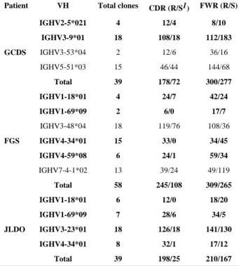
amplification of H and L chain V genes during APD library constructions (47). To further determine whether there are any characteristic mutations of anti-Dsg1 and anti-LJM11 IgG4 Abs in FS during their evolution, the somatic mutations of five lineages of mAb clones were identified using IgBlast and aligned (Figure 2). All mAbs isolated from the libraries are extensively mutated, both in their CDRs and FWRs (Figure 2). Similar to those phylogenic analyses shown in Figure 1, the different mutations among different clones in the same lineages of mAbs (using the same VH genes) demonstrate clear step-wise mutation during their evolutions, consistent with continuous antigenic stimulation. One striking consistency is that despite extensive different FWR mutations among those different clones of the same clonal lineage, almost all the mutations at CDR regions are preserved. For example, clones from IGHV3-23 lineage of JLDO shown in Figure 2 (bottom panel highlighted in red), all mutations at CDR1 and CDR2 are identical (C63T, G89C, and G92A in CDR1, and G15157A, G158A, G161A, T165C, and A172G in CDR2). In contrast, there is at least one mutation difference at FWRs for these listed clones using the same VH gene (IGHV3-23). In addition, as shown in Figure 3 using GCDS-L3 as an example, except one mutation at CDR2 (T-C mutation), all other mutations at CDR1 and CDR2 are replacement mutations which result in the change of amino acids compared to the germline VH (IGHV3-9). Those highly preserved replacement mutations at CDRs suggest that they were specifically favored during affinity maturation of these mAbs during their evolution. The average ratios of replacement mutations versus silent mutations (R/S) in CDRs are 2.47, 2.27, and 7.92, respectively, for the three FS patients, which is significantly higher than that in FWR (1.08, 1.17, and 1.26) (χ2 test, p<0.001) (Table II). The CDR R/S ratios of 10 clones (in bold in Table II), out of
total of 14 independent clones are higher than 2.9, which also indicates antigen selection (54). Highly diverse nucleotide mutations on the same VH clone clusters suggests that these
Author Manuscript
Author Manuscript
Author Manuscript
mAbs underwent multiple rounds of clonal expansion, consistent with our hypothesis that chronic antigenic stimulation (by either self or environmental antigen, or both) is a feature for FS Ab development.
Shared mutations have been found previously in CDRs of the same VH genes encoding anti-Dsg1 mAbs among FS patients and an individual before the onset of FS (40). To determine whether particular shared mutations are necessary for the development of the Dsg1/LJM11 specificity, the three shared VH genes (IGHV1-18, IGHV1-69, and IGHV4-34) from two FS patients (FGS and JLDO) were aligned to identify shared mutation among them. However, no shared mutation is present among all these VH gene-sharing mAbs between those from two patients (Figure 4). Furthermore, the CDR3 sequences of these VH gene-sharing mAbs from two patients are also different as shown in Table I. In general, H chain CDR3 is important for Ab specificity (55, 56). Though our result suggests that the Dsg1/LJM11 specificity of cross-reactive Abs from FS may be governed by the combination of all three CDR sequences rather than by a single CDR such as CDR3. This suggests that the Ab response in different individuals is flexible and may have multiple routes to achieve the same specificity during the positive selection of their respective Ab evolution. The extensive mutations in FWRs among those FS autoAbs (Table II) suggest that those mutations may also aid in improving the affinity of these autoAbs to their target antigens in FS, as found previously in broadly neutralizing Abs to HIV-1 (57).
Germline VH and VL encoded revertant mAbs from FS recognize both Dsg1 and LJM11
The wide presence of Dsg1 and LJM11 cross-reactive Abs in FS suggests that either Dsg1 or LJM11, or both may participate in driving the development of these Abs. However, it does not reveal whether Dsg1 or LJM11 could be the initial target for the naïve B cells in FS. It is also possible that some other antigens (either environmental or self), but not Dsg1 or LJM11 is the initial target that activates the naïve B cells which are the precursors for the eventual plasma cells that secrete these autoreactive Abs in FS. Therefore, it would be interesting to determine if naïve B cells expressing germline V genes can bind Dsg1 and LJM11.
The mutations in the VH and VL genes of two cross-reactive mAbs, namely FGS-4E4 and GCDS-2D11 (their VH are in bold in Table I) which were found using both hybridoma method (13, 40) and APD method, were identified using IgBlast (45) and reverted back to the germline configurations (Figure 5A). The DNA of the germline VH and VL genes of these two revertant mAbs were synthesized, subcloned into the original modified pComb3xSS vector (Methods), produced and purified as scFv (4E4-R and 2D11-R). The reactivity of these two revertant mAbs to Dsg1 and LJM11 was tested by ELISA and compared to that of their mutated counterpart scFv. As shown in Figure 5B, these two revertant scFv react to both Dsg1 and LJM11. This suggests that either Dgs1 or LJM11 (or both) may be the initial target(s) of autoimmune responses in FS, further implicating the environmental contribution to the development of FS.
Discussion
Our previous studies regarding the presence of cross-reactive mAbs in FS patients (13) suggest that there might be an overlap between the immune responses towards these two
Author Manuscript
Author Manuscript
Author Manuscript
antigens. In this investigation we seek to disclose the presence of an overlap between IgG4 responses against either Dsg1 or LJM11 in FS. Based on our hypothesis, the cross-reactive epitope(s) on Dsg1 and LJM11 would drive the development of cross-reactive Abs in FS. Due to the polyclonal nature of the Abs in patients’ sera, the cross-reactivity of those IgG4 Abs can only be determined using monoclonal IgG4 Abs. The construction of IgG4 specific APD libraries and the subsequent panning of these libraries against either autoantigen or environmental antigen, i.e. LJM11 sand fly antigen make it possible to identify these antigen-specific IgG4 Abs. Compare to the hybridoma method utilized in our previous investigation on FS Ab development (40), our newly developed IgG4 APD method modified from original APD method (41) has a distinct advantage over the hybridoma method. For example, only two IgG4 mAbs were isolated using hybridoma method even several thousands of hybridoma clones were screened from 8 FS patient B cell samples ((40) and (unpublished data)). However, 14 IgG4 independent mAb clones are isolated from three FS patients using this IgG4 APD method. It indicates that IgG4 APD method has broader coverage of the Ab repertoire than hybridoma method does. Moreover, this newly developed IgG4 APD method may have a wider application to the study of IgG4 Ab repertoires of other IgG4-related diseases in the future.
The presence of cross-reactive mAbs in FS (13) also suggests the presence of cross-reactive epitope(s) between autoantigen Dsg1 and environmental antigen LJM11. Notably, these two molecules are evolutionary distant and share little primary sequence homology. The cross-reactive mAbs from FS patients bind both molecules via conformational epitope (13) which does not rely on the similarity of the linear sequences of them. This explains the possible presence of a “cross-reactive” epitope on both molecules and also suggests that there are other epitopes on both molecules which are not cross-reactive, such as those epitopes that consist of continuous amino acids (linear epitopes). Thus, besides these Abs derived against the cross-reactive epitope(s) on both molecules, it should be expected that there are Abs from FS patients that are either Dsg1- or LJM11-specific which do not cross-react to the other. It is quite surprising that all mAbs identified from our investigation are cross-reactive, regardless of whether they are obtained from panning against Dsg1 or LJM11. Even though it might be unlikely, we cannot rule out that these two molecules share more than one conformational epitopes. Hence it is possible that these mAbs bind different “cross-reactive” epitopes shared by these two molecules. Still, there might be other explanations. For example, the current screening method for mAb clones identified by panning of APD libraries only allows us to examine a limited number of clones. Thus, it is possible that only the most abundant clones were captured while those rare clones, such as Dsg1- or LJM11-specific, escaped the detection. Nevertheless, the detection of identical and clonally related clones indicates that the most abundant IgG4 mAbs in these FS patients have been disclosed and the presence of these cross-reactive IgG4 demonstrates the possible contribution of the environmental antigen in FS development. The rapid development of next generation sequencing method for high throughput Ab sequence analysis will make it possible to identify those rare clones if the need arises in the future (58).
Another implication of our finding is that the cross-reactive epitope(s) might be the most immunogenic (or most dominant) that induces the strongest immune response against this epitope(s), as the conservation of the CDR mutations among most VH sharing mAbs
Author Manuscript
Author Manuscript
Author Manuscript
identified from the same patient is remarkably stable. Thus, most IgG4 Abs from FS patients might be developed against this epitope. LJM11 is known to be the most immunogenic component in sand fly saliva (35) and it might be due to this cross-reactive epitope. In this regard, the cross-reactive epitope on Dsg1 must be a predominant epitope for the
development of anti-Dsg1 Abs in FS. The presence of a predominant epitope is corroborated by other investigations on pemphigus. For instance, it has been shown that one predominant pathogenic epitope on Dsg1 may be targeted by most PF patients, as the binding of an anti-Dsg1 pathogenic mAb can be inhibited by sera from these patients (48). Similarly, there is also an immunodominant epitope on Dsg3 in mucocutaneous PV (46). Not only is the binding of a pathogenic mAb from a PV patient to Dsg3 inhibited by these tested PV sera, this pathogenic mAb also inhibits PV polyclonal Abs from binding to Dsg3 (46). Therefore, it is possible that all these mAbs isolated from this study target one conformational epitope shared by Dsg1 and LJM11, or epitopes in the vicinity of or overlapping this epitope.
The isolated cross-reactive mAbs could be either pathogenic or not pathogenic. If these IgG4 autoantibodies are pathogenic, it would indicate that an environmental antigen may be directly involved in driving the pathogenic IgG4 autoantibody response in FS. If they are not pathogenic, it would suggest that these IgG4 autoantibodies may play a role in activating an extended autoantibody response, via mechanisms such as epitope spreading in FS, and this subsequent autoantibody response leads to the development of pathogenic autoantibodies in FS as demonstrated before (59). In either case, the inciting antigen(s) (Dsg1 and/or LJM11) would be directly and/or indirectly involve in the development of pathogenic IgG4
autoantibodies in FS.
The current study also demonstrates that a non-infectious environmental antigen may contribute to the initiation of an autoimmune disease. It is possible, as shown in a previous study (36), that these FS-genetically susceptible individuals mount an IgE response to sand fly antigen exposure. They then develop IgG4 Abs in response to the repeated (chronic) exposure to sand fly bites, because of the well-known transition from IgE to IgG4 during chronic antigenic stimulation or allergen-specific immunotherapy in allergy (60–63). These IgG4 Abs turn out to be cross-reactive to Dsg1 autoantigen. Those anti-Dsg1 IgG4 Abs themselves or their triggered subsequent development of additional anti-Dsg1 IgG4 Abs result in the development of FS in those genetically susceptible individuals.
It is noteworthy that reverting mutated IgG4 autoAbs from FS back to their germline VH and VL genes do not abolish their reactivity to both Dsg1 and LJM11. Numerous studies using revertant Abs to study human autoimmune diseases, such as pemphigus vulgaris and Systemic Lupus Erythematosus indicate that the revertant Abs lose their reactivity (46, 64) or have reduced reactivity (65) to their corresponding autoantigens once their H and L chain V regions reverted back to their germline formats. AutoAb development from
non-autoreactive B cells has been proposed (64, 65). Due to the lack of candidate antigenic stimulants for triggering the development of these autoAbs in human autoimmune diseases, the initial targets (except these autoantigens of their corresponding autoimmune diseases) for these naïve B cells could not be further identified using revertant mAb method. In this study we provide evidence that environmental antigens, such as LJM11 from sand fly, could be an
Author Manuscript
Author Manuscript
Author Manuscript
antigenic stimulant that activates the naïve B cells in those individuals who are genetically predisposed to FS disease, and the subsequent development of IgG4 autoreactive Abs.
In summary, the extensive overlap of the IgG4 responses towards both Dsg1 autoantigen and LJM11 environmental antigen in this study is consistent with our hypothesis and thus establishes the possible link between the exposure to a non-infectious environmental antigen and the development of IgG4 autoAbs in FS. Moreover, that revertant mAbs also cross-react to both autoantigen (Dsg1) and environmental antigen (LJM11), provides further evidence that LJM11 could be the environmental antigen that triggers the activation of naïve B cells. All this evidence strongly suggests that LJM11 component from sand fly saliva as an environmental antigen may trigger the development of IgG4 autoAbs in FS susceptible individuals in FS endemic regions. Our findings regarding the role of IgG4 response to an exogenous antigen in FS may be expanded to the understanding of the etiology of other IgG4-related autoimmune diseases.
Supplementary Material
Refer to Web version on PubMed Central for supplementary material.
Acknowledgments
This work was supported by NIH grants, K01-AR056378, R01 AR067315 (to YQ) and R01-AR32599 (to LAD) and was supported in part by the Intramural Research Program of the National Institutes of Health, National Library of Medicine.
Abbreviations used in this paper
Dsg1 desmoglein 1
FS Fogo Selvagem
PF pemphigus foliaceus
APD Ab phage display
scFv single-chain fragment variable
References
1. Diaz LA, Sampaio SA, Rivitti EA, Martins CR, Cunha PR, Lombardi C, Almeida FA, Castro RM, Macca ML, Lavrado C, et al. Endemic pemphigus foliaceus (Fogo Selvagem): II. Current and historic epidemiologic studies. J Invest Dermatol. 1989; 92:4–12. [PubMed: 2642512]
2. Empinotti J, Diaz L, Martins C, Rivitti E, Sampaio S, Lombardi C, Sanches J. Endemic pemphigus foliaceus in western Parana, Brazil (1976–1988). Cooperative Group for Fogo Selvagem Research. Br J Dermatol. 1990; 123:431–437. [PubMed: 2095173]
3. Lever W. Pemphigus. Medicine (Baltimore). 1953; 32:1–123. [PubMed: 13024494]
4. Beutner EH, Jordon RE. Demonstration of Skin Antibodies in Sera of Pemphigus Vulgaris Patients by Indirect Immunofluorescent Staining. Proc Soc Exp Biol Med. 1964; 117:505–510. [PubMed: 14233481]
5. Udey MC, Stanley JR. Pemphigus--diseases of antidesmosomal autoimmunity. JAMA. 1999; 282:572–576. [PubMed: 10450720]
Author Manuscript
Author Manuscript
Author Manuscript
6. Anhalt G, Diaz L. Prospects for autoimmune disease: Research advances in pemphigus. JAMA. 2001; 285:652–654. [PubMed: 11176877]
7. Amagai M. Desmoglein as a target in autoimmunity and infection. J Am Acad Dermatol. 2003; 48:244–252. [PubMed: 12582396]
8. Ding X, Aoki V, Mascaro JM Jr, Lopez-Swiderski A, Diaz LA, Fairley JA. Mucosal and mucocutaneous (generalized) pemphigus vulgaris show distinct autoantibody profiles. J Invest Dermatol. 1997; 109:592–596. [PubMed: 9326396]
9. Diaz L, Sampaio S, Rivitti E, Martins C, Cunha P, Lombardi C, Almeida F, Castro R, Macca M, Lavrado C. Endemic pemphigus foliaceus (fogo selvagem). I. Clinical features and
immunopathology. J Am Acad Dermatol. 1989; 20:657–669. [PubMed: 2654208]
10. Stanley J, Klaus-Kovtun V, Sampaio S. Antigenic Specificity of Fogo Selvagem Autoantibodies Is Similar to North American Pemphigus Foliaceus and Distinct from Pemphigus Vulgaris
Autoantibodies. J Invest Dermatol. 1986; 87:197–201. [PubMed: 3525686]
11. Moraes ME, Fernandez-Vina M, Lazaro A, Diaz LA, Hans-Filho G, Friedman H, Rivitti EA, Aoki V, Stastny P, Moraes JR. An epitope in the third hypervariable region of the DRB1 gene is involved in the susceptibility to endemic pemphigus foliaceus (fogo selvagem) in three different Brazilian populations. Tissue Antigens. 1997; 49:35–40. [PubMed: 9027963]
12. Aoki V, Millikan RC, Rivitti EA, Hans-Filho G, Eaton DP, Warren SJ, Li N, Hilario-Vargas J, Hoffmann RG, Diaz LA. Environmental risk factors in endemic pemphigus foliaceus (fogo selvagem). J Investig Dermatol Symp Proc. 2004; 9:34–40.
13. Qian Y, Jeong JS, Maldonado M, Valenzuela JG, Gomes R, Teixeira C, Evangelista F, Qaqish B, Aoki V, Hans G Jr, Rivitti EA, Eaton D, Diaz LA. Cutting Edge: Brazilian Pemphigus Foliaceus Anti-Desmoglein 1 Autoantibodies Cross-React with Sand Fly Salivary LJM11 Antigen. J Immunol. 2012; 189:1535–1539. [PubMed: 22798673]
14. Goodnow CC. Multistep pathogenesis of autoimmune disease. Cell. 2007; 130:25–35. [PubMed: 17632054]
15. Fierabracci A. Unravelling the role of infectious agents in the pathogenesis of human autoimmunity: the hypothesis of the retroviral involvement revisited. Curr Mol Med. 2009; 9:1024–1033. [PubMed: 19747107]
16. Chervonsky AV. Influence of microbial environment on autoimmunity. Nat Immunol. 2010; 11:28–35. [PubMed: 20016507]
17. Miller FW. Environmental agents and autoimmune diseases. Adv Exp Med Biol. 2011; 711:61–81. [PubMed: 21627043]
18. Miller FW, Alfredsson L, Costenbader KH, Kamen DL, Nelson LM, Norris JM, De Roos AJ. Epidemiology of environmental exposures and human autoimmune diseases: findings from a National Institute of Environmental Health Sciences Expert Panel Workshop. Journal of autoimmunity. 2012; 39:259–271. [PubMed: 22739348]
19. Germolec D, Kono DH, Pfau JC, Pollard KM. Animal models used to examine the role of the environment in the development of autoimmune disease: findings from an NIEHS Expert Panel Workshop. Journal of autoimmunity. 2012; 39:285–293. [PubMed: 22748431]
20. Rock B, Martins CR, Theofilopoulos AN, Balderas RS, Anhalt GJ, Labib RS, Futamura S, Rivitti EA, Diaz LA. The pathogenic effect of IgG4 autoantibodies in endemic pemphigus foliaceus (fogo selvagem). N Engl J Med. 1989; 320:1463–1469. [PubMed: 2654636]
21. Warren S, Arteaga L, Rivitti E, Aoki V, Hans-Filho G, Qaqish B, Lin M, Giudice G, Diaz L. The Role of Subclass Switching in the Pathogenesis of Endemic Pemphigus Foliaceus. J Invest Dermatol. 2003; 120:104–108. [PubMed: 12535205]
22. Qaqish BF, Prisayanh P, Qian Y, Andraca E, Li N, Aoki V, Hans-Filho G, dos Santos V, Rivitti EA, Diaz LA. Development of an IgG4-based predictor of endemic pemphigus foliaceus (fogo selvagem). J Invest Dermatol. 2009; 129:110–118. [PubMed: 18704107]
23. Sitaru C, Mihai S, Zillikens D. The relevance of the IgG subclass of autoantibodies for blister induction in autoimmune bullous skin diseases. Arch Dermatol Res. 2007; 299:1–8. [PubMed: 17277959]
24. Kamisawa T, Okamoto A. Autoimmune pancreatitis: proposal of IgG4-related sclerosing disease. J Gastroenterol. 2006; 41:613–625. [PubMed: 16932997]
Author Manuscript
Author Manuscript
Author Manuscript
25. Yamamoto M, Takahashi H, Sugai S, Imai K. Clinical and pathological characteristics of Mikulicz's disease (IgG4-related plasmacytic exocrinopathy). Autoimmun Rev. 2005; 4:195–200. [PubMed: 15893711]
26. Stone JH, Zen Y, Deshpande V. IgG4-related disease. N Engl J Med. 2012; 366:539–551. [PubMed: 22316447]
27. Stone JH, Khosroshahi A, Deshpande V, Chan JK, Heathcote JG, Aalberse R, Azumi A, Bloch DB, Brugge WR, Carruthers MN, Cheuk W, Cornell L, Castillo CF, Ferry JA, Forcione D, Kloppel G, Hamilos DL, Kamisawa T, Kasashima S, Kawa S, Kawano M, Masaki Y, Notohara K, Okazaki K, Ryu JK, Saeki T, Sahani D, Sato Y, Smyrk T, Stone JR, Takahira M, Umehara H, Webster G, Yamamoto M, Yi E, Yoshino T, Zamboni G, Zen Y, Chari S. Recommendations for the nomenclature of IgG4-related disease and its individual organ system manifestations. Arthritis and rheumatism. 2012; 64:3061–3067. [PubMed: 22736240]
28. Tokura Y, Yagi H, Yanaguchi H, Majima Y, Kasuya A, Ito T, Maekawa M, Hashizume H. IgG4-related skin disease. Br J Dermatol. 2014; 171:959–967. [PubMed: 25065694]
29. Roscoe J, Diaz L, Sampaio S, Castro R, Labib R, Takahashi Y, Patel H, Anhalt G. Brazilian Pemphigus Foliaceus Autoantibodies Are Pathogenic to BALB/c Mice by Passive Transfer. J Invest Dermatol. 1985; 85:538–541. [PubMed: 3905977]
30. Anhalt GJ, Labib RS, Voorhees JJ, Beals TF, Diaz LA. Induction of pemphigus in neonatal mice by passive transfer of IgG from patients with the disease. N Engl J Med. 1982; 306:1189–1196. [PubMed: 7040962]
31. Gourley M, Miller FW. Mechanisms of disease: Environmental factors in the pathogenesis of rheumatic disease. Nat Clin Pract Rheumatol. 2007; 3:172–180. [PubMed: 17334340]
32. Cooper GS, Gilbert KM, Greidinger EL, James JA, Pfau JC, Reinlib L, Richardson BC, Rose NR. Recent advances and opportunities in research on lupus: environmental influences and
mechanisms of disease. Environ Health Perspect. 2008; 116:695–702. [PubMed: 18560522] 33. Munz C, Lunemann JD, Getts MT, Miller SD. Antiviral immune responses: triggers of or triggered
by autoimmunity? Nat Rev Immunol. 2009; 9:246–258. [PubMed: 19319143]
34. Francis L, Perl A. Infection in systemic lupus erythematosus: friend or foe? Int J Clin Rheumtol. 2010; 5:59–74. [PubMed: 20209114]
35. Teixeira C, Gomes R, Collin N, Reynoso D, Jochim R, Oliveira F, Seitz A, Elnaiem DE, Caldas A, de Souza AP, Brodskyn CI, de Oliveira CI, Mendonca I, Costa CH, Volf P, Barral A, Kamhawi S, Valenzuela JG. Discovery of markers of exposure specific to bites of Lutzomyia longipalpis, the vector of Leishmania infantum chagasi in Latin America. PLoS Negl Trop Dis. 2010; 4:e638. [PubMed: 20351786]
36. Qian Y, Jeong JS, Abdeladhim M, Valenzuela JG, Aoki V, Hans-Filhio G, Rivitti EA, Diaz LA. IgE Anti-LJM11 Sand Fly Salivary Antigen May Herald the Onset of Fogo Selvagem in Endemic Brazilian Regions. J Invest Dermatol. 2015; 135:913–915. [PubMed: 25285921]
37. McClain MT, Heinlen LD, Dennis GJ, Roebuck J, Harley JB, James JA. Early events in lupus humoral autoimmunity suggest initiation through molecular mimicry. Nat Med. 2005; 11:85–89. [PubMed: 15619631]
38. Cusick MF, Libbey JE, Fujinami RS. Molecular mimicry as a mechanism of autoimmune disease. Clin Rev Allergy Immunol. 2012; 42:102–111. [PubMed: 22095454]
39. Joseph CG, Darrah E, Shah AA, Skora AD, Casciola-Rosen LA, Wigley FM, Boin F, Fava A, Thoburn C, Kinde I, Jiao Y, Papadopoulos N, Kinzler KW, Vogelstein B, Rosen A. Association of the autoimmune disease scleroderma with an immunologic response to cancer. Science. 2014; 343:152–157. [PubMed: 24310608]
40. Qian Y, Clarke SH, Aoki V, Hans-Filhio G, Rivitti EA, Diaz LA. Antigen selection of anti-DSG1 autoantibodies during and before the onset of endemic pemphigus foliaceus. J Invest Dermatol. 2009; 129:2823–2834. [PubMed: 19571823]
41. Barbas, CFI.; Burton, DR.; Scott, JK.; Silverman, GJ. Phage display: a laboratory manual. Cold Spring Harbor, New York, USA: Cold Spring Harbor Laboratory Press; 2001.
42. Efremov DG, Batista FD, Burrone OR. Molecular analysis of IgE H-chain transcripts expressed in vivo by peripheral blood lymphocytes from normal and atopic individuals. J Immunol. 1993; 151:2195–2207. [PubMed: 8345203]
Author Manuscript
Author Manuscript
Author Manuscript
43. Xu X, Oliveira F, Chang BW, Collin N, Gomes R, Teixeira C, Reynoso D, My Pham V, Elnaiem DE, Kamhawi S, Ribeiro JM, Valenzuela JG, Andersen JF. Structure and function of a "yellow" protein from saliva of the sand fly Lutzomyia longipalpis that confers protective immunity against Leishmania major infection. J Biol Chem. 2011; 286:32383–32393. [PubMed: 21795673] 44. Qian Y, Prisayanh P, Andraca E, Qaqish BF, Aoki V, Hans-Filhio G, Rivitti EA, Diaz LA. IgE,
IgM, and IgG4 anti-desmoglein 1 autoantibody profile in endemic pemphigus foliaceus (fogo selvagem). J Invest Dermatol. 2011; 131:985–987. [PubMed: 21191415]
45. Ye J, Ma N, Madden TL, Ostell JM. IgBLAST: an immunoglobulin variable domain sequence analysis tool. Nucleic Acids Res. 2013; 41:W34–W40. [PubMed: 23671333]
46. Di Zenzo G, Di Lullo G, Corti D, Calabresi V, Sinistro A, Vanzetta F, Didona B, Cianchini G, Hertl M, Eming R, Amagai M, Ohyama B, Hashimoto T, Sloostra J, Sallusto F, Zambruno G, Lanzavecchia A. Pemphigus autoantibodies generated through somatic mutations target the desmoglein-3 cis-interface. J Clin Invest. 2012; 122:3781–3790. [PubMed: 22996451]
47. Payne AS, Ishii K, Kacir S, Lin C, Li H, Hanakawa Y, Tsunoda K, Amagai M, Stanley JR, Siegel DL. Genetic and functional characterization of human pemphigus vulgaris monoclonal
autoantibodies isolated by phage display. J Clin Invest. 2005; 115:888–899. [PubMed: 15841178] 48. Ishii K, Lin C, Siegel DL, Stanley JR. Isolation of pathogenic monoclonal anti-desmoglein 1
human antibodies by phage display of pemphigus foliaceus autoantibodies. J Invest Dermatol. 2008; 128:939–948. [PubMed: 18007588]
49. Yamagami J, Kacir S, Ishii K, Payne AS, Siegel DL, Stanley JR. Antibodies to the desmoglein 1 precursor proprotein but not to the mature cell surface protein cloned from individuals without pemphigus. Journal of immunology. 2009; 183:5615–5621.
50. Yamagami J, Payne AS, Kacir S, Ishii K, Siegel DL, Stanley JR. Homologous regions of autoantibody heavy chain complementarity-determining region 3 (H-CDR3) in patients with pemphigus cause pathogenicity. The Journal of clinical investigation. 2010; 120:4111–4117. [PubMed: 20978359]
51. Snow RE, Chapman CJ, Holgate ST, Stevenson FK. Clonally related IgE and IgG4 transcripts in blood lymphocytes of patients with asthma reveal differing patterns of somatic mutation. Eur J Immunol. 1998; 28:3354–3361. [PubMed: 9808205]
52. Link JM, Schroeder HW Jr. Clues to the etiology of autoimmune diseases through analysis of immunoglobulin genes. Arthritis Res. 2002; 4:80–83. [PubMed: 11879542]
53. Foreman AL, Van de Water J, Gougeon ML, Gershwin ME. B cells in autoimmune diseases: insights from analyses of immunoglobulin variable (Ig V) gene usage. Autoimmun Rev. 2007; 6:387–401. [PubMed: 17537385]
54. Shlomchik MJ, Marshak-Rothstein A, Wolfowicz CB, Rothstein TL, Weigert MG. The role of clonal selection and somatic mutation in autoimmunity. Nature. 1987; 328:805–811. [PubMed: 3498121]
55. Mahmoud TI, Schroeder HW Jr, Kearney JF. Limiting CDR-H3 diversity abrogates the antibody response to the bacterial polysaccharide alpha 1-->3 dextran. Journal of immunology. 2011; 187:879–886.
56. Silva-Sanchez A, Liu CR, Vale AM, Khass M, Kapoor P, Elgavish A, Ivanov II, Ippolito GC, Schelonka RL, Schoeb TR, Burrows PD, Schroeder HW Jr. Violation of an Evolutionarily Conserved Immunoglobulin Diversity Gene Sequence Preference Promotes Production of dsDNA-Specific IgG Antibodies. PLoS One. 2015; 10:e0118171. [PubMed: 25706374]
57. Klein F, Diskin R, Scheid JF, Gaebler C, Mouquet H, Georgiev IS, Pancera M, Zhou T, Incesu RB, Fu BZ, Gnanapragasam PN, Oliveira TY, Seaman MS, Kwong PD, Bjorkman PJ, Nussenzweig MC. Somatic mutations of the immunoglobulin framework are generally required for broad and potent HIV-1 neutralization. Cell. 2013; 153:126–138. [PubMed: 23540694]
58. Ravn U, Didelot G, Venet S, Ng KT, Gueneau F, Rousseau F, Calloud S, Kosco-Vilbois M, Fischer N. Deep sequencing of phage display libraries to support antibody discovery. Methods. 2013; 60:99–110. [PubMed: 23500657]
59. Li N, Aoki V, Hans-Filho G, Rivitti EA, Diaz LA. The role of intramolecular epitope spreading in the pathogenesis of endemic pemphigus foliaceus (fogo selvagem). J Exp Med. 2003; 197:1501– 1510. [PubMed: 12771179]
Author Manuscript
Author Manuscript
Author Manuscript
60. Aalberse RC, van der Gaag R, van Leeuwen J. Serologic aspects of IgG4 antibodies. I. Prolonged immunization results in an IgG4-restricted response. J Immunol. 1983; 130:722–726. [PubMed: 6600252]
61. Gleich G, Zimmermann E, Henderson L, Yunginger J. Effect of immunotherapy on
immunoglobulin E and immunoglobulin G antibodies to ragweed antigens: a six-year prospective study. J Allergy Clin Immunol. 1982; 70:261–271. [PubMed: 6811645]
62. Rossi R, Monasterolo G, Coco G, Silvestro L, Operti D. Evaluation of serum IgG4 antibodies specific to grass pollen allergen components in the follow up of allergic patients undergoing subcutaneous and sublingual immunotherapy. Vaccine. 2007; 25:957–964. [PubMed: 17045368] 63. Golden D, Lawrence I, Hamilton R, Kagey-Sobotka A, Valentine M, Lichtenstein L. Clinical
correlation of the venom-specific IgG antibody level during maintenance venom immunotherapy. J Allergy Clin Immunol. 1992; 90:386–393. [PubMed: 1527321]
64. Wellmann U, Letz M, Herrmann M, Angermuller S, Kalden JR, Winkler TH. The evolution of human anti-double-stranded DNA autoantibodies. Proc Natl Acad Sci U S A. 2005; 102:9258– 9263. [PubMed: 15968001]
65. Zhang J, Jacobi AM, Wang T, Diamond B. Pathogenic autoantibodies in systemic lupus erythematosus are derived from both self-reactive and non-self-reactive B cells. Mol Med. 2008; 14:675–681. [PubMed: 18677426]
Author Manuscript
Author Manuscript
Author Manuscript
Figure 1.
Phylogenetic tree of clonally related IgG4 antibodies isolated from panning of IgG4 ADP libraries. Clone names start with D indicate these clones were identified from Dsg1 panning while those start with L indicate they were identified from LJM11 panning.
Author Manuscript
Author Manuscript
Author Manuscript
Figure 2.
Alignments of clonally related IgG4 Abs identified from IgG4 ADP libraries. CDR1, CDR2, and partial CDR3 sequences are boxed in red. Mutated nucleotides are highlighted in red. One representative clone is selected for those clones that have the identical VH gene sequences.
Author Manuscript
Author Manuscript
Author Manuscript
Figure 3.
Cross-reactive IgG4 monoclonal antibody genes are extensive mutated, especially in CDRs (red boxes) compared to FWRs (blue boxes). The H chain gene of a representative clone is aligned with its germline gene. Amino acids from germline sequence changed as a results of replacement mutations are shown in purple.
Author Manuscript
Author Manuscript
Author Manuscript
Figure 4.
Alignments of IgG4 Ab clusters that use the same VH genes identified from two different patients. CDR1, CDR2, and partial CDR3 sequences are boxed in red. Clones from different patients, as well as the germline genes (bottom of each alignment panel) are separated by black lines. All nucleotide mutations are in red.
Author Manuscript
Author Manuscript
Author Manuscript
Figure 5.
Revertant mAbs derived from two cross-reactive anti-Dsg1 mAbs from FS also recognize both Dsg1 and LJM11. (A) H and L chain amino acid sequences of 4E4 (upper panel) and 2D11 (lower panel) and their corresponding revertant counterparts (encoded by germline VH and VL genes). Only mutated amino acids are shown for the VH and VL chains of 4E4 and 2D11. The amino acid sequences of CDR1, 2, and 3 of VH and VL are boxed. (B) Similar to mutated anti-Dsg1 IgG4 scFv mAbs, their revertant scFv mAbs also react to both Dsg1 (left panel) and LJM11 (right panel). The negative control scFv (TT1) does not react with either Dsg1 or LJM11. The reactivity of each scFv Abs to either Dsg1 (left panel) or LJM11 (right panel) is measured by relative light units (RLU) as previously described (36). The results shown are representative of three independent experiments with triplicates in each experimental group.
Author Manuscript
Author Manuscript
Author Manuscript
Author Manuscript
Author Manuscript
Author Manuscript
Author Manuscript
Table I
IgG4 scFv isolated from panning of IgG4 antibody phage display libraries Patient
VH
CDR3(FWR4)
Clones identified from panning
Cross-reactivity confirmed by
Dsg1 LJM11 GCDS IGHV2-5*02 1 AHRRLGLRYCSTSSCFG DFDY(W GQGTLVTVSS) 3 1 ELISA 3 IGHV3-9*01 VKDSDTGSSIRDASIFDS(WGQGSLVTVSS) 9 9 Alternate panning
4, ELISA
IGHV3-53*04 ARSLLSVGRYFDL(WGRGTLVTVSS) 0 2 2 ELISA IGHV5-51*03 ARSYNSS EYYIW FEA(WGQGTLVTVSS) 11 4
Alternate panning, ELISA
Total 23 16 FGS IGHV1-18*01 ARDRNMVRGLINKK FYYYY GMDV(WGQGTTLTVSS) 4 0 ELISA IGHV1-69*09 ASNNLTGTTVTYSYYGMDV(WGQGTTVTVSS) 2 0 ELISA IGHV3-48*04 ARGRTTFGGEGQLFDY(WGQGTLVTVSS) 9 9
Alternate panning, ELISA
IGHV4-34*01
ARGLDFRSGYSSSDGDYDSYYVDV(WGKGTTVTVSS)
7
8
Alternate panning, ELISA
IGHV4-59*08 AGGYCNSTSCYNQAFYS YYYY VDL(WGTGTTVTVSS) 2 4 ELISA IGHV7-4-1*02 AKGGDYGDFD(WGPGTLVTVSS) 7 6
Alternate panning, ELISA
Total 31 27 JLDO IGHV1-18*01 ARDFLPTVVTPAPFDY(WGQGTLVTVSS) 3 3
Alternate panning, ELISA
IGHV1-69*09 ASPYSGYDFLWAY(WGQGTLVTVSS) 5 2 ELISA IGHV3-23*01 AKWDHYPTRAFDI(WGQGTMVTVPS) 12 6
Alternate panning, ELISA
IGHV4-34*01
ASRPLYCSGGNCYSYYYQYGMNV(WGQGTTVTVSS)
5
3
Alternate panning, ELISA
Total
25
14
1 Those mAbs with VH in bold are also identified previously using hybridoma method (
40
).
Author Manuscript
Author Manuscript
Author Manuscript
Author Manuscript
Table II
Replacement(R) vs silent(S) mutation in CDRs and FWRs of VH genes of the IgG4 scFv from FS patients
Patient VH Total clones CDR (R/S1) FWR (R/S)
GCDS
IGHV2-5*021 4 12/4 8/10
IGHV3-9*01 18 108/18 112/183
IGHV3-53*04 2 12/6 36/16
IGHV5-51*03 15 46/44 144/68
Total 39 178/72 300/277
FGS
IGHV1-18*01 4 24/7 42/24
IGHV1-69*09 2 6/0 17/7
IGHV3-48*04 18 119/76 108/36
IGHV4-34*01 15 33/0 34/45
IGHV4-59*08 6 24/1 59/34
IGHV7-4-1*02 13 39/24 49/119
Total 58 245/108 309/265
JLDO
IGHV1-18*01 6 12/0 18/20
IGHV1-69*09 7 28/6 34/5
IGHV3-23*01 18 126/18 141/130
IGHV4-34*01 8 32/1 17/12
Total 39 198/25 210/167
1