WEAR PERFORMANCE OF MONOLITHIC DENTAL CERAMICS AGAINST ENAMEL
Taneet Ghuman
A thesis submitted to the faculty at the University of North Carolina at Chapel Hill in partial fulfillment of the requirements for the degree of Master of Science in the School of Dentistry (Operative Dentistry)
Chapel Hill 2016
Approved by:
Terence E. Donovan André V. Ritter
iii
ABSTRACTTaneet Ghuman: Wear Performance of Monolithic Dental Ceramics against Enamel (Under the direction of Terence E. Donovan)
Wear of human enamel and the restorative materials is often a functional and anatomical concern when selecting a restorative material for clinical application. Ceramic restorations have been known to cause wear of opposing enamel. Demands for more biocompatible ceramics have led to the development of new generations of monolithic ceramic materials, but their wear performance has not been fully understood.
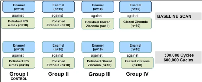
Objective: To measure and compare enamel wear against monolithic ceramic materials (Lithium Disilicate and Zirconia) as a function of surface treatment (polished, glazed, polished then glazed). Additionally, the surface roughness (Ra) of the ceramic materials was measured before wear testing to evaluate the effect of surface treatment on the surface roughness.
iv
test was conducted at 37°C using a continuous solution of 33% glycerin + 66% water for lubrication. Volume and depth loss (mm3) were determined, the 3D scans were superimposed with PROFORM software. Data was analyzed by ANOVA and Tukey post-hoc tests (p=0.05).
Results: Wear Volume: Ceramics: there was no measurable wear loss on polished zirconia, significant material loss wear values were seen with other groups. Enamel demonstrates significantly greater wear when opposed with polished lithium disilicate, polished and then glazed zirconia and glazed zirconia (p<0.0001) than polished zirconia specimens.
v
I dedicate my thesis to my parents, Mrs. Simran and Mr. Gurvinder Ghuman.
Thank you for your unconditional love and devotion with which you have raised me into this world. Thank you for giving me a chance to prove and improve myself through all walks of life. I am honored to
have you as my parents.
I dedicate my thesis to my sister and brother, Ameet and Jaikaran Ghuman.
To my wonderful fiancé Mark C. Milburn,
vi
ACKNOWLEDGEMENTS
I am grateful to the many individuals who contributed to my education, culminating in achieving this Master’s degree.
First, I wish to recognize Dr. Terence E. Donovan, Chair of my graduate committee. Without his vision and guidance, this work could not have been completed. I convey my earnest appreciation and gratitude for his invariable support, patience and motivation. I am highly honored to have worked under his guidance and would love to work with him in the future.
I would like to express my sincere thankfulness to my committee members, Dr. Andre V. Ritter, Dr. Lee W. Boushell for their invaluable advice over the period of my research project. They provided helpful assistance and advice for this work.
I wish to extend my utmost gratitude to Dr. John O. Burgess, Dr. Nathaniel Lawson, Mr. Preston Beck, without whose expertise, I would not have been able to finish this study in time. I cannot thank them enough for all that they have done for me.
Finally, I thank all my friends and the Operative Dentistry residents, without the moral support and encouragement of whom I would not have been able to achieve this.
I wish to thank Ivoclar Vivadent who generously donated the materials for the study.
vii
TABLE OF CONTENTS
LIST OF TABLES ... x
LIST OF FIGURES ... xi
LIST OF ABBREVIATIONS ... xiii
CHAPTER 1: LITERATURE REVIEW ... 1
1.1 Introduction ... 1
1.2 Ceramics Background ... 2
1.3 Dental Ceramic ... 4
1.3.1 IPS e.max ... 4
1.3.2 Zirconia ... 6
1.3.2.1 Properties ... 7
1.3.2.2 Transformation Toughening ... 7
1.3.2.3 Configuration ... 8
1.4 Wear ... 11
1.4.1 Clinical Significance of wear ... 13
1.4.2 Mechanisms of Wear ... 14
1.4.3 Mechanical Properties Related to Wear ... 16
1.4.3.1 Flexural Strength ... 17
1.4.3.2 Elastic Modulus ... 17
1.4.3.3 Fracture Toughness ... 17
1.4.3.4 Surface Roughness (Ra) ... 17
viii
1.6 Contributing factors for in-vitro wear simulation ... 19
16. Wear debris ... 23
1.7 Wear of Ceramic Restorative Materials ... 23
CHAPTER 2: MANUSCRIPT ... 26
2.1 Introduction ... 26
2.2 Hypothesis and Aims ... 27
2.3 Materials and Methods ... 28
2.3.1 Preparation of ceramic and enamel specimens ... 28
2.3.1.1 Ceramic Specimen preparation ... 28
2.3.1.2 Enamel styli preparation ... 29
2.3.1.3 Roughness Measurement ... 29
2.3.2 Cyclic loading of specimens ... 29
2.3.2.1 Wear simulation: Cyclic Loading in the newly modified UAB-Chewing Simulator ... 29
2.3.3. Wear determination ... 30
2.3.3.1 Determination of volumetric wear & depth loss ... 30
2.3.4 Surface imaging... 31
2.4 Statistical Analysis ... 31
2.5 Results ... 32
2.5.1 Wear of the Ceramic Materials ... 32
2.5.2 Enamel Wear ... 32
2.6 Discussion ... 33
2.7 Conclusion ... 37
Tables ... 38
ix
x
LIST OF TABLES
Table 1. Classification of commonly used Dental Ceramics………...38
Table 2. Study Protocol...39
Table 3. Materials Used...40
Table 4. Ceramic Wear with the means and Standard deviation of the groups...41
xi
LIST OF FIGURES
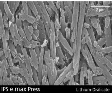
Figure 1. Lithium disilicate crystals in IPS e.max...43
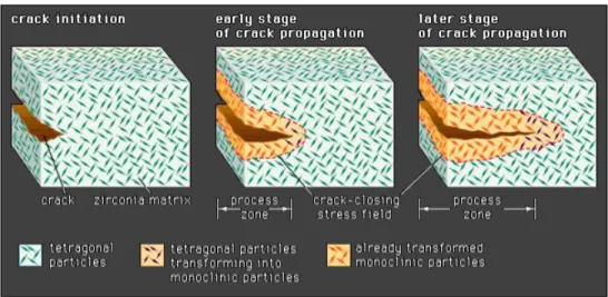
Figure 2. Phase Transformation in Zirconia...44
Figure 3. Schematic Description of Abrasion and Attrition...45
Figure 4. Two Body Wear...46
Figure 5. Three Body Wear...47
Figure 6. Fatigue Wear...48
Figure 7. Milling of Lithium Disilicate...49
Figure 8. Milling of Zirconia...50
Figure 9. Milled Flat Specimens...51
Figure 10. Specimen preparation for wear testing...52
Figure 11. Enamel styli preparation for wear testing...53
Figure 12. Pre scan of Enamel styli before wear testing...54
Figure 13. Modified UAB Wear Testing Device...55
Figure 14. Samples scanned using Proscan 2000 non-contact profilometer...56
Figure 15. Proform Wear Difference View...57
Figure 16. Proform Superimposed Wear View...58
Figure 17. Proform Before Scan...59
Figure 18. Proform After Scan...60
Figure 19. Material and enamel wear volume of Monolithic materials at 600,000 Cycles...61
Figure 20. Enamel wear volume of Monolithic materials...62
Figure 21. SEM image of IPS e.max (100x magnification) ...63
Figure 22. SEM image of IPS e.max (500x magnification) ...64
Figure 23. SEM image of IPS e.max (500x magnification) ...65
Figure 24. SEM image of Polished Zirconia (50x magnification) ...66
xii
Figure 26. SEM image of Polished Glazed Zirconia (50x magnification) ...68
Figure 27. SEM image of Polished Glazed Zirconia (500x magnification) ...69
Figure 28. SEM image of Polished Glazed Zirconia (500x magnification) ...70
Figure 29. SEM image of Glazed Zirconia (50x magnification) ...71
Figure 30. SEM image of Glazed Zirconia (300x magnification) ...72
xiii
LIST OF ABBREVIATIONS PFM: Porcelain fused to metal
MPa: Mega Pascal
CAD: Computer assisted design
CAD-CAM: Computer aided design - Computer aided manufacturing CTE: Coefficient of thermal expansion
FPD: Fixed partial denture
USPHS: United States public health service SEM: Scanning Electron Microscope
UCLA: University of California at Los Angeles TMJ: Temporomandibular Joint
PMMA: Polymethylmethacrylate CFOA: Contact-free occlusal area OCA: Wear at the occlusal contact area
FDA: Food and Drug Administration of the United States of America ISO: International Standard Organization
N: Newton nm: Nanometer
UAB: University of Alabama at Birmingham µm: Micrometer
1
CHAPTER 1: LITERATURE REVIEW 1.1 Introduction
In the recent decades, increased patient demands for esthetic dentistry has generated interest in all ceramic dental restorations. Increased development of newer generations and innovative techniques of fabrication have led many dentist to use all ceramic restorations, and this is likely to continue to rise and as more natural teeth are retained into old age.
Dental ceramics are known for their excellent chemical and optical properties. The wear of human enamel and the restorative material are often a functional, esthetic and anatomical concern when selecting a restorative material for clinical restorative treatment. However, ceramics are brittle and susceptible to fatigue fractures when subject to repetitive function and cause wear of opposing enamel.
2
1.2 Ceramics BackgroundThe first use of ceramics may have been in use more than 10,000 years ago during the Stone Age.1 In 1723, Pierre Fauchard described the use of ceramic in the “enameling” of metal denture bases.2 Earliest documentation on the use of ceramic “porcelain” dental reconstruction materials goes back to the
late 1700s in France by de Chemant. The first porcelain tooth in United States was fabricated only in 1817.3 These porcelain teeth were embedded in vulcanized rubber bases. Land introduced the first feldspathic porcelain crown in 1886.4 It was only at the beginning of the 1900s that the basic principles of individual ceramic crowns came in to existence. However, it took until early 1960 before any more significant technology developments were made, when vacuum firing technology and techniques to bond porcelain to metal were introduced. Alumina Reinforced ceramics were introduced in the 1970s as a result of McLean and Hughes research, glass ceramics in 1968, and Leucite-reinforced glass ceramics with pressure casting technology at the beginning of 1990. The bonding of ceramics to tooth structure was introduced in 1973 after the introduction of the acid-etch technique.5
Porcelain fused to metal, cast metal crowns and gold restorations were used in the past with considerable success. In the recent years, there is an increased concern for tooth colored restorations across the globe.6 Though porcelain based materials are still a major component of the market there have been efforts to replace metal core systems with all ceramic systems. Additionally, the increasing costs of base metal alloys and gold emphasized the need for economical and durable alternatives. Technical, nano-technical and design developments have resulted in high strength ceramics with improved marginal quality, esthetics and wear properties that can be pressed or machined.7-9 Today, ceramic materials are rapidly becoming common-place in dentistry, available in both naturally based and partially synthetic formulas.10
Ceramics used in dental restorations are essentially oxide based glass-ceramic systems. They have four fundamental features:
3
2. Sufficient mechanical properties and corrosion resistance 3. Appropriate aesthetic appeal.4. Biocompatibility
As the world of dentistry was beginning to embrace all ceramic restorations, leucite reinforced glass ceramics were introduced for veneers, onlays and single crowns.11 This was followed by the introduction of the lithium disilicate materials, which had improved mechanical properties.12 In Ceram alumina and zirconia were introduced as high strength cores that were indicated for single crowns and 3 unit anterior bridges.13 This glass ceramic core, which was prepared by a slip casting technique was veneered with any over-layering of porcelain.14
More recently, densely sintered high strength ceramics with mechanical properties superior to conventional ceramics have been developed for restorative dentistry.15 High strength ceramics such as alumina and Zirconia with metal oxides were used as the core material in the high load bearing areas.16 Zirconia, the most stable of these high strength ceramics was found to have flexural strength and fracture toughness values of 900-1400 MPa and 9-12 MPa/m1/2, respectively. Zirconium oxide–based materials, especially yttria-tetragonal zirconia polycrystals (Y-TZP), were introduced for prosthetic rehabilitations as a core material for single crowns, conventional and resin-bonded fixed partial dentures (FPDs), and, in dental implantology, as abutments or implants.16 The raw materials of the zirconia are the minerals zircon (ZrSiO4) and baddelyite (β-ZrO2), whose mines are located in South Africa, Australia and USA. Zirconia
was discovered by the German chemist Martin Heinrich Klaproth in 1789.17 The term zirconium refers to the metal, while zirconia ceramic (“zirconia”) refers to zirconia-dioxide-ceramic (ZrO2).17 Zirconia was
first introduced in the biomedical sciences in early 1960s. Its application further extended to orthopedics in 1980s, 15, 16 and then to dentistry in 1990.18
Previously, ceramic crowns because of their brittleness and low tensile strength were mostly used to restore only anterior teeth. Introduction of zirconia as a high strength core material, for all ceramics crowns has increased their use for posterior teeth. When selecting a restorative material for dental
4
missing tooth structure, it must have adequate strength to withstand the forces generated during mastication.
1.3 Dental Ceramic
Dental Ceramics consist of silicate glasses, porcelains, glass-ceramics and highly crystalline solids. Dental ceramics are nonmetallic, inorganic structures that mainly contain compounds of oxygen with one or more metallic or semi-metallic elements (aluminium, boron, sodium, titanium and
zirconium).2 Ceramics are undoubtedly the best materials available for matching the esthetics of a complex human tooth.
Glass-ceramics are particularly suitable for fabricating inlays and crowns, as these materials achieve very strong, esthetic results. High-strength ceramics are preferred in situations where the material is exposed to high masticatory forces. A well-designed and fabricated ceramic crown is often
indistinguishable from the adjacent nature tooth. Although commonly used to replace decayed tooth structure, the esthetic ceramic material is also used to cover pathological conditions of the enamel and dentin such as unsightly stains, malformations of the teeth, or improper calcification. Etchable-ceramic materials are used to close spaces (diastemae) existing between teeth and as enamel/dentin bonded partial or total coverage restorations without macro-retention. Different types of glass-ceramics and ceramics are available and necessary today to fulfill the needs of patients, dentists and dental technicians.
1.3.1 IPS e.max
In 2001, Ivoclar Vivadent released IPS e.max Press, which is a pressable lithium disilicate glass ceramic with the improved mechanical and optical properties. Four years later, IPS e.max CAD was introduced for CAD-CAM restoration in the dental clinic.19
5
lithium disilicate crystals forming within this material are needle-shaped and comprise about two thirds of the volume of the glass ceramic. The shape and volume of the crystals contribute increased flexural strength and fracture toughness of this material as compared with its predecessor Empress 2. The material comes in a pressable and machinable form. IPS e.max CAD blocks are manufactured in millable partially crystalized (Blue stage) ceramic blocks that can be milled and shaped by CAD/CAM equipment. After the IPS e.max CAD blocks are milled, the crystallization of the material is finalized via sintering in a ceramic furnace. During this time the fabricated ceramic restoration achieves full density and increased strength. At the same time, the initially bluish color changes to a tooth-like shade with improved translucency and brightness. There is very minimal shrinkage (approx. <1%) associated with lithium disilicate glass ceramics. The crystallization process causes the microstructure to change through controlled growth of lithium disilicate crystals. This material can be very translucent due to the high crystalline content and relatively low refractive index of the lithium disilicate crystals.21-23
IPS e.max is translucent enough to be used for monolithic full-contour restorations or for improved esthetics, as layered veneers, partial crowns and anterior and posterior crowns. IPS e.max materials may also be used as cores for all-ceramic restorations. In this case, the porcelain veneering materials for lithium disilicate glass ceramics are alumino-silicate glasses with fluoroapatite rather than leucite crystals. The fluoroapatite crystals contribute to the veneering porcelain’s optical properties and
coefficient (CTE) of thermal expansion to produce a veneering material that matches the CTE of lithium disilicate pressable or machinable material. Both the veneering material and lithium disilicate material are etchable due to the presence of the amorphous glass (non-crystalline) phase inherently present. Initial clinical data for single restorations are promising with this material, especially if bonded to the underlying tooth structure.21-23
6
posterior) IPS Empress 2 crowns & 20 3-unit FPDs (anterior or posterior) in 15 patients using the USPHS criteria. Single unit IPS Empress 2 all-ceramic crowns exhibited a satisfactory clinical performance over 2-year period. Ten (50%) catastrophic failures of FPD were reported, with 5 (25%) failures within 1-year and (25%) by 2 years.25
In another 2 year clinical study by Fasbinder et al, 62 IPS e.max crowns were cemented with 2 types of adhesive resin cements, Multilink sprint & experimental self-adhesive cement by Ivoclar Vivadent. The crowns evaluated at baseline, 6 months, 1 year and 2 yrs. There were no clinically identified cases of crown fracture or surface chipping and no reported sensitivity at 1 or 2 years with either cement.26 Early results indicate that IPS e.max crowns may be an effective option for all-ceramic crowns.26 Etman et al clinically evaluated 90 posterior teeth crowns in 48 patients for 3 years using modified USPHS criteria. Ceramic systems included IPS e.max, Procera AllCeram & metal ceramic veneered with IPS Classic Porcelain. Crowns that developed visible cracks were sectioned and removed, and the surfaces were analyzed using a scanning electron microscope (SEM). Data was analyzed using the Kruskal-Wallis nonparametric statistical test, followed by the Mann-Whitney test with Bonferroni
correction (α=0.05). IPS e.max & metal ceramic crowns showed fewer clinical changes than Procera
AllCeram. Visible roughness, wear & deformity were noticed in occlusal contact areas of Procera AllCeram crowns. SEM images showed well defined wear facets in both ceramic crown systems. IPS e.max's clinical behavior was comparable to Procera AllCeram & metal ceramic crowns but wear resistance was superior compared to the Procera AllCeram crowns.27 Early ceramics had a failure mechanism of bulk fracture when used as frameworks. Newer ceramic materials that use zirconia-based frameworks exhibit promising clinical data and superior esthetics.
1.3.2 Zirconia
7
alternative for single crowns and three-unit bridges anywhere in the mouth. Larger bridges have been discussed but no large sample clinical documentation exists for this application. Solid-sintered, monolithic ceramics are materials that are formed by directly sintering crystals together without any intervening amorphous glass matrix to form a dense, glass-free, polycrystalline structure.
1.3.2.1 Properties
Extensive research has been performed since the discovery of the transformation toughening capabilities of this material. The different stages of polymorph zirconia are temperature dependent: at ambient pressure, unalloyed zirconia can assume three crystallographic forms. At room temperature and upon heating up to 1170°C, the symmetry is monoclinic. The structure is tetragonal between 1170 and 2370°C and cubic above 2370°C and up to the melting point.18 The transformation from the tetragonal (t) phase to the monoclinic (m) phase upon cooling is accompanied by a substantial increase in volume (∼4.5%), sufficient to lead to catastrophic failure18. The ceramic shows a hysteretic martensic t → m transformation during heating and cooling. This transformation is reversible and begins at ∼950◦C on cooling. Alloying pure zirconia with stabilizing oxides such as CaO, MgO, Y2O3 or CeO2 allows the retention of the tetragonal structure at room temperature. Zirconia has a high temperature stability and melting point (2680°C), high hardness (1200-1350 HVN), high thermal expansion (>10 x 10-6 1/K), low thermal conductivity (<1 W/mK) and good thermo shock resistance (ΔT=400-500°C).18
1.3.2.2 Transformation Toughening
However, the metastability of tetragonal zirconia could increase the susceptibility to aging because some stress-generating surface treatments such as grinding or sandblasting can trigger the t→m transformation with volume increase and formation of compressive stresses on the surface, thereby modifying the phase integrity though increasing flexural strength.28 The increase of volume effectively stops the crack propagation. This process is called “transformation toughening”, with the resistance
8
1.3.2.3 ConfigurationZirconia in dentistry is milled in the pre-sintered stage. This configuration is a soft, chalk-like stage that is called the “green” stage. During the sintering process, the material shrinks and results in
volume shrinkage of about 20-25%.
It’s very important to know the exact volume shrinkage information for the individual zirconia
blank blocks in order to optimize fit of the restoration. The zirconia is called hipped (hot isostatic pressed) when the material is industrially sintered, and then is CAD-milled at its final high strength. Hipped zirconia has a constant grading and thus a more homogeneous quality. As expected, milling time and wear of the milling tools is higher in comparison to the pre-sintered variants. Zirconia for dental applications is stabilized at room temperature with the addition of 3 mol% yttria.30
Solid-sintered ceramics (polycrystalline glass-free) have the highest potential for strength and toughness and find application as high-strength frameworks for crowns and fixed partial dentures. Techniques for the accomplishment of high firing temperatures and sintering demands as well as
compensation for shrinkage have recently been developed. These configurations reach high strength (900-1200 MPa) and presses good fracture toughness (9-14 MPa. m1/2).31
It is important to note there is no direct correlation between flexural strength (modulus of rupture) and clinical performance. However, with all things being equal, it is better to have an inherently stronger material than a weaker one. A more important physical property of ceramics is fracture toughness, a measure of a material’s ability to resist crack growth. The fracture toughness of zirconia is significantly
higher, than any previous reported ceramic ranging from 1-3 MPa, and roughly twice the amount reported for the alumina materials (4-6 MPa). Clinical studies for zirconia fixed partial dentures have not
demonstrated a problem with fractures of the zirconia framework, but rather, with fractures of the veneering porelain.31-33
9
framework surface treatments, types of porcelain and zirconia, the fabrication process, relatively low thermal conductivity of zirconia and relatively low elastic modulus of zirconia.34-39 McLaren et al and his team did a pilot study testing the cracking resistance of porcelain fired to zirconia. Using a slow-cooling protocol at the glaze bake to equalize the heat dissipation from the zirconia and porcelain increased the fracture resistance of the porcelain by 20%.30
More than 1, 200 Lava or Vita YZ restorations have been placed at the UCLA Center for Esthetic Dentistry over the last 5 years, with less than < 1% per year failure rate for core fracture. Chipping of the porcelain resulted in a need for restoration replacement was noted in > 6% of the restorations that were examined. It was reported that many more restorations showed minimal chipping not requiring
replacement. The slow-cooling firing treatment on the glaze bake has minimized or almost eliminated this problem. McLaren et al concluded from his clinical data that if the proper porcelain firing protocol is used, single restorations and three-unit bridges (specifically Lava and Vita YZ) anywhere in the mouth have performed well as a PFM substitute.30 The primary limiting component of these restorations has been the inherent brittleness, low flexural strength & fracture toughness of conventional glass, alumina ceramics and veneering materials. While still in its infancy, zirconia technology advances the fabrication of new biocompatible all-ceramic restorations with improved physical properties for a wide range of promising clinical applications. In clinical studies, zirconia core showed an overall success rate of about 90-100% where as zirconia veneer material has problems with the veneer chipping or cracking with minor loss of material after only 1–2 years.34-39 Schmitt et al conducted a 3 year clinical evaluation of 3 or 4 unit Lava fixed partial dentures for 30 patients and reported veneering fractures of 4% and no framework fracture.40 Raigrodski et al reported veneer fracture of 25% at 3 years, and Sailer et al reported 13% veneer chipping at 3 years & 15.2% at 5 years.33, 41, 42 To overcome the chipping complications, monolithic zirconia crowns have been marketed.
10
improved strength, abrasiveness and long term wear the opposing dentition compared to other materials. An esthetic restoration should not wear an opposing enamel surface. Particle-size distribution and amount of polishing or glazing performed on zirconia surface, may result in rough surface that can eventually produce wear of opposing enamel.43
In 1986 a survey by Christensen at the American Academy of Esthetic Dentistry found "less wear on opposing teeth" to be the single most desirable need for change in posterior tooth-colored crowns.44 In 1971 Mahalick et al. reported enamel-porcelain wear, in vitro, to be 2.4 times greater than enamel-acrylic resin wear and 17 times greater than enamel-gold.45 Monasky and Taylor tested a variety of surface finishes of porcelain against tooth substance and concluded that the rate of enamel wear was a function of porcelain roughness.46 Ekfeldt and 0ilo et al, using a bruxing subject, studied occlusal wear of porcelain, gold, and resin in vivo.47 They also verified that enamel surfaces exhibited the greatest loss when opposed by feldspathic porcelain. These and other studies (Rosenstiel et.al, 1988; Wiley, 1989) have led some clinicians to caution against the use of porcelain occlusal surfaces where rapid enamel attrition might be predicted, such as for a bruxer or complete-denture wearer having porcelain opposed by natural teeth.
A ceramic restorative material that combines good strength without the disadvantage of increased enamel wear would be a significant addition to clinical dental practice. Lambrechts et al. found that in vivo enamel vertical wear, when opposing enamel was between 20 m to 40 m per year in the premolar and molar regions, respectively.48 Dental wear, at first considered a pathological condition, is now
regarded as a physiological mechanism of tooth adaptation to continuous masticatory stresses.49 Enamel is the main tissue subjected to wear; however, advanced enamel wear exposes dentin which wears at a faster rate. Enamel hardness (ability to withstand development of microfractures during cyclic loading) is subject to inherent levels of anisotropy. Anisotropy is mainly related to the different orientation of prism bundles and of hydroxyapatite crystals. Enamel wear development is also related to differences in
11
dental materials may be grouped in four different categories: metal alloys, ceramics, composites and unfilled polymers. Clinical studies have shown that metal alloys and ceramic materials are generally very wear-resistant, whereas composites and unfilled polymers have lower wear resistance.50, 51
Ceramic materials may damage the opposing enamel. In vivo studies have shown that the enamel wear depends on the type of ceramic material used 52, 53 and internal porosities and surface defects.54 Staining ceramic materials on the occlusal surface can influence the wear of a ceramic material, as metal oxides used in the ceramic stains are abrasive to enamel.54 Furthermore, patient-related factors such as dietary habits, dysfunctional occlusion, biting force and bruxism contribute to accelerated enamel antagonist loss.
Ceramic wear testing remains difficult to assess both in vitro and in vivo. Ceramics are often studied using flat ceramic specimens opposing either human cusps in their natural anatomic state or flattened (ground) enamel. It has been noted that the sum of the vertical loss of enamel and of the restorative material can be a key to evaluating wear characteristics relative to its clinical performance. Evaluating the wear of restorative materials requires that both the material of interest and the opposing material be considered. Clinically, it is the combined wear that is important; especially if the opposing material is enamel. In an in vitro study conducted using an artificial oral environment developed at the University of Minnesota, significant differences were found in the ranking of material wear depending on whether the material alone or the combined wear of the material and enamel were considered. Enamel wear was measured when it opposed enamel, amalgam, and porcelain. If only the material was
considered, then enamel wore more than porcelain, which wore more than amalgam. Combining material wear with the opposing enamel wear found that the enamel–porcelain combination wore more than the enamel–enamel combination and that both wore significantly more than the enamel–amalgam
combination.53, 54
1.4 Wear
12
perspective, teeth that were heavily worn were found in human skulls dated as early as 160,000 years ago. 56 Tooth wear can be attrition, abrasion, adhesive, fatigue, or a combination of all.57
Attrition or two body wear and abrasion (three body wear) are experienced by teeth over time.57 Levels of attrition and abrasion are influenced by a variety of factors such as the thickness of enamel, abrasiveness of food, patient’s oral habits, musculo-skeletal and neuromuscular control.57-59 The nature of
the physiological occlusal contacts of teeth may be described as point-point, edge-edge, Point-Area, and Edge-area. This presence of an abundant number of spillways adjacent to areas of contact is more efficient for mastication.60. But, due to their very high roughness, ceramic restorations are more prone to adjacent wear and antagonist tooth wear leading to non-physiologic area to area contacts.60, 61 Similar to the tooth, restorations are also subjected to wear, and the material loss can be in the form of
microploughing, microcracking, microcutting and microfatigue. 62 The systemic complications of ingestion of worn particles have are yet to be determined. Experiences from the past decade reveal that tooth wear occurs in an increasing number of cases in dental practice. Tooth surface losses or ‘tooth wear’
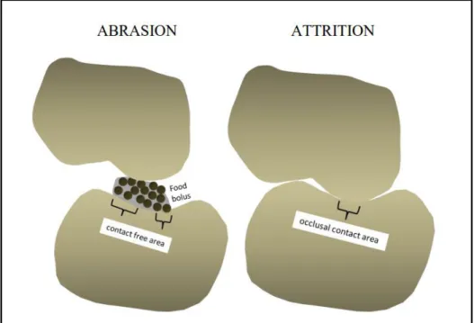
refers to the pathological loss of tooth tissue by a disease process other than dental caries. The relative amount of attrition and abrasion are influenced by biomechanical factors of mastication.
Mastication involves two processes that affect wear, abrasion and attrition. Abrasion occurs in the presence of food as the jaw closes. It begins when both mandibular and maxillary teeth contact the food bolus and ends when the two teeth contact each other. Because the teeth do not come in direct contact during abrasion, this stage is termed the contact free area (CFA) region of wear.63 This stage of wear involves abrasive, adhesive and corrosive wear. Attrition begins when the mandibular and maxillary teeth directly contact and ends when they separate. This is termed the occlusal contact area (OCA) region of wear.63 This stage of mastication involves abrasive; adhesive and fatigue wear.
13
teeth begin to separate. Of the three phases, only the second two phases place force on the teeth and contribute to wear. During the crushing phase, food particles are interposed between the two teeth and contribute to the wear process. The mode of wear occurring during this chewing phase has been termed abrasion. During the gliding phase, wear occurs by the contact of the opposing teeth. This mode of wear is termed attrition.64, 65
1.4.1 Clinical Significance of wear
Clinical wear has a multifactorial etiology and is a major concern in the practice of dentistry. Leinfelder discussed two types of wear.66 One of these is wear initiated by generalized conditions (the type of wear generated by a food bolus during mastication) and the other is wear generated under localized conditions (represented by direct tooth to materials contact). Some authors have suggested that localized wear may be a more important contributor to the breakdown of a material and contact wear may be more than two times as great as that in non-contact areas.66 Clinical studies offer the most meaningful data on the performance of the material. However, such studies are rare because of the time involved and costs associated with clinical studies. This has caused manufacturers to have a strong interest in the use of wear simulators for the purpose of testing prototype materials during development. Initially it was hoped that wear simulators might be useful in predicting clinical performance of a material. An ideal wear simulation would incorporate both abrasion and attrition.
The primary factors affecting clinical wear are understanding the mechanism of action, properties of the two contacting materials, and the surrounding and interfacial media are important in an appropriate restoration material selection. Individual factors may enhance the wear rates: aggressive tooth brushing, parafunction, diet, acidic/aqueous environment, surface geometry, and diminished tooth support. Failure of ceramic restoration due to creation of micro cracks/flaws, poor masticatory function67, impaired aesthetic appearance 63, sensitivity, secondary caries and systemic effects through ingested wear products.62
14
wear is surface degradation that results in progressive, but very slow loss of convexity of the cusps, which manifests as a flattening of cusp tips on the posterior teeth and incisal edges of mammelons on the
anterior teeth. Excessive wear results in unacceptable damage to the occluding surfaces and alteration of the functional path of masticatory movement.54 It may also destroy anterior tooth structure that is essential to acceptable anterior guidance function or esthetics, resulting in increased horizontal stresses on the masticatory system and associated temporomandibular joint remodeling. If wear continues unabated, the
enamel will eventually be breached. Once breached, both the enamel and exposed dentin wear at accelerated rates. Excessive wear on multiple teeth can have disastrous consequences. Biological
consequences are related to pain of the temporomandibular joint (TMJ), elongation of antagonists, loss of periodontal ligament and tilting and movement of adjacent teeth.54
However, the time involvement and costs associated with clinical studies have driven dental manufacturers to have a strong interest in the use of wear simulation of prototype materials as a screening tool and predictor of clinical performance. Leinfelder et al developed a laboratory simulator capable of evaluating both generalized and localized wear.66 This system transfers masticatory stresses to a composite specimen by means of a flattened polyacetal stylus (generalized wear) or a stainless steel conical stylus (localized wear) in the presence of slurry of polymethylmethacrylate (PMMA) beads. This device enabled in vitro studies capable of providing somewhat improved ability to predict in vivo
performance. Previous work showed that there may be a correlation between in vitro wear and in vivo generalized wear of dental restorative materials. 57, 58
1.4.2 Mechanisms of Wear
Wear takes place at two surfaces of a tooth: occlusal surface and proximal surface. Wear
15
While chewing, opposing dentition traps a layer of food and grinds it as the teeth move past one another. The chewing forces produced during this phase have been are modeled in the range of 10-20N.66 At the end of chewing cycle, sliding motion stops as the teeth reach maximum intercuspation. Masticatory muscle loading of the teeth during maximum intercuspation results in a force in the range of 50-150N.67
Wear is influenced by at least 5 inter-related processes. 1) Two-body wear, 2) Three-body Wear, 3) Fatigue Wear, 4) Tribochemical Wear, 5) Adhesive Wear
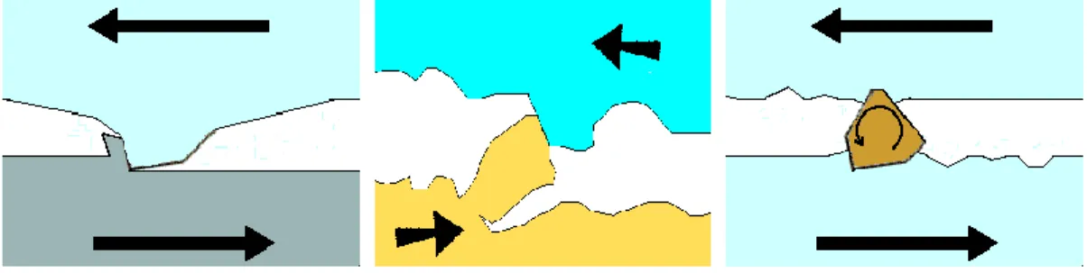
1) Two-body Wear
Two-body wear occurs when surfaces are rubbed away by direct contact. During this movement, the asperities must either fracture or deform. At a microscopic level, no surfaces are smooth and hence they contact by the reunion of their asperities. If both surfaces are brittle, there is fracture of the asperities. If one surface is ‘soft’, then the harder surface will plough into it, rising up ‘chips’, which eventually
fracture away. Gradually, all the asperities fracture and the cumulative effect of loss manifests as wear.68 In the oral cavity, these conditions predominantly occur during ‘non- masticatory tooth movement’.
Attrition is a form of two-body abrasion tooth wear that can be considered physiological. Attrition of a tooth surface may occur as a compensatory mechanism of occlusal prematurities (interferences to normal mastication). It is the physiological wearing away of dental hard tissues as a result of tooth-to-tooth contact without the intervening foreign substances that causes localized wear of occlusal contacts.69 The
wear rate of enamel at occlusal contact areas in molars is about 41 μm per year.69
2) Three Body Wear
16
of the masticatory slurry in the escape route of the groove. This process tends to hollow out the softer regions on a surface producing the chipping defects seen in occlusal molar dentin.68 As the teeth begin to approximate during the later stages of mastication, the remaining slurry particles get trapped between the asperities, in pits and in surface grooves. If both surfaces have similar morphology then the abrasive
particles may transfer between scratches and cause more or less equal loss of both opposing surfaces. 68
3) Fatigue wear
Some of the movement of the surface molecules is transferred to the subsurface causing rupture of intermolecular bonds and a zone of subsurface damage. Micro cracks form within the subsurface and coalesce to the surface, therefore causing loss of a fragment of material inducing fatigue wear.68
4) Tribochemical wear (dental erosion)
Tribochemical wear, clinically referred to as dental erosion occurs when chemicals weaken the inter-molecular bonds of the surface and potentiate the wear processes. There is an interplay between erosion, attrition and three-body abrasion. A primary example of tribochemical wear is loss of tooth structure from low pH (acidic) conditions. Extrinsic acids (from diet) and intrinsic acids (gastric reflux) result in weakening of the tooth surface intermolecular bonds. These surface molecules are then
physically dislodged by antagonistic movement of the opposing surfaces. The newly exposed underlying surface is then immediately attacked by the acid, which results in further wear.68
5) Adhesive wear
Adhesive wear occurs when there is a high attraction between surfaces such that cold welds form between the asperities. As the mandibular movement continues, these micro-welds fracture but not along their original line of fusion. This type of wear is normally seen in metals.68
1.4.3 Mechanical Properties Related to Wear
17
their tendency for clinical wear. Several studies have proposed equations to calculate wear based on its ceramic hardness, flexural strength, fracture toughness, and modulus of elasticity.63, 70 A brief
description of these properties will be given.
1.4.3.1 Flexural Strength
Flexural strength is defined as the resistance to plastic deformation. In crystalline materials, such as metals and ceramics, plastic deformation occurs by slip, kinking, twinning or phase transformation.71
1.4.3.2 Elastic Modulus
Elastic modulus is defined as the measure of the flexibility of a material. The elastic modulus is derived from the slope of a stress-strain curve.72 As noted earlier, the elastic modulus of a material will affect its tendency to wear by fatigue or abrasion. Under the same stress, a material with a low modulus will be more likely to undergo elastic deformation leading to fatigue than a high modulus material, which may experience abrasion.73
1.4.3.3 Fracture Toughness
Fracture toughness is defined as the amount of energy a material can absorb before fracture. The fracture of a material is derived by calculating the area under a stress strain curve. Therefore, it is a function of the both the strength of a material and its deformation before failure.72 In ceramic research, fracture toughness measures the amount of stress required to propagate an existing crack.
1.4.3.4 Surface Roughness (Ra)
Surface roughness, is also known as the “center line average” of a material. This parameter is
most frequently used for the purposes of general quality control. It is defined as the average absolute deviation of the roughness irregularities from the mean line over one sampling length. As such, this parameter is easy to define, easy to measure, efficient and gives a good description of height variations. Additionally, it is not sensitive to small changes.86
1.5 Wear simulation devices
18
of dental materials by means of expensive time-consuming clinical trials. Therefore, many research centers have developed a variety of wear testing devices, each with different degrees of complexity. As more and more materials are developed, and is difficult to evaluate them by expensive time-consuming clinical trials to obtain a meaningful data. Several mechanical testing systems (machines) have been created to simulate in vivo occlusal wear on the influence of test materials on the occlusal wear of natural enamel. The Food and Drug Administration of the United States of America (FDA) has established guidelines for non-clinical laboratory studies. Included in the guidelines is the statement that equipment should be calibrated and its maintenance defined and ensured so that the generation, measurement, and assessment of data is adequately tested, calibrated, and/or standardized. 74
The International Standards Organization (ISO) published a 2001 technical specification for wear titles “Dental Material, Guidance on testing wear, Part 2, Wear by two-and/or three-body contact”. This
specification discusses the following eight wear testing systems methods: Acta, Zurich, Alabama, Frieburg, DIN, Minnesota, OSHU and New Castle.74 Many wear simulator research centers are trying to mimic the oral environment and biological variables with the goal of ranking restorative material according to their wear resistance in comparison to reference materials.
All these simulators have been developed to measure the in vitro wear of dental materials; each of these machines has their advantages and limitations and new simulators are being designed (Willytec). In 2006, Heintze conducted a round robin study correlating the results of the different wear simulating devices with ACTA, OHSU, Willytec and Zurich wear devices on eight different composites. Specimens were prepared at the Ivoclar Vivadent laboratories, and sent to different testing sites for testing and the resultant data was analyzed.62 Heinz concluded that the relative ranks of the materials differed
19
An accurate comparison among testing devices may be possible if the load profiles and machine wear movements are the same. However, this is exactly what differentiates the different wear devices. For example, the Minnesota wear device is more expensive and complex than the other devices and is located in only one test site. The BIOMAT wear device simulates only two body wear. The Alabama wear simulating device has been used prolifically in the United States. Existing oral wear devices have varying methods of simulating the abrasion and attrition phases of wear. The BIOMAT, OHSU, Minnesota and Willytec devices have a stylus that impacts the specimen and slides a specific distance. The Leinfelder wear testing device stylus impacts the specimen and rotates 30 degrees. These devices all incorporate both the abrasion and attrition phases of mastication. The ACTA device has two wheels, a wheel containing composite specimens and one steel counter surface wheel that rotate next to each other at slightly different speeds. As such, this device measures only the abrasion phase of mastication. Further analysis has revealed that the ACTA, OHSU, and Willytec revealed that these devices measure different wear mechanisms 62. Despite prolific wear testing by industry and academia with these devices, the International Standards Organization (ISO) has not specified a standard wear testing system.
Generally speaking, oral wear simulating devices incorporate three methods of producing wear: sliding, sliding with impact, and rotation with impact. The effects of sliding wear (abrasive and adhesive mechanisms) were compared with impact wear (abrasive, adhesive and fatigue mechanisms) using a BIOMAT simulator.62 The comparative rankings of seven restorative materials (including two composites) differed significantly between the two methods, and the study concluded that “there is no correlation
between impact-cum-sliding wear and non-impact sliding wear”. Evaluation of the current literature reveals that there has been no published study that has compared the effect of wear produced with impact and rotation to wear produced with impact and sliding.
1.6 Contributing factors for in-vitro wear simulation 1. Standardization of the antagonist: Counter sample materials
20
factors including hardness, wear surface evolution and frictional coefficients have to be considered, relative to the tribology of the in vivo situation. Assessment of potential counter sample materials should be based on the essential tribological simulation supported by investigations of mechanical, chemical and structural properties.75 Antagonists standardized for shape and size and according to materials should show mean values similar to those found in natural, non-standardized cusps. Krejci et al. measured the shapes and sizes of palatal cusps of non-erupted human upper third molars. Natural enamel antagonists are preferable for the simulation of wear in the occlusal contact area.75
2. Composition of the antagonist
A variety of antagonists have been used which include enamel, gold, ceramics, stainless steel, annealed chromium-steel counter bodies, alumina ball: 10 mm ceramic (alumina) and dental porcelain. A study by Heintze concluded that enamel provided similar wear results as two different ceramic
antagonists and produced no more variation in the wear data.68 3. Shape of the antagonist
A variety of antagonist such as flat, ball or rounded, flattened enamel surfaces, enamel harvested from extracted human third molars and machined into cusps with a 5 mm spherical radius or hemi- spherically and standardized human enamel cusps with a uniform contact area have been used. 68 4. Load/force
In the load/force diagram several variations are possible. These include static and/or sinusoidal cyclic and dynamic motions. Contact loads may range anywhere from 1, 10, 20, 25, 50, 75, 100 N, contact loads ranging from 1.7, 3.2, 4, 6.7, 9.95, 16.2 kgf/cm. Chewing force may range from 53 to a maximum of 75.6 N force. The abrasion load may be set at 20 N and the attrition load 90 N. The wear devices may not be able to reproduce the resiliency of the periodontal ligament.76
5. Contact area size: force per unit surface area.
21
In different studies, the number of cycles has ranged from 5000, 10,000, 25,000, 50,000, 100,000 to 120,000 and so on. In order to compare results different studies, number of cycles should be taken into consideration.
7. Chewing frequency: frequency of load cycles
The chewing frequency used in vitro studies varies from 1.2 to 1.7 Hz 8. Duration of tooth contact
The duration of tooth contact during the in vitro loading should mimic the in vivo situation. Load and time significantly influence wear.
9. Sliding speed: relative speed of opposing surfaces
The sliding speed (2.5 mm/s) during the in vitro simulation should be comparable with the in vivo situation.54
10. Temperature
Temperature plays a plasticizing effect. Constant temperature (20, 37 ◦ C) or thermocycling (5–55 ◦ C) should be maintained.
11. Food bolus during mastication
Variety of food bolus or slurry can be used during mastication movement simulating such as slurry of water and unplasticized PMMA beads, PMMA powder, hydroxyapatite slurry and millet-seed/PMMA-beads mixture.
12. Lubricant and friction
22
liquids are incorporated in the three-body wear machines, such as water, alcohol, acids, olive oil, olive oil/CaF slurry and artificial saliva. Some systems include bacteria in the lubricant, others do not.77 13. pH
pH conditions seem to influence dramatically the wear conditions and therefore they should be controlled carefully during in vitro wear testing. pH levels (1.2, 3.3, and 7.0) are frequently used during wear simulation so as to mimic plaque acids, gastric acids and dietary acids. If human enamel is used as counter body, acidity of the medium has an impact on the wear behavior. The effect of acidic pH on abrasion, attrition and erosion of human enamel under several different pH conditions has been tested. During acid conditions, the combination of erosion and abrasion resulted in significantly greater wear than erosion alone. Simultaneous erosion and abrasion resulted in about 50% more wear than alternating erosion and abrasion. Chewing of acidic foods with some abrasive properties might cause enhanced tooth wear. Dentin is more susceptible than enamel to erosion and abrasion alone or combined. Load and time significantly influence enamel wear both in acid and neutral conditions. Depth of dentin erosion
significantly increases non-linearly with time and significantly decreases with increasing pH. Dentin is susceptible to erosion even at relatively high pH. Furthermore, in addition to a higher critical pH than enamel, the dentinal tubule system is readily exposed and dentine, unlike enamel, shows little propensity to remineralize.77
14. Enzymes
Proteolytic enzymes are present in saliva and generated by bacterial metabolism. Research suggests that enzymatic activity contributes to degradation of samples during in vitro testing. de Gee et al. used esterase solution in the ACTA wear machine. Chemical cycling can induced a generalized swelling of the composite samples and a modified wear curve.58
15. Enamel structure and physiology related to microwear
23
structure at the level of organization of crystallites rather than prisms per se. Variation in crystallite orientation in prismatic enamels may contribute to optimal dental function through the property of differential wear in functionally distinct regions of teeth.79
16. Wear debris
Little has been reported with regard to the potential effect of wear debris (and associated friction) at the zone of impact.
1.7 Wear of Ceramic Restorative Materials
The wear rate of an ideal restorative material should approximate that of enamel.1 Lambrechts et al. reported vertical wear of enamel to be between 20 μm to 40 μm per year when opposing enamel in the premolar and molar regions, respectively.69 Surface texture and surface hardness have each been
investigated as possible determinants of wear rate. 68 However, surface hardness has been shown to be a poor indicator of wear.68 Laboratory technicians utilize various approaches when seeking to finish ceramic restorations. These finishing approaches include glaze, autoglaze, natural glaze, overglaze and polish. Finishing of ceramic restorations
Definitions:
Glaze: 1: To cover with a glossy, smooth surface or coating 2: the attainment of a smooth and reflective surface 3: the final firing of porcelain in which the surface is vitrified and a high gloss is imparted to the material 4: a ceramic veneer or a dental porcelain restoration after it has been fired, producing a
nonporous, glossy or semi-glossy surface.80
Autoglaze: the production of a glazed surface by raising the temperature of a ceramic to create surface flow.80
Natural glaze: The production of a glazed surface by the vitrification of the material itself and without addition of other fluxes or glasses.80
24
Polishing: to make smooth and glossy usually by friction.80Tooth wear caused by ceramic restorative materials wear remains difficult to assess in both in vitro and in vivo controlled evaluations. In vitro ceramic investigations are most often studied by the use of flat ceramic specimens opposing either human cusps in their natural anatomic state or flattened
(ground) enamel. Jacobi et al. used human canines opposing flat specimens of ceramics and of gold. They found that type III dental gold to be less abrasive than any of the six ceramic surfaces tested.81 In this study, wear of the enamel was measured and correlated to weight loss of the enamel samples. Jacobi et al. also showed the outer cerammed layer of cast Dicor (Dentsply Intl. Inc., York, Pa.) specimens had abrasiveness similar to that of conventional porcelain.81 Jagger investigated the in vitro wear effects of glazed, unglazed, and polished porcelain (Vita, Vitadur N) against human enamel in the laboratory by use of a wear machine designed to simulate the masticatory cycle. The results from this study suggested that glazed and unglazed porcelain produced similar amounts of enamel wear.82 During the wear test, the glaze was worn away after a relatively short period of wear (2 hours). Polished porcelain produced substantially less enamel wear. This study indicated the potential damage porcelain can produce upon enamel and suggested that porcelain should be polished instead of reglazed after chairside adjustment.82 The main benefits of finishing and polishing of restorative materials are thought to be better gingival health, chewing efficiency, patient comfort, esthetics and wear. A smoother surface provides less retention of plaque, and it is easier to maintain by the patient and the dentist. Also, oral function is enhanced with a well-polished restoration since food can glide more freely over the occlusal and through embrasure areas during mastication. Furthermore, smooth restoration surfaces minimize wear rates on opposing and adjacent teeth. This becomes particularly important when considering restorative materials (ceramics) that are harder than tooth enamel and dentin such as ceramics.
25
Additionally, the subjective nature of qualitative wear assessment and the technical difficulty of quantitative wear measurement reduce the reliability of in vivo wear analysis. In response, both industry and academia have developed in vitro wear testing methods. Several analyses have been performed in an attempt to correlate the results of in vitro wear testing methods with in vivo results. Despite these
26
CHAPTER 2
:
MANUSCRIPT
2.1 Introduction
27
2.2 Hypothesis and AimsNull Hypothesis:
There is no difference in enamel wear produced by (1) polished, (2) polished and glazed, and (3) glazed zirconia compared to polished lithium disilicate material.
Specific Aims:
1. To measure the wear of human enamel against polished, polished and glazed, and glazed zirconia; and compare it with polished commercially used lithium disilicate ceramic material.
2. To measure the wear of ceramic materials in polished, polished and glazed, and glazed states opposing enamel specimens.
28
2.3 Materials and MethodsStudy Design:
1. Preparation of ceramic and enamel specimens 2. Cyclic loading of specimens
3. Wear determination
2.3.1 Preparation of ceramic and enamel specimens
2.3.1.1 Ceramic Specimen preparation
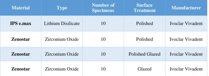
Ceramic blocks were cut in the dimensions of 7x11x6mm±0.3 mm using an Isomet diamond-wafering saw. Ten flat specimens were milled using the IPS e.max and zirconia firing parameters. For Polishing IPS e.max. and Zirconia:
The specimens were wet ground to a flat surface with a series of SiC abrasive paper (320-, 600-, 1200- and 2000-grit) for 1 min each using a rotational polishing device (Ecomet Buehler Ltd). A final finish with 0.05µm alumina slurry and a polishing cloth was applied. After polishing, the specimens were steam cleaned for 5 minutes (Branson 1200) to remove the polishing debris.
For Glazing Zirconia:
The specimens were steam cleaned for 5 minutes (Branson 1200) to remove the debris. FCZ Glaze was mixed to a creamy consistency and painted onto specimen and baked according manufacturer’s instructions:
Apply the glaze material in an even layer in the usual manner
Glaze firing should be conducted using the stipulated firing parameters and with the firing
equipment belonging to the furnace.
After completion of the firing process, remove the restoration from the furnace.
Allow it to cool at room temperature. Don't touch the hot surface with metal tongs.
29
2.3.1.2 Enamel styli preparationFourty extracted caries free premolars were selected and randomly divided for this study (n=10/ group). Caries free enamel cusps of maxillary premolars were sectioned using isomet wafering saw and the roots were removed. The dimensions of their buccal cusps were standardized with a cone shaped diamond bur (Brasseler, USA) in a straight handpiece (NSK, Japan). The standardized cusp was mounted on a metal stylus and stabilized with self -cure acrylic. The enamel styli were scanned using a non-contact 3D surface profilometer (PROSCAN 2000, Scantron Industrial Products Ltd. Taunton, England) with a 20µm resolution.
2.3.1.3 Roughness Measurement
Surface roughness (Ra) of all the specimens was determined using a non-contact light
profilometer (Proscan 2000). As the pretest surface was assumed to be homogenous, an area in the middle of each specimen was selected for testing. A 0.7 µm length was measured with a 0.8 mm cutoff length and a 40 surface filter number selected for polished surface and 125 surface filter number selected for all other groups (based on ISO 4288-1996).
2.3.2 Cyclic loading of specimens
2.3.2.1 Wear simulation: Cyclic Loading in the newly modified UAB-Chewing Simulator
The in vitro study was carried out on a newly modified UAB chewing simulator. The Alabama wear method has proved its reliability for the past two decades. Its earliest publication was in 1989 by Drs. Leinfelder and Suzuki and hence it is also known as Leinfelder-Suzuki wear method.66 This was modified from the basic Roulet method.87 The Alabama wear method later included modifications such as integration of a 30º clockwise rotations as the stylus hits the surface, and replacing the metal stylus with a fiber glass stylus. According to a recent review, it has the highest citation frequency, followed by ACTA, OHSU, Zurich and MTS wear simulators.62 The Alabama wear method is also included in the ISO Technical Specification on the wear by two/three body contact.88
30
disk, tooth brush simulators for determining wear. Their results varied and the correlation to clinical wear was not established. 4 This can be attributed to the variations in the force, motion and the testing
conditions. Due to the considerable attention given to the validation of existing wear methods, a more clinically relevant wear device, a chewing axis simulator was redesigned to improve the reliability. The eight station machine is a highly precise instrument applying designed load along the longitudinal axis. It is designed to allow lateral movement up to 8mm. A variable amount of load from 5N-400N can be applied during the wear testing. A custom made thermocycler runs in conjugation with the wear-testing machine. It maintains the constant flow of hot/cold fluids on the specimens.
Wear testing was conducted at 37°C using a slurry (3 parts Distilled water (66%) +1 part Glycerin (33%)) (Fischer Scientific, Pittsburgh, PA). The formulation of solution is maintained by an electo-magnetic stirrer, circulated at a rate of 3ml/min at 37ºC with continuous stirring. (Corning, USA). A longitudinal force of 20 N was applied by enamel styli on the flat ceramic specimens, which were positioned on a 2mm horizontal sliding platform. These enamel styli contacted the lower flat specimens with a frequency of 1.2 Hz and continued for 300,000 and then another 300,000 cycles. After the enamel styli contacted the ceramic specimen, a 2mm slide across the opposing surface under a constant 20N load was produced. A timer automatically alters the hot and cold cycles in specified time. The medium has produced a reproducible wear in controlled conditions when compared to others.89
2.3.3. Wear determination
2.3.3.1 Determination of volumetric wear & depth loss
31
150mm x 100mm. It uses a focal multiplexing sensor with up to 0.005μm resolution. Safe white light is
transmitted through a lens, which has a built in spectral aberration. Takes the white light and divides it into the full spectral field, focusing each different color frequency at a slightly different point through a defined measuring range. When an object is placed within this range, only one particular color frequency reflects back from the surface. Information passes back into processor where a spectrometer analyzes the signal and converts it to a measurement. Proscan combines these measurements with the precise location of a moving X and Y linear table, giving three co-ordinates from which a three dimensional profile is created. Results of the surface profile appear immediately on the computer monitor and an image of the graphical 3-D representation can be saved on the computer.
Ceramic specimens and enamel styli were scanned at baseline, 300,000 and 600,000 cycles using a non-contact 3D surface measurement instrument PROSCAN 2000 (Scantron Industrial Products Ltd. Taunton, England). A step size of 0.01 mm was used to scan both material and enamel specimens. Wear volumetric loss differences between baseline, 300,000 and 600,000 cycles were obtained using the Proform software by superimposition of the original (baseline) scans over the modified (300,000 & 600,000 cycles) scans. The measurements were repeated two times on the same specimen and recorded to check the operators’ reliability.
2.3.4 Surface imaging
One specimen from each group was imaged with SEM. The specimens were cleaned in ethanol, secured to SEM tabs with gold conducting tape, and gold-coated in a vacuum sputter coater (Desk-I). The wear tracks were examined in an SEM (Quanta FEG 650; FEI) with the secondary electron imaging mode.
2.4 Statistical Analysis
32
means between multiple groups. Tukey–Kramer post hoc test was used for pair wise comparison of group means (p=0.05).
For ceramics, there was no measurable wear loss on polished zirconia, significant material loss
wear values were seen with other groups.
For enamel, polished zirconia showed less material and enamel wear values compared to other
groups.
Surface Roughness ranking:
Glazed Zirconia > Polished Glazed Zirconia > Polished Zirconia > Polished Lithium Disilicate
2.5 Results
2.5.1 Wear of the Ceramic Materials
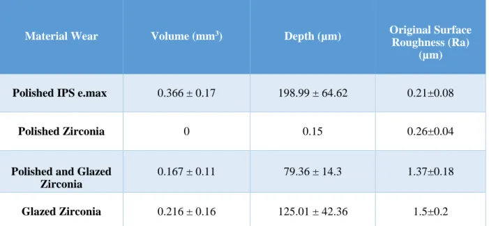
The polished zirconia showed no signs of wear after 600,000 cycles and was significantly
different than rest of the groups. The glazed zirconia had the highest wear among the zirconia groups with a mean volume loss of 0.216 ± 0.16 mm3 and a depth loss of 125.01 ± 42.36 μm at 600,000 cycles. The polished and glazed zirconia had less wear than the glazed zirconia with a mean volume loss of 0.167 ± 0.11 mm3 and a depth loss of 79.36 ± 14.3 μm at 600,000 cycles. The polished lithium disilicate reported the highest amount of volume and the depth loss of 0.366 ± 0.17 mm3 and 198.99 ± 64.62 μm at 600,000 cycles.
2.5.2 Enamel Wear
The enamel opposing the polished zirconia showed a very minimal wear with a mean volume and depth loss of 0.07 ± 0.01 mm3 and 199.49 ± 48.05 μm at 300,000 cycles and 0.118 ± 0.03 mm3 and 244.24 ± 67.2 μm after 600,000 cycles. The enamel opposing glazed zirconia showed the highest amount
of wear of all the groups with a mean volume and depth loss of 0.209 ± 0.09 mm3 and 262.75 ± 140.89 μm at 300,000 cycles and 0.288 ± 0.09 mm3 and 310.59 ± 164.9 μm after 600,000 cycles. The polished
33
cycles and 0.282± 0.09 mm3 and 286.68 ± 103.2 μm after 600,000 cycles. The lithium disilicate had significantly higher wear than the polished zirconia with a mean volume and depth loss of 0.177 ± 0.06 mm3 and 265.96-± 65.8 μm at 300,000 cycles but it increased 0.274 ± 0.07 mm3 and 342.46 ± 81.8 μm after 600,000 cycles.
The ceramic based materials experienced abrasive wear as evidenced by the scratches seen at higher resolution on the surface of their wear tracks. The wear tracks were imaged at an original magnification of 50X, 100x and 500x with scanning electron microscope (SEM).
2.6 Discussion
The tested groups of ceramics have each been introduced to dentistry to restore the anatomic form and clinical function of human teeth. The groups of zirconia that are polished, glazed and polished and glazed samples were included to simulate clinical situations.90 A group of commonly used polished lithium disilicate samples served as the control. Before wear simulation, the surface roughness obtained for all the groups by obtaining the preliminary scans with a 3D non-contact profilometer (PROSCAN, UK). In this study, the application of glaze made the surface rougher making it more abrasive to the opposing tooth or restorative material.
34
homogeneity of the solution is maintained by an electromagnetic stirrer (Corning, USA). The study was carried out for 300,000 cycles and 600,000 cycles accounting for a clinical service of approx. 24 and 48 months.93 Ideally, the wear rate of the restoration should match that of posterior tooth enamel, which was estimated to be approximately 20 to 40 µm per year.69
There were significant different materials wear and opposing enamel wear values for all materials tested. The results of this study suggest that the polished zirconia experienced less material wear and produced less opposing enamel wear than the other groups. The ceramic specimens of the polished zirconia showed no traces of wear. The glazed zirconia specimens showed a considerable amount of wear with a loss of surface glaze within the first 300,000 cycles exposing substructure zirconia which
continued to wear the opposing enamel. There is still a considerable amount of specimen loss at 600,000 cycles exposing the underlying rough surface and making it susceptible to wear. Greater wear of glazed zirconia can be explained by loss of the soft gloss surface, and opposing enamel contacting the rough surface. The glazed zirconia showed a considerable amount of wear of the antagonist. The surface loss of glaze created rougher areas during the wear testing and causing an exposure of the harder crystalline phases aggravating the loss of the opposing enamel. This can be well established with some clinical studies that show the loss of the glaze within 6 months of clinical function.93 This can get exaggerated by the presence of the worn particles leading to the formation of the wear tracts as the particles plough the surface.
The amount of the ceramic wear is slightly less in the polished then glazed zirconia. This could be due to wear process slowing as the superficial glaze wears out exposing the underlying polished
subsurface zirconia. This exposed polished layer of zirconia may limit the progression of zirconia and enamel wear.