Cytosolic DNA Promotes Signal Transducer and Activator of
Transcription 3 (STAT3) Phosphorylation by TANK-binding
Kinase 1 (TBK1) to Restrain STAT3 Activity
*
□SReceived for publication, December 13, 2016, and in revised form, January 31, 2017Published, JBC Papers in Press, February 10, 2017, DOI 10.1074/jbc.M116.771964
Hung-Ching Hsia‡§, Jessica E. Hutti§1, and Albert S. Baldwin‡§2
From the‡Department of Cell Biology and Physiology and the§Lineberger Comprehensive Cancer Center, University of North Carolina, Chapel Hill, North Carolina 27599
Edited by Alex Toker
Cytosolic DNA can elicit beneficial as well as undesirable immune responses. For example, viral or microbial DNA trig-gers cell-intrinsic immune responses to defend against infec-tions, whereas aberrant cytosolic accumulation of self-DNA results in pathological conditions, such as autoimmunity. Given the importance of these DNA-provoked responses, a better understanding of their molecular mechanisms is needed. Cyto-solic DNA engages stimulator of interferon genes (STING) to activate TANK-binding kinase 1 (TBK1), which subsequently phosphorylates the transcription factor interferon regulatory factor 3 (IRF3) to promote interferon expression. Recent studies have reported that additional transcription factors, including nuclear factorB (NF-B) and signal transducer and activator of transcription 6 (STAT6), are also activated by cytosolic DNA, suggesting that cytosolic DNA-induced gene expression is orchestrated by multiple factors. Here we show that cytosolic DNA activates STAT3, another member of the STAT family, via an autocrine mechanism involving interferon  (IFN) and IL-6. Additionally, we observed a novel cytosolic DNA-induced phosphorylation at serine 754 in the transactivation domain of STAT3. Upon cytosolic DNA stimulation, Ser754is directly phos-phorylated by TBK1 in a STING-dependent manner. Moreover, Ser754phosphorylation inhibits cytosolic DNA-induced STAT3 transcriptional activity and selectively reduces STAT3 target genes that are up-regulated in response to cytosolic DNA. Taken together, our results suggest that cytosolic DNA-induced STAT3 activation via IFNand IL-6 is restrained by Ser754 phos-phorylation of STAT3. Our findings reveal a new signaling axis downstream of the cytosolic DNA pathway and suggest poten-tial interactions between innate immune responses and STAT3-driven oncogenic pathways.
Double-stranded DNA (dsDNA) in the cytosol is a danger-associated molecular pattern that triggers inflammation and
immune responses. Cytosolic DNA can be derived from viral or intracellular microbial infections, undigested phagocytosed materials, and activated self-retroelements (1). The presence of cytosolic DNA is detected by several cellular sensors, which in turn initiate signaling cascades to induce inflammatory response and type I interferon production (2). Despite the redundancy between these cytosolic DNA sensors, cyclic GMP-AMP synthase (cGAS)3is the predominant sensor relaying the presence of cytosolic DNA to downstream signaling cascades (3). Upon binding to dsDNA, cGAS produces cyclic 2⬘-3⬘ GMP-AMP (cGGMP-AMP), which serves as a second messenger to activate the endoplasmic reticulum adaptor protein stimulator of inter-feron genes (STING) (3– 6). Activation of STING by cGAMP leads to STING oligomerization, followed by recruitment and activation of TANK-binding kinase 1 (TBK1) (7, 8). Subse-quently, TBK1 phosphorylates interferon regulatory factor 3 (IRF3) to promote expression of interferons (IFNs), thereby ini-tiating immune responses to establish an antiviral state (7, 9, 10). In addition to IRFs, other transcription factors, including nuclear factorB (NF-B) and signal transducer and activator of transcription 6 (STAT6), are also activated by STING and TBK1 downstream of cytosolic DNA (11–14).
The transcription factor STAT3 is activated by tyrosine 705 phosphorylation downstream of a variety of cytokines, such as epidermal growth factor (EGF) and IL-6 (15). Phosphorylation at tyrosine 705 leads to nuclear accumulation of STAT3 homodimers and expression of target genes containing a␥ -ac-tivated site (GAS) in their promoters (16). STAT3 drives the expression of prosurvival and inflammatory genes, and sus-tained activation of STAT3 has been shown to promote prolif-eration, enhance survival of neoplastic cells, and facilitate inflammation-driven tumorigenesis (17–19). In addition to its role in promoting tumorigenesis, STAT3 also represses the anti-tumor activity of hematopoietic cells, making it a key can-didate for targeted cancer therapy and immunotherapy (20, 21). STAT1, another STAT family member, predominantly func-tions downstream of interferons. STAT1 homodimers induced *This work was supported by National Institutes of Health (NIH) Grants
AI35098 and R35CA197684 (to A. S. B.). The authors declare that they have no conflicts of interest with the contents of this article. The content is solely the responsibility of the authors and does not necessarily represent the official views of the National Institutes of Health.
□S This article containssupplemental Figs. 1–3.
1Present address: AbbVie, Inc., 1 N. Waukegan Rd., North Chicago, IL 60064.
2To whom correspondence should be addressed: 450 West Dr., Lineberger
Comprehensive Cancer Center, UNC-Chapel Hill, Chapel Hill, NC 27599. Tel.: 919-966-3652; Fax: 919-966-8212; E-mail: albert_baldwin@med. unc.edu.
3The abbreviations used are: cGAS, cyclic GMP-AMP synthase; cGAMP, cyclic
2⬘-3⬘GMP-AMP; STING, stimulator of interferon genes; GAS,␥-activated site; ISRE, IFN-stimulated response element; IKK, IB kinase; TAD, transac-tivation domain; TLR, Toll-like receptor; SH2, Src homology 2; RIPA, radio-immune precipitation assay; RLU, relative luciferase unit(s); bis-tris, 2-[bis(2-hydroxyethyl)amino]-2-(hydroxymethyl)propane-1,3-diol; qRT-PCR, quantitative RT-PCR; TK, thymidine kinase.
by IFN␥recognize almost identical GAS sites as STAT3 dimers
doin vitro, but STAT1 and STAT3 have different, albeit
over-lapping, target genes in vivo(22). On the other hand, type I IFNs, including IFN␣and IFN, induce the formation of not only the STAT1 homodimer but also the interferon-stimulated gene factor 3 (ISGF3) complex comprising STAT1, STAT2, and IRF9. The ISGF3 complex promotes the expression of genes containing an IFN-stimulated response element (ISRE) in their promoters (23). Reciprocal antagonizing effects between STAT1 and STAT3 can be observed in certain scenarios (24). For example, STAT3 inhibits STAT1-dependent induction of ISRE genes in response to type I IFN stimulation presumably through STAT1-STAT3 heterodimerization (25).
Whereas dimerization and activity of STAT proteins are controlled by tyrosine phosphorylation, recent studies have demonstrated that the function of STATs can also be modu-lated by TBK1 and the closely remodu-lated kinase IB kinase ⑀ (IKK⑀). Phosphorylation of STAT1 at Ser708by IKK⑀disrupts STAT1 homodimerization and favors ISGF3 formation, thereby shifting the type I IFN-induced gene expression profile from GAS-driven genes to ISRE-driven genes (26). Cytosolic nucleic acids and viral infections engage the STING-TBK1 pathway, leading to TBK1-mediated phosphorylation of STAT6 at Ser407 and STAT6 activation (14). Interestingly, functional loss of cGAS or STING has been observed in colo-rectal cancer and melanoma and correlates with disease pro-gression and elevated STAT3 activation (27–29), suggesting a role of STING in restricting STAT3 activity and tumor progres-sion. Thus, we sought to determine whether activity of STAT3 can be regulated by the STING-TBK1 pathway downstream of cytosolic DNA. Here we show that STAT3 is activated by cyto-solic DNA through an autocrine mechanism involving IFN and IL-6. At the same time, cytosolic DNA activates TBK1 in a cGAS- and STING-dependent manner to directly phosphory-late STAT3 at serine 754 in the transactivation domain (TAD). This TBK1-mediated phosphorylation at Ser754 is inhibitory and restrains cytosolic DNA, IL-6, and IFN-induced activa-tion of STAT3. Our finding provides a possible explanaactiva-tion for the role of STING in limiting STAT3 activation and further emphasizes the complex signaling cascades and gene expres-sion initiated by cytosolic DNA.
Results
TBK1 Directly Phosphorylates STAT3 at Serine 754 —
Previ-ous studies demonstrated that TBK1 and IKK⑀, respectively, regulate the function of STAT6 and STAT1 by direct phosphor-ylation (14, 26). To determine whether TBK1 and IKK⑀ also phosphorylate other STATs, we examined the sequences of STATs against the optimal substrate motif for TBK1 and IKK⑀ (30, 31) and identified serine 754 in the TAD of STAT3 as a potential TBK1/IKK⑀ phosphorylation site (Fig. 1A). To test whether STAT3 is a substrate of TBK1 or IKK⑀, IKK⑀ and STAT3 were overexpressed in HEK293T cells, and immuno-precipitated STAT3 was blotted with an IKK family phospho-substrate motif antibody (see “Experimental Procedures”). Overexpression of wild-type IKK⑀strongly induced phosphor-ylation of STAT3 at Tyr705as well as a site recognized by the IKK phosphosubstrate motif antibody (Fig. 1B). We then
gen-erated an antibody specific for phospho-Ser754-STAT3 and repeated the experiment with TBK1. Similarly, overexpression of wild-type, but not kinase-dead, TBK1 induced STAT3 phos-phorylation at multiple sites, and S754A mutation of STAT3 abolished the signal of Ser(P)754-STAT3-specific antibody (Fig. 1C), suggesting that TBK1 kinase activity is critical for the phos-phorylation of STAT3 at Ser754. Overexpression of TBK1 and IKK⑀ in HEK293T may lead to activation of other kinases, which in turn phosphorylate STAT3. To determine whether TBK1 is capable of phosphorylating STAT3 directly, we per-formed anin vitrokinase assay with purified TBK1 and recom-binant GST-STAT3 from bacteria. Autoradiography showed that incubation with wild-type but not kinase-dead TBK1 led to strong phosphorylation on wild-type STAT3 and that S754A mutation of STAT3 abolished the phosphorylation (Fig. 1D). This TBK1-mediated phosphorylation on STAT3 was also rec-ognized by the Ser(P)754-STAT3-specific antibody (Fig. 1D). These data show that TBK1 is capable of directly phosphorylat-ing STAT3 at Ser(P)754.
STAT3 Is Phosphorylated at Ser754in Response to Cytosolic
dsDNA—TBK1 is activated downstream of Toll-like receptors
TBK1 activators, cytosolic DNA induces the most robust STAT3 activation and phosphorylation at Ser754.
TBK1 Is Required for Cytosolic DNA-induced STAT3
Phos-phorylation at Ser754
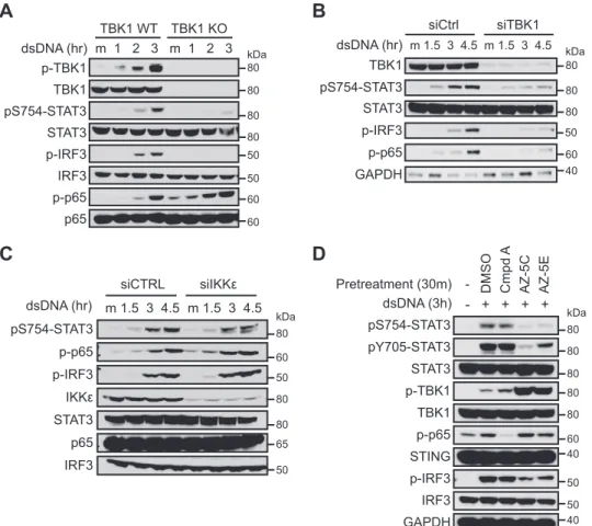
—Given that cytosolic DNA induces robust TBK1 activation, STAT3 phosphorylation, and associa-tion between TBK1 and STAT3, we asked whether TBK1 is required for cytosolic DNA-induced Ser754phosphorylation of STAT3. We found that STAT3 Ser754 phosphorylation and IRF3 phosphorylation were abrogated with genetic ablation of TBK1 or siRNA-mediated TBK1 knockdown (Fig. 3,AandB). On the other hand, IKK⑀knockdown had negligible effects on STAT3 Ser754 or IRF3 phosphorylation (Fig. 3
C), indicating that TBK1 but not IKK⑀is required for cytosolic DNA-induced STAT3 phosphorylation. It has been shown that cytosolic DNA also activates IKK␣and IKK(Fig. 2B) (13) and that the optimal substrate motif of IKK␣and IKKshares a partial homology to that of TBK1 and IKK⑀(30, 31, 34, 35). To further investigate the role of individual IKKs in promoting STAT3 phosphoryla-tion upon cytosolic DNA challenge, we used an IKK␣/IKK -specific inhibitor compound A (36) and two TBK1/IKK⑀ -specific inhibitors, AZ-5C and AZ-5E (37). The TBK1/IKK⑀ inhibitors blocked the induction of phospho-IRF3 and Ser(P)754 -STAT3, whereas phospho-p65 was mostly unaffected (Fig. 3D). Tyr705phosphorylation of STAT3 was also dependent on TBK1
and/or IKK⑀(Fig. 3D). In contrast, the IKK␣/IKKinhibitor potently blocked the induction of phospho-p65 but had mini-mal effect on phospho-IRF3, Ser(P)754-STAT3, or Tyr(P)705 -STAT3 in THP-1 cells (Fig. 3D). This indicates that phos-phorylation of STAT3 Ser754 is mediated by TBK1 and is independent of IKK␣and IKK, whereas activation of NF-B p65 is mediated by IKK␣and/or IKK. Taken together, our data demonstrate that TBK1, rather than IKK␣, IKK, or IKK⑀, is the principle kinase that mediates Ser754phosphorylation of STAT3 upon cytosolic DNA challenge.
The cGAS-STING-TBK1 Pathway Induces STAT3 Ser754
Phosphorylation in Response to Cytosolic DNA—Because the
endoplasmic reticulum membrane protein STING is indispens-able for cytosolic DNA-induced TBK1 activation (10, 38), we tested whether STING is also required for STAT3 Ser754 phosphorylation in response to cytosolic DNA. Indeed, knock-down of STING significantly reduced cytosolic DNA-induced Ser(P)754-STAT3 (Fig. 4A). Moreover, Ser(P)754-STAT3 can be induced by ectopic expression of STING in HEK293T cells in a dose-dependent manner, suggesting that Ser754 phosphoryla-tion occurs downstream of STING activaphosphoryla-tion (Fig. 4B). We also tested whether the cytosolic dsDNA sensor cGAS is required in this setting. Knockdown of cGAS led to reduced activation of TBK1 and phosphorylation of IRF3 and a moderate
tion in the levels of Ser(P)754-STAT3 (Fig. 4C). The moderate reduction of Ser(P)754-STAT3 may be due to incomplete knockdown of cGAS or the redundancy of other cytosolic DNA sensors. Activation of cGAS by cytosolic DNA leads to produc-tion of cGAMP, which subsequently promotes STING activa-tion and activaactiva-tion of the TBK1-IRF3 signaling axis. We found that transfection of cGAMP is sufficient to induce Ser754 phos-phorylation of STAT3 (Fig. 4D), further supporting the model in which Ser754phosphorylation of STAT3 occurs downstream of STING. These data demonstrate that cytosolic DNA engages cGAS and STING to activate TBK1 and induce STAT3 Ser754 phosphorylation.
Secreted Cytokines Induce Activation of STAT3 in Response to
Cytosolic DNA—We next set out to determine the mechanism
of STAT3 activation (as marked by Tyr705phosphorylation) in response to cytosolic DNA. In THP-1 cells, activation of STAT3 was suppressed by TBK1/IKK⑀inhibitors (Fig. 3D), suggesting that TBK1 kinase activity is required for STAT3 activation. Because TBK1 is not a tyrosine kinase, activation of STAT3 is
probably mediated by tyrosine kinases that are directly or indi-rectly activated by TBK1. It is well established that Janus kinases (JAKs) activate STAT3 by phosphorylating Tyr705. We thus asked whether JAKs are involved in STAT3 activation in response to cytosolic DNA. Treating the cells with a pan-JAK inhibitor, pyridine 6 (39), effectively blocked STAT3 phosphor-ylation at Tyr705, but TBK1 activation and phosphorylation of STAT3 at Ser754was unaffected (Fig. 5A). These data and the TBK1 inhibitor data (Fig. 3D) suggest that cytosolic DNA-in-duced STAT3 activation is primarily mediated by JAKs, the activation of which is dependent on TBK1. Because JAKs usu-ally function downstream of cytokine receptors, we hypothe-sized that cytosolic DNA induces production of cytokines, lead-ing to activation of the JAKs and STAT3 through an autocrine mechanism. An alternative hypothesis would be that TBK1 activates JAKs directly or through a cascade of kinase activation in a cell-autonomous manner. To test these hypotheses, we asked whether conditioned media from dsDNA-transfected cells are able to activate STAT3 in naive recipient cells. We
pY705-STAT3 STAT3 p-IRF3 IRF3 p-TBK1 TBK1 Actin pS754-STAT3 TBK1 STAT3 IP: STAT3
Input
mock 1.5h
dsDNA 1.5h 3h 4.5h 6h
A B
C
pS754-STAT3
pS727-STAT3
pY705-STAT3
STAT3
p-TBK1
TBK1
pY-STAT1
STAT1
p-IRF3
IRF3
p-p65
p65
STING
Mock t/f
Untreated poly (I:C) poly (dA:dT) dsDNA Serum starv LPS
pS754-STAT3 IKKε
IKKε TBK1
pTBK1 pY705-STAT3
pIRF3
IRF3
STING pY-STAT1
TBK1 STAT3
p-IKKα/β
p65 p-p65 STAT3 IP:STAT3
Input
Untreated Mock t/f poly (I:C) poly (dA:dT) dsDNA Flagellin LPS
STAT1 pS727-STAT3
80
80
80
80
50
50
60
60 kDa
80
80
80
80
40
80
80
80
80
80
50
50 kDa
80
80
80
80
40 80
80
80
80
60
60 80
80
80
80
80
40 kDa
80
50
50
80
found that conditioned media from dsDNA-transfected THP-1 strongly induced Tyr(P)705-STAT3 but not Ser(P)754-STAT3 in the recipient cells (Fig. 5B, lanes 10 –14). When cells were treated with cycloheximide immediately before dsDNA trans-fection to block protein synthesis, the ability of conditioned media to induce Tyr(P)705-STAT3 in the recipient cells was abrogated (Fig. 5B,lanes 15–18). Although the possibility of cell-autonomous TBK1-mediated JAK activation cannot be completely ruled out because a weak STAT3 activation was still observed in cycloheximide-treated cells (Fig. 5B, lanes 6 –9), these results argue that STAT3 activation is primarily medi-ated by de novo synthesized secreted factors. We noticed that expression of several STAT3-activating cytokines, includ-ing IL-6 and IFN, was induced by cytosolic DNA, and the induction of IFNwas blunted by TBK1 inhibitor ( supplemen-tal Fig. 1). Therefore, we tested the involvement of IL-6 and IFN in STAT3 activation. Indeed, an IFN-neutralizing antibody reduced the ability of conditioned media to activate STAT3 in the recipient cells, and an IL-6-neutralizing antibody also had a modest effect (Fig. 5C). Taken together, our data show that cytosolic DNA-induced Ser754 phosphorylation of STAT3 is strictly cell-autonomous, whereas Tyr705 phosphor-ylation, and thus activation of STAT3, is primarily mediated
by secreted factors, such as IFN, through an autocrine mechanism.
Ser754 Phosphorylation of STAT3 Restricts Cytosolic
DNA-induced STAT3 Target Gene Expression—Next, we sought to
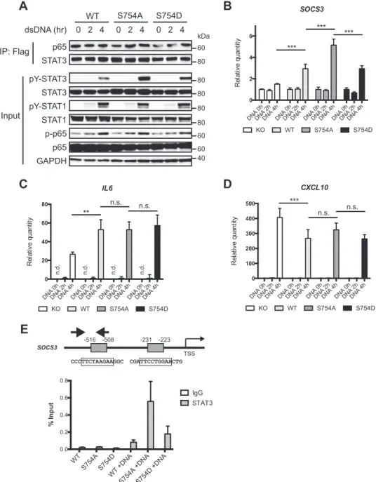
determine the effect of cytosolic DNA-induced Ser754 phos-phorylation on STAT3 activation and target gene expression. THP-1 cells reconstituted with wild-type or mutant STAT3 following CRISPR-mediated knock-out (supplemental Fig. 2) were transfected with dsDNA to induce STAT3 activation and Ser754 phosphorylation. Activation of wild-type and S754D STAT3 by cytosolic DNA was significantly lower than that of the S754A mutant, suggesting that Ser754 phosphorylation inhibits STAT3 activation (Fig. 6A). STAT1 and p65 were also activated by cytosolic DNA, but their activation was not affected by the status of STAT3, and p65 showed constitutive association with STAT3 (Fig. 6A). We also examined the gene expression induced by cytosolic DNA. STAT3 target gene
SOCS3was up-regulated in the presence of STAT3 (Fig. 6B),
and its expression was further elevated in the S754A cells, con-sistent with increased activation of the S754A mutant (Fig. 6A). The NF-B target geneIL6was also up-regulated in the pres-ence of STAT3, but Ser754phosphorylation did not have any measurable effect on its expression (Fig. 6C). This suggests that
pY705-STAT3 pS754-STAT3
STAT3
p-TBK1
TBK1
p-p65
STING
p-IRF3
IRF3
GAPDH Pretreatment (30m)
dsDNA (3h) - + + + + - DMSO Cmpd A AZ-5C AZ-5E
pS754-STAT3
p-p65
p-IRF3
IKKε
STAT3
p65
IRF3
siCTRL siIKKε
m 1.5 3 4.5 m 1.5 3 4.5 dsDNA (hr)
TBK1
pS754-STAT3
STAT3
p-IRF3
p-p65
GAPDH
siCtrl siTBK1
dsDNA (hr) m 1.5 3 4.5 m 1.5 3 4.5
A
B
C
D
p-TBK1
TBK1
pS754-STAT3
STAT3
p-IRF3
IRF3
dsDNA (hr) m 1 2 3 m 1 2 3
TBK1 WT TBK1 KO
p-p65
p65
80
80
80
80
50
50
60
60 kDa
80
80
80
50
60 40 kDa
80
60
50
80
80
65
50 kDa
80
80
80
80
80
60 40
50 kDa
50 40
there may be cooperative binding and transcriptional activa-tion between STAT3 and NF-B in regulating IL-6 transcrip-tion, but Ser754phosphorylation of STAT3 does not affect the gene expression mediated by this complex. In agreement with this hypothesis, the interaction between p65 and STAT3 was not affected by cytosolic DNA or the status of Ser754 phosphor-ylation (Fig. 6A). We also asked whether Ser754 phosphoryla-tion of STAT3 modulates the inhibitory effect of STAT3 on ISGF3 target genes (25) and found that CXCL10was down-regulated by wild-type and mutant STAT3 to similar levels (Fig. 6D), suggesting that Ser754phosphorylation is not involved in the inhibition of ISGF3 target genes by STAT3. Finally, chro-matin immunoprecipitation demonstrated that upon cytosolic DNA challenge, STAT3 was recruited to the predicted binding sites inSOCS3promoter (Fig. 6E) and that the increased abun-dance of S754A STAT3 at theSOCS3promoter correlated with gene expression levels (Fig. 6B). These data demonstrate that cytosolic DNA-induced Ser754 phosphorylation of STAT3 dampens the expression of STAT3 target gene without affect-ing the expression of NF-B or ISGF3 genes.
Ser754Phosphorylation Suppresses the Transcriptional
Activ-ity of STAT3—Because Ser754is located in the transactivation
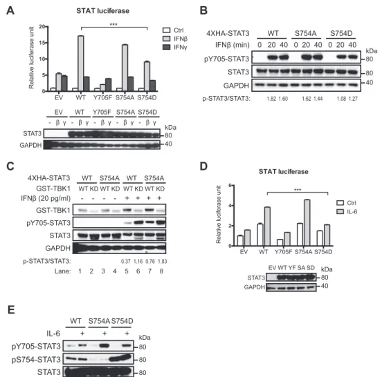
domain of STAT3, we hypothesized that phosphorylation at Ser754 modulates the transcriptional activity of STAT3 in response to IL-6 and IFN. We asked whether Ser754 phosphor-ylation of STAT3 affects the expression of a STAT reporter containing tandem GAS sites in response to IFNand IL-6 by using a phosphomimetic mutant (S754D), because Ser754 phos-phorylation is not induced by IFNor IL-6 alone. To avoid interference from endogenous STAT3, the reporter assay was carried out using CRISPR-mediated STAT3 knock-out HEK293T cells (supplemental Fig. 2) or STAT3-null MEFs
sta-bly expressing wild-type or mutant STAT3. We found that IFNinduced a much higher expression of the STAT report-er in the presence of wild-type STAT3, but the transcriptional inactive Y705F mutant inhibited reporter expression (Fig. 7A), indicating that STAT3 contributes to GAS-driven gene expres-sion in response to IFN. The reporter expression was reduced with the S754D mutant, suggesting an inhibitory role for Ser754 phosphorylation in the transcriptional activity of STAT3 (Fig. 7A). In contrast, STAT3 did not affect GAS-driven gene expres-sion in response to IFN␥(Fig. 7A), which predominantly acti-vates STAT1 but not STAT3 (15). The S754D mutant also showed decreased activation upon IFNstimulation (Fig. 7B), consistent with the results from reporter assays. We then asked whether TBK1-induced Ser754phosphorylation affects STAT3 activation in response to IFN. Because overexpression of TBK1 leads to significant production of cytokines, resulting in elevated basal STAT3 activation (40) (Fig. 1C), here we expressed a moderate amount of TBK1 before treating the cells with IFN. We found that TBK1 expression suppressed IFN -induced STAT3 activation, but the S754A mutant was more refractory to this TBK1-mediated inhibition (Fig. 7C,lanes 5 and7). These data show that Ser754phosphorylation inhibits IFN-induced activation of STAT3. We also probed the possi-bility that Ser754phosphorylation of STAT3 may affect STAT3-mediated inhibition on ISGF3 target genes. Consistent with
CXCL10 expression in THP-1 cells (Fig. 6D), wild-type and
mutant STAT3 inhibited IFN-induced ISRE reporter expres-sion to comparable levels (supplemental Fig. 3), indicating that Ser754phosphorylation of STAT3 does not affect ISGF3 activ-ity. Similar to the IFNreporter assays, S754D mutant was also less active in response to IL-6 as measured by STAT reporter assays and by the levels of Tyr705 phosphorylation, which
pS754-STAT3
STAT3
GST
Flag
2 1.6 1.2 0.2 1.8
-- 0.4 0.8 1.8
--
-0.2
-GST-TBK1 (μg) Flag-STING (μg) pcDNA3 (μg)
p-TBK1
pS754-STAT3 p-IRF3
p-p65
TBK1
STAT3
IRF3
STING
cGAMP - m 1.5h 3h
p-TBK1
TBK1
pS754-STAT3
STAT3
p-IRF3
IRF3
siCTRL sicGAS
dsDNA (3h) m + m +
siCtrl sicGAS
0.0 0.5 1.0 1.5
R
e
la
ti
v
e
quant
it
y
cGAS expression
pS754-STAT3
STAT3
p-TBK1
TBK1
p-IRF3
IRF3
STING
dsDNA (hr) m 1 2 3 m 1 2 3
siCtrl siSTING
A B
C D
80
80
80
80
50
50
40 kDa
80
80
110
40 kDa
80
50
60
80
80
50 40 kDa 80
80
80
80
50
50 kDa 80
inversely correlated with the levels of Ser754phosphorylation or phosphomimetic mutation (Fig. 7,DandE). Collectively, these data demonstrate that Ser754phosphorylation suppresses the transcriptional activity of STAT3 induced by IL-6 and IFN.
Discussion
In this study, we identified STAT3 as a novel substrate of TBK1 downstream of the cytosolic DNA pathway. In the pres-ence of cytosolic DNA, TBK1 phosphorylates STAT3 at Ser754 to limit STAT3 activity induced by cytokines, such as IL-6 and IFN. Previously, it has been shown that IKK⑀regulates STAT1 dimerization and that TBK1 regulates STAT6 activity by direct phosphorylation (14, 26). Our finding places a third STAT member under the control of IKK⑀/TBK1. Interestingly,
the IKK⑀/TBK1-mediated phosphorylation sites in STAT1, STAT3, and STAT6 differ in their location within the proteins (Fig. 1A). In the case of STAT1, phosphorylation of Ser708, which resides between the SH2 domain and the TAD, disrupts SH2 domain-mediated STAT1 homodimerization by steric hindrance (26). How TBK1-mediated Ser407phosphorylation regulates the activity of STAT6 is less clear. Ser407 resides within a highly conserved region of the STAT DNA binding domain, and structural analysis demonstrated that mutations in this region abolish the DNA binding ability of STATs (41). Thus, it is plausible that Ser407 phosphorylation affects the DNA binding affinity of STAT6. It is also worth noting that TBK1 induces a reduced but still significant phosphorylation on STAT6 S407A mutant (14), suggesting the existence of addi-tional TBK1 phosphorylation sites in STAT6. In fact, we iden-tified another IKK⑀/TBK1 substrate motif in STAT6 TAD, in which Ser733 is the residue that corresponds to Ser754of STAT3. Our preliminary data suggest that TBK1 overexpres-sion also leads to STAT6 phosphorylation at Ser733.4For future investigations, it would be of interest to determine whether this phosphorylation serves as an additional mechanism by which TBK1 regulates STAT6 activity in a manner similar to what we discovered with STAT3.
The two IKK-related kinases TBK1 and IKK⑀are structurally similar and prefer almost identical substrate sequencesin vitro (30, 31). However, they appear to have distinct yet partially overlapping roles in vivo (42). Studies using TBK1 or IKK⑀ knock-out cells showed that TBK1 is the principle kinase that phosphorylates IRF3 to initiate interferon production in response to innate immune stimuli and pathogens, whereas IKK⑀has a minor or negligible role in activating IRF3 and inter-feron production (11, 43, 44). Similarly, in our model, although overexpression of TBK1 and IKK⑀both induced Ser754 phos-phorylation of STAT3 (Fig. 1,BandC), endogenous IKK⑀did not have a measurable impact on STAT3 phosphorylation in response to VACV70mer (dsDNA with 33% GC content) trans-fection (Fig. 3C). However, it is worth noting that whereas VACV70mer only induced interaction between STAT3 and TBK1, poly(dA:dT) transfection induced interaction of STAT3 with TBK1 and IKK⑀(Fig. 2B), suggesting that IKK⑀may con-tribute to the signaling cascades and STAT3 phosphorylation downstream of AT-rich cytosolic DNA. AT-rich cytosolic DNA not only activates the STING-TBK1 pathway but also engages the cytosolic dsRNA sensor RIG-I by a polymerase III-dependent mechanism (45). Thus, the differential interactions between STAT3 and IKK⑀/TBK1 in response to VACV70mer and poly(dA:dT) may be due to the activation of cytosolic dsRNA pathway specifically downstream of poly(dA:dT). It is also conceivable that IKK⑀will play a more dominant role in scenarios where its expression is highly induced (46). Whether IKK⑀contributes to STAT3 Ser754phosphorylation and regu-lation under these conditions remains to be tested.
The NF-B pathway is also activated by cytosolic DNA, but the roles of different IKKs in this context remain controversial (11–13). Ishiiet al.(11) suggested that TBK1 is dispensable for
4H.-C. Hsia, unpublished observation.
STAT3
GAPDH
IgG Anti-IFNβ Anti-IL6 Both pY705-STAT3
IgG Anti-IFNβ Anti-IL6 Both IgG Anti-IFNβ Anti-IL6 Both
Ctrl
2h CM 3h CM 4h CM
pS754-STAT3
pY705-STAT3
STAT3
p-TBK1
TBK1
p-IRF3
IRF3
STING
1.5 3 4.5 6 1.5 3 4.5 6
dsDNA (hr) - m
0.5h Pretreatment DMSO Pyridone 6
1h pretreatment
dsDNA (hr)
Untreated CHX
m 1 2 3 4
Naive cells, treated with CM from:
pS754-STAT3
pY705-STAT3
STAT3
p-IRF3
IRF3
Untreated CHX
1 2 3 4 m 1 2 3 4 1 2 3 4 Cells transfected with dsDNA
A
B
C
+/- CHX + dsDNA, 0-4h
CM
Lane: 1 2 3 4 5 6 7 8 9 10 11 12 13 14 15 16 17 18
80
80 80 50
50 40 kDa 80
80
80
50
50 kDa 80
80
80 40 kDa 80
cytosolic DNA-induced NF-B activation, whereas Abeet al. (12, 13) demonstrated a significant dependence of NF-B acti-vation on TBK1. In MEFs and THP-1 cells, we observed that IKK␣/IKKand TBK1 are each responsible for cytosolic DNA-induced p65 NF-B activation or IRF3 activation (Fig. 3,Aand D), indicating that the signaling events dictating NF-B and IRF3 activation diverge at or above the level of these kinases. Intriguingly, p65 activation in L929 is mostly dependent on TBK1 (Fig. 3B), consistent with the observation made by Abeet al.(13). It is unclear why such differences exist. One possible
explanation may be the availability of different signaling mole-cules and the formation of different complexes. For instance, depending on the cell type, TBK1 may localize to the mitochon-dria or the endoplasmic reticulum in response to cytosolic DNA (47). Therefore, it is likely that signaling pathways down-stream of cytosolic DNA and STING may be influenced by the availability of cell type-specific machinery and platforms as well as the subcellular localization of TBK1.
Although it is unclear why Ser754phosphorylation dampens the activity of STAT3, studies on a natural occurring STAT3
STAT1 pY-STAT1 STAT3 pY-STAT3
p65 p-p65
GAPDH
WT S754A S754D
dsDNA (hr) 0 2 4 0 2 4 0 2 4
A
B
C
D
DNA 0hDNA 2 h
DNA 4hDNA 0hDNA 2hDNA 4hDNA 0hDNA 2 h DNA 4
h DNA 0hDNA 2hDNA 4
h 0
2 4 6
R
e
la
ti
v
e
q
u
a
n
ti
ty
SOCS3
KO WT S754A S754D
*** ***
***
DNA 0hDNA 2hDNA 4hDNA 0hDNA 2hDNA 4hDNA 0hDNA 2 h DNA 4
h DNA 0hDNA 2
h DNA 4h 0
20 40 60 80
R
e
la
ti
v
e
quant
it
y
IL6
n.
d.
n.
d.
n.
d.
n.
d.
**
n.s. n.s.
KO WT S754A S754D
DNA 0hDNA 2hDNA 4 h
DNA 0hDNA 2 h DNA 4
h DNA 0hDNA 2
h DNA 4hDNA 0hDNA 2hDNA 4
h 0
100 200 300 400 500
R
e
la
ti
v
e
quant
it
y
CXCL10
***
n.s. n.s.
KO WT S754A S754D
STAT3 p65 IP: Flag
Input
80
80
80
80
60
60 kDa
60
80
40
CCCTTCTAAGAAGGC -516 -508
CGATTCCTGGAACTG -231 -223
TSS
SOCS3
E
WT S754A S754D
WT + DN
A
S754A +DNAS754D +DNA
0.0 0.2 0.4 0.6 0.8
%
Input
IgG STAT3
isoform STAT3 provide a plausible hypothesis. Alternative splicing of the STAT3 transcript results in the truncated STAT3isoform that is 48 amino acids shorter than the full-length STAT3 (48). STAT3shows strong Tyr705 phosphor-ylation and DNA binding activity even in the absence of stim-ulation, but it lacks intrinsic transcriptional activity due to the lack of TAD (48, 49). Interestingly, STAT3dimers are more stable than dimers formed by full-length STAT3, and deletion of 19 residues at the C terminus (amino acids 752–770) of full-length STAT3 is sufficient to significantly enhance Tyr705 phos-phorylation, DNA binding activity, and dimer stability (50). Because the C terminus of STAT3 is rich in acidic amino acids that are negatively charged, it was therefore proposed that the cluster of negative charges in the TAD interferes with STAT3 dimerization and makes phosphorylated Tyr705more accessi-ble to phosphatases (50). Given this mechanism,
phosphoryla-tion or phosphomimetic mutaphosphoryla-tion of Ser754will introduce more negative charges to the region and may destabilize STAT3 dimers, thereby reducing its transcriptional activity. Alterna-tively, it is also possible that Ser754phosphorylation affects the interaction between STAT3 and its co-activators to modulate STAT3 activity. Future studies will focus on testing these hypotheses.
There is mounting evidence of the critical role of cytosolic DNA in tumorigenesis and anti-tumor immune responses. Functional loss of the cGAS-STING cytosolic DNA pathway has been observed in some commonly used cell lines and high passage immortalized MEFs (5) as well as colorectal cancer and melanoma, in which loss of the cytosolic DNA pathway corre-lates with tumor progression (27, 28). In a mouse model of colitis-associated colorectal cancer, STING-deficient mice showed elevated NF-B and STAT3 activity and developed STAT3
GAPDH
- β γ - β γ - β γ - β γ - β γ EV WT Y705F S754A S754D
STAT3 pY705-STAT3
GAPDH
4XHA-STAT3 WT S754A S754D IFNβ (min) 0 20 40 0 20 40 0 20 40
GAPDH pY705-STAT3 GST-TBK1
STAT3
GST-TBK1 WT KD WT KD WT KD WT KD
4XHA-STAT3 WT S754A WT S754A
IFNβ (20 pg/ml) - - - - + + + +
STAT3
GAPDH
EV WT YF SA SD
pY705-STAT3
pS754-STAT3 STAT3
IL-6 + + +
WT S754A S754D
A
B
C
D
E
EV WT Y705F S754A S754D
0 5 10 15 20
R
e
la
ti
v
e
l
u
c
ife
ra
s
e
u
n
it
STAT luciferase
Ctrl IFN IFN
***
EV WT Y705F S754A S754D 0
2 4 6
R
e
la
ti
v
e
l
u
c
ife
ra
s
e
u
n
it
STAT luciferase
Ctrl IL-6
***
Lane: 1 2 3 4 5 6 7 8
p-STAT3/STAT3: 1.82 1.60 1.62 1.44 1.081.27
p-STAT3/STAT3: 0.37 1.16 0.76 1.03
80 kDa 80
80 40 kDa 80
80 kDa 80
40
40 kDa 80
advanced disease, suggesting a role of STING in controlling inflammatory responses and tumorigenesis (29). According to our model, loss of the cytosolic DNA sensing pathway will pre-dict loss of the TBK1-mediated restraint on STAT3 activity, allowing the tumor cells to have elevated STAT3 activation in response to cytokines such as IL-6. Given the well established role of STAT3 in promoting tumorigenesis, our finding sug-gests a possible mechanism in which tumor cells without the cytosolic DNA pathway may have a survival advantage due to unchecked STAT3 activation. On the other hand, activation of dendritic cells in the tumor microenvironment by engaging the cGAS/STING pathway leads to significant T cell recruitment and anti-tumor immunity (51, 52). Because STAT3 negatively regulates dendritic cell activity and the anti-tumor response of hematopoietic cells (20, 53), cytosolic DNA-mediated restraint on STAT3 activity may serve as an additional mechanism to strengthen the anti-tumor immunity of hematopoietic cells in the tumor microenvironment.
In summary, we identified a novel signaling axis in which TBK1 modulates STAT3 activity in response to cytosolic DNA. Our findings reveal a new mechanism by which the activity of STAT3 can be fine-tuned by a single phosphorylation and shed light on the possible cross-talk between innate immune responses and STAT3-driven oncogenic pathways.
Experimental Procedures
Plasmids and Viruses—Mouse STAT3 was cloned into
pBabe-puro (Addgene, catalog no. 1764) (54) using BamHI and EcoRI. N-terminal HA-tagged STAT3 was cloned into 3XHA-pEBB vector using BamHI and NotI. N-terminal FLAG-tagged STAT3 was cloned into pENTR-3C plasmid using BamHI and EcoRI, followed by Gateway recombination into pLenti6/ UbC/V5 destination vector (Thermo Fisher). The C terminus of STAT3 (amino acids 700 –770) was cloned into pGEX-4T-1 in frame with N-terminal GST using BamHI and EcoRI. STAT3 mutant plasmids were generated by site-directed mutagenesis and confirmed by sequencing. STAT3 CRISPR plasmid was con-structed using lentiCRISPRv2 (Addgene, catalog no. 52961) (55) with a single guide RNA targeting the 5⬘-UTR of human STAT3 (supplemental Fig. 2). The primer sequences for single guide RNA cloning are as follows: 5⬘-CAC CGT GCC GGA GAA ACA GGT GAA G-3⬘and 5⬘-AAA CCT TCA CCT GTT TCT CCG GCA C-3⬘.
Protein Purification and in Vitro Kinase Assay—Rosetta cells
(Novagen) harboring pGEX-4T-1-STAT3 plasmid were grown to log phase and treated with 0.5 mMisopropyl-D
-1-thioga-lactopyranoside at 30 °C for 16 h. The cells were lysed (50 mM
Tris, pH 7.6, 150 mMNaCl, 1 mMEDTA, 5 mMDTT, 25g/ml
lysozyme, protease inhibitor mixture (Promega)) at room tem-perature, followed by the addition of 1.25g/ml sodium deoxy-cholate, 1.25 M MgCl2, and 62.5 g/ml DNase I. Cleared lysates (13,000 rpm, 15 min, 4 °C) were incubated with glutathi-one-agarose beads (Amersham Biosciences) at 4 °C for 2 h. The beads were washed twice with modified RIPA buffer (150 mM
NaCl, 1% Nonidet P-40, 0.25% sodium deoxycholate, 50 mM
Tris, pH 7.6, 1 mM-glycerol phosphate), twice with high salt
(500 mMNaCl) modified RIPA, and twice with kinase buffer (1
mM-glycerol phosphate, 20 mMTris, pH 7.4, 12 mMMgCl2).
Purified protein was eluted by incubating the beads with 30 mM
glutathione in kinase buffer with 0.1% Tween 20 for 20 min at room temperature with shaking.
Wild-type and K38A GST-TBK1 were purified from HEK293T transfected with pEBG-GST-TBK1 plasmids as described previously (31). Forin vitrokinase assays, 1–2l of purified GST-STAT3 was incubated with 2– 4l of wild-type GST-TBK1 or K38A GST-TBK1 in kinase buffer with 1 mM
ATP and 10Ci of [␥-32P]ATP at room temperature for 2 h. The reaction was resolved by SDS-PAGE, followed by immu-noblotting or autoradiography.
Cells—THP-1 cells were maintained in RPMI1640 with 10%
FBS, whereas HEK293T, L929, and MEFs were maintained in DMEM with 10% FBS and kept at 37 °C with 5% CO2. STAT3-null MEFs were a gift from Dr. Hua Yu (City of Hope), and TBK1 null MEFs were a gift from Amgen. Retroviruses were produced by co-transfection of pBabe-puro-STAT3 and pCL10A1 plasmids into HEK293T cells, and lentiviruses were produced by co-transfection of pLenti6-STAT3 with psPAX2 and pMD2.G into HEK293T cells. The supernatants containing viruses were 0.45m-filtered before being used for transduc-tion. To reconstitute STAT3 expression in STAT3-null MEFs, MEFs were transduced with pBabe-puro-STAT3 retroviruses and selected for puromycin resistance. To generate STAT3 CRISPR knock-out cells, THP-1 or HEK293T cells were trans-duced with STAT3 CRISPR virus and selected for puromycin resistance, and single clones were analyzed for STAT3 knock-out efficiency (supplemental Fig. 2). To reconstitute STAT3 in THP-1,⌬STAT3-THP-1 cells were transduced with pLenti6-STAT3 viruses and selected for blasticidin resistance.
To activate TBK1, cells were transfected with poly(I:C), poly-(dA:dT), VACV70mer, or 2⬘-3⬘cGAMP using Lipofectamine 2000 (Thermo Fisher) at a 1:1 ratio (g/l), and the final con-centration of nucleic acids was 2g/ml unless otherwise noted. THP-1 cells were treated with 25 ng/ml phorbol 12-myristate 13-acetate (Sigma, P1585) overnight to induce adherence before cytosolic DNA transfection. Poly(I:C) (tlrl-pic) poly(dA: dT) (tlrl-patn), cGAMP (tlrl-cga23), and LPS (tlrl-eblps) were from Invivogen. VACV70mer was prepared by annealing com-plementary 70-nucleotide primers as described previously (33). For experiments with conditioned media, supernatants from nucleic acid transfected cells were collected and 0.2 m-fil-tered; mixed with 20 g/ml normal mouse IgG (Millipore, 12-371), 20 g/ml human IL-6-neutralizing antibody (R&D Systems, MAB206), or 40g/ml human IFNneutralizing anti-body (BioLegend, catalog no. 514004); and incubated at room temperature for 20 min before being added to recipient cells.
Transfection of siRNA—Transfection of siRNA was carried
out using Lipofectamine 2000 according to the manufac-turer’s protocol. Mouse Tbk1 (M-063162-01-0005), mouse
Ikbke(IKK⑀) (M-040798-01-0005), mouseTmem173(STING)
(M-055528-01-0005), and control siRNAs (D-001210-03-05) were from Dharmacon. MouseMb21d1 (cGAS) siRNA was from Sigma (SASI_Mm01_00129826)
Reporter Assay—The STAT reporter 4xM67 pTATA
with 4xHA-STAT3 plasmids. Twenty-four hours after trans-fection, cells were treated with 25 pg/ml mouse IFN(R&D Systems, 8499-IF-010) or 200 pg/ml mouse IFN␥(R&D Sys-tems, 285-IF-100). Cells were lysed with passive lysis buffer (Promega) for the Dual-Luciferase assay (Promega) at 16 –24 h after treatment. The relative luciferase units (RLU) were calcu-lated by normalizing the reading of firefly luciferase to that of
Renillaluciferase, and RLU of the control cells was set to 1. For
IL-6 reporter assays, STAT3 reconstituted MEFs were trans-fected with reporters in the same manner and treated with 100 ng/ml mouse IL-6 (BioLegend, catalog no. 575704) for 24 h.
Immunoblotting and Immunoprecipitation—For
immuno-blotting, cells were lysed in RIPA buffer with protease inhibitor mixture (Promega), phosphatase inhibitor mixture (Sigma), and 1 mMNa3VO4. Cleared lysates were resolved by SDS-PAGE (NuPAGE bis-tris gels, Thermo Fisher), transferred to PVDF membranes (Millipore, IPVH00010), and blocked in 5% nonfat milk in TBST. The membranes were incubated in primary anti-bodies (1:1000 –1:5000) in TBST at 4 °C overnight, washed with TBST, and incubated in appropriate HRP-conjugated second-ary antibodies (Promega) (1:10,000). Pierce ECL (Thermo Fisher) was added to the blots, which were then exposed to films and developed or imaged using ChemiDoc (Bio-Rad). For immunoprecipitation, cells were lysed in 0.5% Nonidet P-40 buffer (0.5% Nonidet P-40, 150 mMNaCl, 20 mMTris, pH 7.6, 1
mM EGTA, 1 mM EDTA, 1 mM -glycerol phosphate, and
0.5% glycerol) with protease and phosphatase inhibitors and Na3VO4. Cleared lysates were incubated with M2 FLAG (Sigma) or STAT3 antibodies at 4 °C overnight, followed by incubation with Dynabead protein G (Thermo Fisher) for 1 h at 4 °C and washing with 0.5% Nonidet P-40 buffer, and resolved by SDS-PAGE and immunoblotting. The following antibodies were from Cell Signaling Technology: GST (catalog nos. 2624 and 2625), phospho-IKK␣/IKK (Ser176/Ser180) (catalog no. 2697), IKK(catalog no. 2684), IKK⑀(catalog no. 3416), phos-pho-IRF3 (Ser396) (catalog no. 4947), IRF3 (catalog no. 4302), phospho-TBK1 (Ser172) (catalog no. 5483), TBK1 (catalog no. 3013), phospho-STAT3 (Tyr705) (catalog nos. 9145 and 9132), phospho-STAT3 (Ser727) (catalog no. 9134), phospho-p65 (Ser536) (catalog no. 3033), p65 (catalog no. 8242), STING (cat-alog no. 13647), phospho-STAT3 (Ser754) (BL14578; catalog no. 5163), and IKK phosphosubstrate motif (G9108). The IKK phosphosubstratemotifantibodywasgeneratedagainstthephos-phorylated IKK consensus sequenceX(Y/F)XpSLX, where pS is the phosphoserine targeted by IKKs (30, 31, 34, 35). GAPDH (sc-25778) and-tubulin (sc-9104) antibodies were from Santa Cruz Biotechnology, Inc. Densitometry analyses were carried out using the gel analysis function of ImageJ.
Chromatin Immunoprecipitation—Chromatin
immunopre-cipitation (ChIP) was performed as described previously (57). Briefly, cells were fixed by formaldehyde, lysed, and sonicated to yield DNA fragments of 200 –500 bp. Lysates were diluted to 0.1% of SDS and precleared by incubating with BSA and salmon sperm DNA-blocked Dynabead magnetic protein G beads (Thermo Fisher). Lysates corresponding to 5⫻106cells were used for each ChIP with 5g of rabbit IgG (Cell Signaling Tech-nology, catalog no. 2729) or rabbit anti-STAT3 antibody (Cell Signaling Technology, catalog no. 12640), followed by capture
with Dynabead magnetic protein G beads. DNA-protein-anti-body complexes were eluted, and DNA was uncross-linked and purified by phenol-chloroform. The quantity of input DNA was determined by Nanodrop. The quantity of DNA corresponding to the STAT3 binding site in theSOCS3promoter in immuno-precipitated chromatin was determined by qRT-PCR with three technical repeats using SYBR Green (Thermo Fisher) and the following primers: 5⬘-TAA GAA GGC TGA TTT CTG GCA GAG G-3⬘and 5⬘-CCA GGT CGG CCT CCT AGA ACT-3⬘. Data are shown as mean with S.D. and are representative of two independent experiments.
Quantitative Real-time PCR—Total RNA from cells were
purified using the Qiagen RNeasy Plus kit according to the manufacturer’s protocol. 1–2g of RNA was used to synthesize cDNA using Moloney murine leukemia virus reverse transcrip-tase (Thermo Fisher). Quantitative real-time PCR (qRT-PCR) was carried out using synthesized cDNA and standard TaqMan probes, primers, and reagents (Thermo Fisher). The expression level of target genes was calculated by the⌬⌬Ctmethod relative to the level ofGUSB. Data shown are the relative quantity, with the relative quantity of the control cells set to 1.
Statistical Analysis—For reporter assays and qRT-PCR, data
are shown as mean with S.D. or mean with 95% confidence intervals, respectively. Each data point was from three technical replicates. Analyses were done by Prism (GraphPad Software, Inc., La Jolla, CA) using attest with false discovery rate con-trolled at 1%.
Inhibitors and Reagents—The IKK␣/IKK-specific inhibitor
Compound A was a generous gift from Dr. Karl Ziegelbauer (Bayer). The TBK1/IKK⑀-specific inhibitors AZ-5C and AZ-5E were synthesized by Dr. Stephen Frye’s group at the University of North Carolina (Chapel Hill, NC). The pan-JAK inhibitor pyridone 6 was from Millipore (catalog no. 420099), and cyclo-heximide was from Sigma (C7698).
Author Contributions—J. E. H. and A. S. B. conceived the study. J. E. H. designed the experiments in Fig. 1 and performed the exper-iments in Fig. 1 (AandB). H. C. H. performed the experiments in Fig. 1 (CandD) and all other experiments and analyzed the data. H. C. H. and A. S. B. wrote the paper. All authors reviewed the results and approved the final version of the manuscript.
Acknowledgments—We thank the members of the Baldwin laboratory and Dr. Blossom Damania for constructive feedback. We also thank Dr. Hua Yu (City of Hope) for STAT3-null MEFs, Dr. Jenny P.-Y. Ting (University of North Carolina, Chapel Hill, NC) for FLAG-STING plasmid, and Dr. Stephen Frye (University of North Carolina, Chapel Hill, NC) for TBK1 inhibitors. The IKK phosphosubstrate motif and Ser(P)754-STAT3 antibodies were kindly provided by Cell Signaling
Technology, and TBK1-null MEFs were kindly provided by Amgen.
References
1. Paludan, S. R., and Bowie, A. G. (2013) Immune Sensing of DNA. Immu-nity38,870 – 880
2. Broz, P., and Monack, D. M. (2013) Newly described pattern recognition receptors team up against intracellular pathogens.Nat. Rev. Immunol.13, 551–565
4. Diner, E. J., Burdette, D. L., Wilson, S. C., Monroe, K. M., Kellenberger, C. A., Hyodo, M., Hayakawa, Y., Hammond, M. C., and Vance, R. E. (2013) The innate immune DNA sensor cGAS produces a noncanonical cyclic dinucleotide that activates human STING.Cell Rep.3,1355–1361 5. Sun, L., Wu, J., Du, F., Chen, X., and Chen, Z. J. (2013) Cyclic GMP-AMP
synthase is a cytosolic DNA sensor that activates the type I interferon pathway.Science339,786 –791
6. Ablasser, A., Goldeck, M., Cavlar, T., Deimling, T., Witte, G., Röhl, I., Hopfner, K.-P., Ludwig, J., and Hornung, V. (2013) cGAS produces a 2⬘ -5⬘-linked cyclic dinucleotide second messenger that activates STING. Na-ture498,380 –384
7. Tanaka, Y., and Chen, Z. J. (2012) STING specifies IRF3 phosphorylation by TBK1 in the cytosolic DNA signaling pathway.Sci. Signal.5,ra20 8. Liu, S., Cai, X., Wu, J., Cong, Q., Chen, X., Li, T., Du, F., Ren, J., Wu, Y.-T.,
Grishin, N. V., and Chen, Z. J. (2015) Phosphorylation of innate immune adaptor proteins MAVS, STING, and TRIF induces IRF3 activation. Sci-ence347,aaa2630
9. Zhong, B., Yang, Y., Li, S., Wang, Y.-Y., Li, Y., Diao, F., Lei, C., He, X., Zhang, L., Tien, P., and Shu, H.-B. (2008) The adaptor protein MITA links virus-sensing receptors to IRF3 transcription factor activation.Immunity
29,538 –550
10. Ishikawa, H., and Barber, G. N. (2008) STING is an endoplasmic reticulum adaptor that facilitates innate immune signalling.Nature455,674 – 678 11. Ishii, K. J., Coban, C., Kato, H., Takahashi, K., Torii, Y., Takeshita, F.,
Ludwig, H., Sutter, G., Suzuki, K., Hemmi, H., Sato, S., Yamamoto, M., Uematsu, S., Kawai, T., Takeuchi, O., and Akira, S. (2006) A Toll-like receptor: independent antiviral response induced by double-stranded B-form DNA.Nat. Immunol.7,40 – 48
12. Abe, T., Harashima, A., Xia, T., Konno, H., Konno, K., Morales, A., Ahn, J., Gutman, D., and Barber, G. N. (2013) STING recognition of cytoplasmic DNA instigates cellular defense.Mol. Cell50,5–15
13. Abe, T., and Barber, G. N. (2014) Cytosolic-DNA-mediated, STING-de-pendent proinflammatory gene induction necessitates canonical NF-B activation through TBK1.J. Virol.88,5328 –5341
14. Chen, H., Sun, H., You, F., Sun, W., Zhou, X., Chen, L., Yang, J., Wang, Y., Tang, H., Guan, Y., Xia, W., Gu, J., Ishikawa, H., Gutman, D., Barber, G.,et al.(2011) Activation of STAT6 by STING is critical for antiviral innate immunity.Cell147,436 – 446
15. Zhong, Z., Wen, Z., and Darnell, J. E. (1994) Stat3: a STAT family member activated by tyrosine phosphorylation in response to epidermal growth factor and interleukin-6.Science264,95–98
16. Reich, N. C. (2013) STATs get their move on.JAKSTAT2,e27080 17. Bromberg, J. F., Wrzeszczynska, M. H., Devgan, G., Zhao, Y., Pestell, R. G.,
Albanese, C., and Darnell, J. E. (1999) Stat3 as an oncogene. Cell98, 295–303
18. Yu, H., Pardoll, D., and Jove, R. (2009) STATs in cancer inflammation and immunity: a leading role for STAT3.Nat. Rev. Cancer9,798 – 809 19. Grivennikov, S. I., and Karin, M. (2010) Dangerous liaisons: STAT3 and
NF-B collaboration and crosstalk in cancer.Cytokine Growth Factor Rev.
21,11–19
20. Kortylewski, M., Kujawski, M., Wang, T., Wei, S., Zhang, S., Pilon-Thomas, S., Niu, G., Kay, H., Mulé, J., Kerr, W. G., Jove, R., Pardoll, D., and Yu, H. (2005) Inhibiting Stat3 signaling in the hematopoietic system elicits multicomponent antitumor immunity.Nat. Med.11,1314 –1321 21. Kortylewski, M., and Yu, H. (2008) Role of Stat3 in suppressing anti-tumor
immunity.Curr. Opin. Immunol.20,228 –233
22. Horvath, C. M., Wen, Z., and Darnell, J. E. (1995) A STAT protein domain that determines DNA sequence recognition suggests a novel DNA-bind-ing domain.Genes Dev.9,984 –994
23. Ivashkiv, L. B., and Donlin, L. T. (2014) Regulation of type I interferon responses.Nat. Rev. Immunol.14,36 – 49
24. Regis, G., Pensa, S., Boselli, D., Novelli, F., and Poli, V. (2008) Ups and downs: the STAT1:STAT3 seesaw of interferon and gp130 receptor sig-nalling.Semin. Cell Dev. Biol.19,351–359
25. Wang, W. B., Levy, D. E., and Lee, C. K. (2011) STAT3 negatively regulates type I IFN-mediated antiviral response.J. Immunol.187,2578 –2585 26. Ng, S.-L., Friedman, B. A., Schmid, S., Gertz, J., Myers, R. M., Tenoever,
B. R., and Maniatis, T. (2011) IB kinase epsilon (IKK⑀) regulates the
balance between type I and type II interferon responses.Proc. Natl. Acad. Sci. U.S.A.108,21170 –21175
27. Xia, T., Konno, H., Ahn, J., and Barber, G. N. (2016) Deregulation of STING signaling in colorectal carcinoma constrains DNA damage re-sponses and correlates with tumorigenesis.Cell Rep.14,282–297 28. Xia, T., Konno, H., and Barber, G. N. (2016) Recurrent loss of STING
signaling in melanoma correlates with susceptibility to viral oncolysis.
Cancer Res.10.1158/0008-5472.CAN-16-1404
29. Zhu, Q., Man, S. M., Gurung, P., Liu, Z., Vogel, P., Lamkanfi, M., and Kanneganti, T.-D. (2014) Cutting edge: STING mediates protection against colorectal tumorigenesis by governing the magnitude of intestinal inflammation.J. Immunol.193,4779 – 4782
30. Hutti, J. E., Shen, R. R., Abbott, D. W., Zhou, A. Y., Sprott, K. M., Asara, J. M., Hahn, W. C., and Cantley, L. C. (2009) Phosphorylation of the tumor suppressor CYLD by the breast cancer oncogene IKK⑀ promotes cell transformation.Mol. Cell34,461– 472
31. Hutti, J. E., Porter, M. A., Cheely, A. W., Cantley, L. C., Wang, X., Kireev, D., Baldwin, A. S., and Janzen, W. P. (2012) Development of a high-throughput assay for identifying inhibitors of TBK1 and IKK⑀.PLoS One7, e41494
32. Chau, T.-L., Gioia, R., Gatot, J.-S., Patrascu, F., Carpentier, I., Chapelle, J.-P., O’Neill, L., Beyaert, R., Piette, J., and Chariot, A. (2008) Are the IKKs and IKK-related kinases TBK1 and IKK-⑀ similarly activated?Trends Biochem. Sci.33,171–180
33. Unterholzner, L., Keating, S. E., Baran, M., Horan, K. A., Jensen, S. B., Sharma, S., Sirois, C. M., Jin, T., Latz, E., Xiao, T. S., Fitzgerald, K. A., Paludan, S. R., and Bowie, A. G. (2010) IFI16 is an innate immune sensor for intracellular DNA.Nat. Immunol.11,997–1004
34. Hutti, J. E., Turk, B. E., Asara, J. M., Ma, A., Cantley, L. C., and Abbott, D. W. (2007) IB kinasephosphorylates the K63 deubiquitinase A20 to cause feedback inhibition of the NF-B pathway.Mol. Cell Biol.27, 7451–7461
35. Marinis, J. M., Hutti, J. E., Homer, C. R., Cobb, B. A., Cantley, L. C., Mc-Donald, C., and Abbott, D. W. (2012) IB kinase␣phosphorylation of TRAF4 downregulates innate immune signaling. Mol. Cell Biol. 32, 2479 –2489
36. Ziegelbauer, K., Gantner, F., Lukacs, N. W., Berlin, A., Fuchikami, K., Niki, T., Sakai, K., Inbe, H., Takeshita, K., Ishimori, M., Komura, H., Murata, T., Lowinger, T., and Bacon, K. B. (2005) A selective novel low-molecular-weight inhibitor of IkappaB kinase-(IKK-) prevents pulmonary inflam-mation and shows broad anti-inflammatory activity.Br. J. Pharmacol.145, 178 –192
37. Wang, T., Block, M. A., Cowen, S., Davies, A. M., Devereaux, E., Gingipalli, L., Johannes, J., Larsen, N. A., Su, Q., Tucker, J. A., Whitston, D., Wu, J., Zhang, H.-J., Zinda, M., and Chuaqui, C. (2012) Discovery of azabenzim-idazole derivatives as potent, selective inhibitors of TBK1/IKK⑀kinases.
Bioorg. Med. Chem. Lett.22,2063–2069
38. Ishikawa, H., Ma, Z., and Barber, G. N. (2009) STING regulates intracel-lular DNA-mediated, type I interferon-dependent innate immunity. Na-ture461,788 –792
39. Pedranzini, L., Dechow, T., Berishaj, M., Comenzo, R., Zhou, P., Azare, J., Bornmann, W., and Bromberg, J. (2006) Pyridone 6, a pan-Janus-activated kinase inhibitor, induces growth inhibition of multiple myeloma cells.
Cancer Res.66,9714 –9721
40. Korherr, C., Gille, H., Schäfer, R., Koenig-Hoffmann, K., Dixelius, J., Eg-land, K. A., Pastan, I., and Brinkmann, U. (2006) Identification of proan-giogenic genes and pathways by high-throughput functional genomics: TBK1 and the IRF3 pathway.Proc. Natl. Acad. Sci. U.S.A.103,4240 – 4245 41. Becker, S., Groner, B., and Müller, C. W. (1998) Three-dimensional
struc-ture of the Stat3homodimer bound to DNA.Nature394,145–151 42. Clément, J.-F., Meloche, S., and Servant, M. J. (2008) The IKK-related
kinases: from innate immunity to oncogenesis.Cell Res.18,889 – 899 43. Hemmi, H., Takeuchi, O., Sato, S., Yamamoto, M., Kaisho, T., Sanjo, H.,
44. Tenoever, B. R., Ng, S.-L., Chua, M. A., McWhirter, S. M., García-Sastre, A., and Maniatis, T. (2007) Multiple functions of the IKK-related kinase IKK⑀in interferon-mediated antiviral immunity.Science315,1274 –1278 45. Chiu, Y.-H., Macmillan, J. B., and Chen, Z. J. (2009) RNA polymerase III detects cytosolic DNA and induces type I interferons through the RIG-I pathway.Cell138,576 –591
46. Shimada, T., Kawai, T., Takeda, K., Matsumoto, M., Inoue, J., Tatsumi, Y., Kanamaru, A., and Akira, S. (1999) IKK-i, a novel lipopolysaccharide-inducible kinase that is related to IB kinases. Int. Immunol. 11, 1357–1362
47. Suzuki, T., Oshiumi, H., Miyashita, M., Aly, H. H., Matsumoto, M., and Seya, T. (2013) Cell type-specific subcellular localization of phospho-TBK1 in response to cytoplasmic viral DNA.PLoS One8,e83639 48. Caldenhoven, E., van Dijk, T. B., Solari, R., Armstrong, J., Raaijmakers,
J. A., Lammers, J. W., Koenderman, L., and de Groot, R. P. (1996) STAT3, a splice variant of transcription factor STAT3, is a dominant negative regulator of transcription.J. Biol. Chem.271,13221–13227
49. Schaefer, T. S., Sanders, L. K., Park, O. K., and Nathans, D. (1997) Func-tional differences between Stat3␣ and Stat3. Mol. Cell Biol. 17, 5307–5316
50. Park, O. K., Schaefer, L. K., Wang, W., and Schaefer, T. S. (2000) Dimer stability as a determinant of differential DNA binding activity of Stat3 isoforms.J. Biol. Chem.275,32244 –32249
51. Woo, S.-R., Fuertes, M. B., Corrales, L., Spranger, S., Furdyna, M. J., Leung, M. Y. K., Duggan, R., Wang, Y., Barber, G. N., Fitzgerald, K. A., Alegre,
M.-L., and Gajewski, T. F. (2014) STING-dependent cytosolic DNA sens-ing mediates innate immune recognition of immunogenic tumors. Immu-nity41,830 – 842
52. Corrales, L., Glickman, L. H., McWhirter, S. M., Kanne, D. B., Sivick, K. E., Katibah, G. E., Woo, S.-R., Lemmens, E., Banda, T., Leong, J. J., Metchette, K., Dubensky, T. W., Jr., and Gajewski, T. F. (2015) Direct activation of STING in the tumor microenvironment leads to potent and systemic tumor regression and immunity.Cell Rep.11,1018 –1030
53. Melillo, J. A., Song, L., Bhagat, G., Blazquez, A. B., Plumlee, C. R., Lee, C., Berin, C., Reizis, B., and Schindler, C. (2010) Dendritic cell (DC)-specific targeting reveals Stat3 as a negative regulator of DC function.J. Immunol.
184,2638 –2645
54. Morgenstern, J. P., and Land, H. (1990) Advanced mammalian gene trans-fer: high titre retroviral vectors with multiple drug selection markers and a complementary helper-free packaging cell line.Nucleic Acids Res.18, 3587–3596
55. Sanjana, N. E., Shalem, O., and Zhang, F. (2014) Improved vectors and genome-wide libraries for CRISPR screening.Nat. Methods11,783–784 56. Besser, D., Bromberg, J. F., Darnell, J. E., Jr., and Hanafusa, H. (1999) A single amino acid substitution in the v-Eyk intracellular domain results in activation of Stat3 and enhances cellular transformation.Mol. Cell Biol.
19,1401–1409