CHEMICAL INHIBITORS OF PHOSPHATIDYLINOSITOL TRANSFER PROTEINS ENABLE HIGHLY SELECTIVE INTERFERENCE WITH SPECIFIC PATHWAYS OF
PHOSPHOINOSITIDE SIGNALING IN CELLS
Aaron Hugh Nile
A dissertation submitted to the faculty of the University of North Carolina at Chapel Hill in partial fulfillment of the requirements for the degree of Doctor of Philosophy in the Department
of Cell and Developmental Biology in the School of Medicine.
Chapel Hill 2014
ABSTRACT
Aaron Hugh Nile: Chemical Inhibitors of Phosphatidylinositol Transfer Proteins Enable Highly Selective Interference With Specific Pathways of Phosphoinositide Signaling in Cells
(Under the direction of Vytas Bankaitis)
Phosphatidylinositol phosphates (PIP) are phosphorylated derivatives of
phosphatidylinositol (PtdIns) that signal to and regulate diverse cellular functions including membrane trafficking, cytokinesis, cell cycle regulation and DNA repair. PIP-signaling is regulated by a variety of proteins through degradation, phosphorylation and dephosphorylation. Members of the Sec14-like phosphatidylinositol transfer protein superfamily (Sec14-PITPs) have at least two functions which include lipid-binding platforms and/or ‘nanoreactors’ that direct PtdIns-OH kinase activity to generate discrete PIP-pools. In Chapter 1, I outline the current literature on the Sec14-superfamily and the structurally unrelated START-like PITPs with special emphasis on mammalian PITPs, and how their disruption results in a number of inherited mammalian diseases.
ACKNOWLEDGEMENTS
I would like to thank my family for supporting all of my endeavors: Hugh Nile, Dody Nile, Amber Nile, Alexis Nile, Brett Hartmann and Mars Hartmann.
To my childhood friends: Billy Springer, Bo Brown, Shane Thomas, Kory Manley, Katelynn Pfeil, Erin Pfeil-McCullough, Brad McCullough, Eric Miller, Lora Brown, Ashley Moore and Brian Birkmire.
My colleagues and friends from current and past institutions: Robert Sons, Brent Hehl, Kelly Watson, Liz Lessey-Morillon, Deepak Jha, Maria Aleman, Dileep Varma, Anita Nair-Varma, Alison Totura, Cheryl Miller, Crystal Neeley, the Recover Room, the Cellar, Tangi Smallwood, Marty Newman, Ashley Clark, Chris Noel, Sarah Nicolson, Rob Davis, Nooshin Lotfi, Bob Fellner, Erin Kirk, Jason Brunton, Stephen Busan, Sandi Wong, Damon Shattuck, Nick Spidale, Matt Geden, Doug Cyr, Pat Brenwald, Con Beckers, Scott Hammond, Aysha Osmani, Stephen Osmani, Jian-Qiu Wu and all of the office staff in the Department of Cell and Developmental Biology.
I would like to thank members of the Bankaitis Lab, in particular: Carl Mousley, Aby Grabon, Ramiro Diz, Mark McDermott, Danish Kahn, Peihua Yuan, Jihui Ren, Ashutosh Tripathi and James Alb.
PREFACE
This Ph.D. thesis was initiated in the Department of Cell Biology at UNC-Chapel Hill and was completed in the Department of Cell and Molecular Medicine at Texas A&M.
Chapter 1’s figures and text were modified with permission from an article published in Clinical Lipidology (2010) entitled “Mammalian diseases of phosphatidylinositol transfer
proteins and their homologs” (Nile, Bankaitis et al. 2010). This article was co-first authored with Aby Grabon.
Chapter 2 is an extended version of work published in Nature Chemical Biology (2014) entitled “PITPs as Targets for Selectively Interfering With Phosphoinositide Signaling in Cells” (Nile, Tripathi et al. 2014).
TABLE OF CONTENTS
LIST OF TABLES…....………...……...…………..xviii
LIST OF FIGURES………...………..………...………..….xviii
LIST OF ABBREVIATIONS AND SYMBOLS………..……….……...xxii
CHAPTER 1: MAMMALIAN DISEASES OF PHOSPHATIDYLINOSITOL TRANSFER PROTEINS AND THEIR HOMOLOGUES...…………...……….1
Overview…....………...…….1
Introduction………...……….1
Operational definitions for the PITPs………4
Sec14-like and START-like PITPs………...….4
The slippery faces of lipid transfer activities………...6
Sec14-Like PITPs as molecules……….9
Sec14 and integration of PtdCho metabolic signals………...9
The anatomy of phospholipid exchange by Sec14-like PITPs………….10
The core engineering of a Sec14 nanoreactor………..…12
A PITP-centric strategy for linking lipid metabolism
to phosphoinositide signaling………...…….…...17
Mammalian Sec14-domain protein disorders………..19
Stand-alone Sec14-like proteins and disease………...21
α-tocopherol transfer protein and vitamin E status………...…21
Caytaxin and cerebellar ataxia……….23
Cellular retinaldehyde binding protein and the vertebrate visual cycle……….25
Multi-domain Sec14-like proteins and disease………28
Rho guanine nucleotide exchange proteins with Sec14 domains………....….29
DBL……….29
Kalirin /Duo……….30
TRIO………31
DBS……….……….32
RhoGAPS……….………33
CDC42GAP/p50RhoGAP………33
Neurofibromin RasGAPs……….34
Type 1 START-like PITPs as molecules……….………38
Cellular functions……….………42
Vertebrate models for type 1 PITP-associated disease………43
PITPα and neurological disease………...44
Anatomy of neurodegenerative disease in PITPα-deficient mice………46
Cell non-autonomous mechanisms for PITPα-dependent neuroprotection…….49
PITPα and cell autonomous signaling……….…….49
PITPα-insufficiencies and chylomicron retention disease……….…..53
pitpα0/0 mice and hepatic steatosis………...58
PITPα--a link between Ins nutrition and lipoprotein metabolism?...59
PITPα and the pancreas………60
Zebrafish type 1 PITPs………..……...61
PITPs and fungal pathogens……….……63
Conclusions and future perspectives……….…...64
Materials and methods……….67
Sequence alignment……….67
CHAPTER 2: CHEMICAL INHIBITORS OF PHOSPHATIDYLINOSITOL TRANSFER PROTEINS ENABLE HIGHLY SELECTIVE
INTERFERENCE WITH SPECIFIC PATHWAYS OF
PHOSPHOINOSITIDE SIGNALING IN CELLS………...………...69
Overview………...…...69
Introduction………..70
Results: NPPM-like SMIs……….…...72
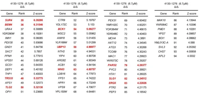
Identification of candidate Sec14-directed SMIs……….72
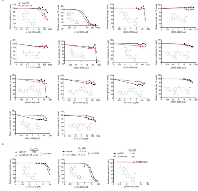
Expansion of the candidate Sec14-directed SMI set………79
Yeast sensitivity to NPPM is a function of Sec14 expression levels…………...84
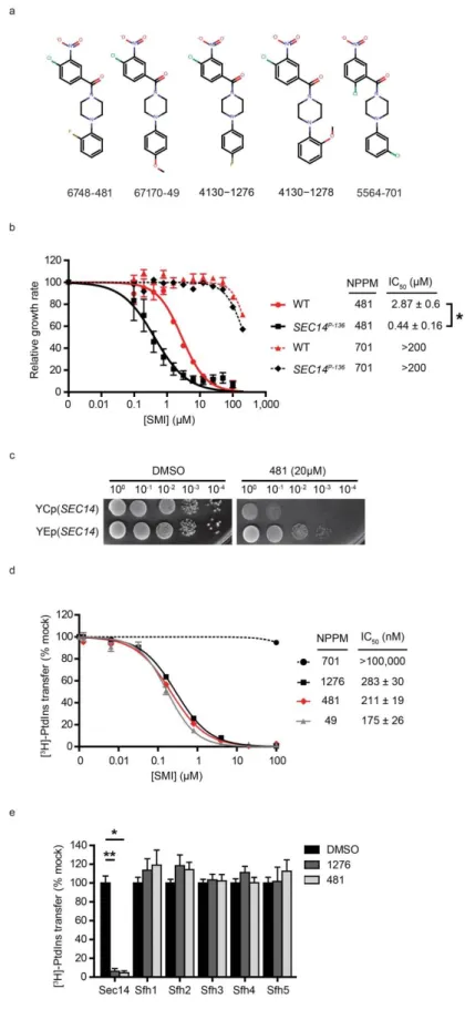
NPPMs directly and selectively inhibit Sec14 in vitro………86
NPPM structure activity relationships……….88
Sec14 inhibition and chemical nature of the NPPM halide……….94
NPPM docking pose within the Sec14 phospholipid-binding pocket…………..95
NPPM-resistant Sec14 proteins………..101
NPPMs and sec14-1ts at non-permissive temperature induce a G2 cell-cycle arrest ………....104
Sec14 is the sole essential NPPM target in cells………106
NPPM intoxication and genetic ablation of Sec14 activity
evoke similar phenotypes……….………..111
NPPMs discriminate between chemically distinct phosphoinositide pools…...124
NPPMs discriminate between local PtdIns(4)P signaling pathways……….….134
Discussion of NPPM-like Sec14-directed inhibitors……….138
Results: alternative Sec14-directed SMIs……….….147
The natural product himbacine is an active Sec14-inhibitor………..…151
Sec14 inhibitors show inhibitory activity against pathogenic yeast in the Candida genus………154
Discussion: alternative Sec14-directed inhibitors………..………160
Materials and methods………...162
Molecular graphics and chemical drawing……….162
Yeast strains, media and reagents………..162
Small molecule inhibitors………..162
Chemogenomic screening………..163
Docking simulations………...163
GOLD docking………...164
Hydropathic scoring………...164
PLIF………166
Hierarchical cluster analysis……….….….……166
FACS sorting………..167
Protein purification……….167
Rat liver microsomes………..168
PtdIns-transfer assays……….168
Statistical analyses……….169
Growth rate analyses………..169
[3H]-serine labeling of yeast cells………...………...170
Choline release assay………...…...171
Homology modeling of Sec14 closed conformation………...…...172
Site-directed mutagenesis………...172
Transmission electron microscopy……….172
Metabolic labeling and immunoprecipitation………173
Phosphoinositide analyses………..174
Fluorescence imaging……….175
Invertase secretion assays………..177
Sequence alignment………...178
NPPM chemogenomic interactions……….……...178
CHAPTER 3: MEASURING AND MODULATING PHOSPHOINOSITIDE SIGNALING IN CELLS………180
Overview………...180
Introduction………181
Methods to monitor phosphatidylinositol phosphate status………..…….184
Direct measurement of phosphatidylinositol phosphates………...184
Mass spectrometry……….186
Isomer-specific PIP antibodies………..….187
PtdIns phosphate binding domains………..…...188
PH domains……….…...191
PX domains……….…...192
GLUE domains……….…..193
Tubby domains……….……..194
BATS domains……….…..196
SYLF domains………...197
EHD domains ………197
FERM domains……….….199
BAR domains……….…200
FYVE domains……….…..201
PIP biosensors and FRET……….…..203
Coincidence detection………....204
Modulation of cellular phosphatidylinositol phosphates………...205
Addition of exogenous phosphatidylinositol phosphates……….…..205
Genetic modulation of PIP modifying enzymes……….…...207
Chemical- and light-induced enzyme targeting………..….…..209
Pharmacological intervention of phosphoinositide signaling……….…...210
Small molecule inhibitor validation in S.cerevisiae………..….211
Inhibitors of PIP signaling pathways……….…212
Akt/PKB inhibitors………213
PtdIns-3-kinases inhibitors………213
Chemical modulators of SHIP phosphatase…………...……...220
Screening for synaptojanin inhibitors……….……...224
Inhibitors of PtdIns-4-kinasess………..226
Inhibitors of phospholipase C………232
Inhibitors of inositol monophosphatase (IMPase)……….236
Conclusions and future directions………..…………239
Utilizing counter-ligand and pocket geometry to identify isomer-specific PITP inhibitors………...…………...…239
Identification of SMIs directed against PITPs………...242
SMIs against ‘bypass Sec14’ proteins and phospholipase D ………242
Closing remarks……….………....244
FUNDING SOURCES………...251
LIST OF TABLES
Table 1. Other selected Sec14-like proteins ... 36
Table 2. Chapter one summary ... 66
Table 3. SMI 741 inhibits dimorphic transitions in Candida albicans ... 159
Table 4. Chapter two summary ... 178
Table 5. List of PtdIns phosphate binding domains ... 189
Table 6. Yeast strains ... 245
Table 7. Protein expression plasmids... 247
LIST OF FIGURES
Figure 1. Transfer vs. nanoreactor models for PITP function... 8
Figure 2. The Sec14-fold. ... 12
Figure 3. Differential phospholipid binding strategies by Sec14-like PITPs. ... 14
Figure 4. The PtdIns-binding bar code in Sec14-like proteins. ... 18
Figure 5. Domain arrangements of Sec14-like proteins ... 20
Figure 6. PITPα Structures. ... 41
Figure 7. Intestinal and hepatic steatosis in PITPα-deficient mice. ... 57
Figure 8. Identification of 4130-1278 and 4130-1276 as candidate Sec14-directed SMIs. ... 73
Figure 9. Chemogenomic interaction-profiles of 4130-1276 and 4130-1278. ... 78
Figure 10. In vivo SAR analyses. ... 82
Figure 11. NPPMs specifically inactivate Sec14. ... 85
Figure 12. In vitro SAR analyses. ... 92
Figure 13. NPPM SAR relationships. ... 93
Figure 14. Homology model of a closed Sec14 conformer. ... 98
Figure 15. In silico docking solutions for 6748-481 binding by Sec14. ... 99
Figure 16. Interaction fingerprints of representative 6748-481 docking poses. ... 100
Figure 17. Sec14 mutants resistant to NPPM inhibition. ... 102
Figure 18. Sec14S173C is resistant to inhibition by NPPMs. ... 103
Figure 19. Active-NPPMs phenocopy sec14-1ts cell cycle arrest ... 105
Figure 20. Sec14 is the sole essential cellular target of bioactive NPPMs. ... 107
Figure 21. NPPM intoxication stimulates phospholipase D activity in vegetative yeast. ... 110
Figure 23. ‘Bypass Sec14’ are resistant to NPPM-induced accumulation of defective
TGN/endosomal compartments. ... 114
Figure 24. SEC14S173C cells are resistant to NPPM-induced accumulation of defective TGN/endosomal compartments. ... 115
Figure 25. NPPMs induce defects in bulk endocytosis. ... 116
Figure 26. Endocytic recycling of GFP-Snc1 is retarded in NPPM-intoxicated cells. ... 117
Figure 27. Sec14-active NPPMs block invertase secretion. ... 118
Figure 28. ‘Bypass Sec14’ mutations correct NPPM-induced trafficking defects. ... 119
Figure 29. Sec14S173C yeast secrete invertase in the face of Sec14-active NPPMs. ... 120
Figure 30. Sec14-active NPPMs induce CPY trafficking defects. ... 121
Figure 31. NPPM induced CPY trafficking defects are poorly reversible. ... 122
Figure 32. NPPM-induced CPY trafficking block is dose-and time-dependent. ... 123
Figure 33. ‘Bypass Sec14’ mutations and Sec14S173C expression alleviate NPPM-induced CPY trafficking defects. ... 124
Figure 34. Sec14-active NPPMs target specific phosphoinositide classes. ... 128
Figure 35. GFP-2xPHPLCδ1 plasma membrane association is unperturbed by challenge with Sec14-active NPPMs. ... 129
Figure 36. PtdIns(4)P biosensors. ... 131
Figure 37. GFP-2xPHOsh2 localization to membranes is unperturbed in kes1Δ ‘bypass Sec14’ mutants by challenge with Sec14-active NPPMs. ... 133
Figure 38. NPPMs discriminate between Sec14- and Sfh4-mediated PtdIns(4)P signaling. ... 136
Figure 39. The Sec14 PtdIns and PtdCho binding barcodes. ... 140
Figure 40. Aromatic nucleophilic substitution of NPPMs. ... 145
Figure 41. Mechanism for NPPM-mediated inhibition of Sec14. ... 146
Figure 42. Hierarchical analysis of SAR reveals 12 structural clusters ... 148
Figure 44. Resistance of Sec14 inhibitors to genetic ‘bypass Sec14’ mutants. ... 151
Figure 45. The structure of himbacine. ... 152
Figure 46. Himbacine inhibits Sec14-mediated PtdIns transfer activity in vitro. ... 153
Figure 47. Mutations in the PtdCho binding site of Sec14 confer Himbacine-resistance. ... 154
Figure 48. Chemical structure of SMI 741 ... 156
Figure 49. Inhibition of Saccharomyces cerevisiae Sec14 in vitro by SMI 741 ... 157
Figure 50. SMI 741 inhibits dimorphic transitions in Candida albicans. ... 158
Figure 51. Structure of phosphoinositides ... 182
Figure 52. PIP diversity in yeast and mammals ... 183
LIST OF ABBREVIATIONS AND SYMBOLS
◦ Degree
Δ Deletion
2μ Episomal plasmid
3AC 3-α-aminocholestane
11-cis-ROL 11-cis-retinol
Å Angstrom (10-10 meter)
aa Amino acid
ADP Adenosine diphosphate
all-trans-RAL All-trans-retinaldehyde
APCI Atmospheric pressure chemical ionization
ATA Aurintricarboxylic acid
ATP Adenosine triphosphate
α-TOH α-tocopherol
αTTP α-tocopherol binding protein
AVED Ataxia with vitamin E deficiency
BiPh(2,3′,4,5′,6)P5 Biphenyl-derived polyphosphate, biphenyl
2,3′,4,5′,6-pentakisphosphate
CEN Centromeric plasmid
CHX Cycloheximide
CID Chemically induced dimerization
ClogP Calculated partition coefficient
for n-octanol/water
CPDA N-[4-(4-chlorobenzyloxy)
pyridin-2-yl]-2-(2,6-difluorophenyl)-acetamide
cpm Counts/minute
CRD Chylomicron retention disease
DHR-1 Dock homology region 1
DIC Differential interference contrast
DMSO Dimethyl sulfoxide
dt Dystonic rat
db Diabetes
dl Decaliter
EDTA Ethylenediaminetetraacetic acid
EHD Eps15 homology domain
ENTH Epsin NH2-terminal homology
ESI Electrospray ionization
ET-18-OCH3 Edelfosine
Etn Ethanolamine
FAB Fast atom bombardment
FERM 4.1/ezrin/radixin/moesin
FM4-64 N-[3-Triethylammoniumpropyl]-4-
[p-diethylaminophenylhexatrienyl] pyridinium dibromide
FYVE Fab 1,YOTB, Vac 1, and EEA1
G6Pase Glucose-6-phosphatase
GAPs GTPase activating proteins
GEFs Guanine nucleotide exchange proteins
GFP Green fluorescent protein
GLUE GRAM-Like Ubiquitin-binding in EAP45
GO Gene ontology
Gro Glycerol
[3H] Tritium
HINT Hydropathic INTeractions
HPLC High-performance liquid chromatography
hr Hour
IC50 Half maximal inhibitory concentration
IMPase Inositol monophosphatase
Ins Soluble inositol
Ins-phosphates Phosphorylated forms of D-myo-inositol
IPs Inositol polyphosphates
ji Jittery mouse
jihes Hesitant mouse
jiswd Sidewinder mouse
kanga Kanga mouse
kDa Kilodalton
KES1 KrE11-1 suppressor
LBDD Ligand-based drug design
LC-MS Liquid chromatography
mass spectrophotometry
LD Lipid droplets
LSB6 Las Seventeen binding protein 1
M Molar
m 10-3
m Meter
MALDI Matrix-assisted laser desorption/ionization
mg Milligram
min Minute
mm Millimolar
MP Methylenephosphonate
mRFP Monomeric red fluorescent protein
ms Metabolically-stabilized
MS Mass spectrometry
MSS4 Multicopy suppressor of Stt4 mutation
mol% Molar %
MW Molecular weight
NaCl Sodium chloride
NaN3 Sodium azide
nM Nanomolar
nm Nanometer
NMR Nuclear magnetic resonance
NPPM nitrophenyl(4-(2-methoxyphenyl)
piperazin-1-yl)methanones
OD Optical density
p 10-12
p Protein
PAGE Polyacrylamide gel electrophoresis
PC Personal computer
PDB Protein databank file
PH Pleckstrin homology
PT Phosphorothioate
PIK1 Phosphatidylinositol kinase 1
PIP Phosphatidylinositol phosphate
PITP Phosphatidylinositol transfer proteins
PLD Phospholipase D
PLIF Protein-ligand
interaction fingerprint
PMSF Phenylmethanesulfonyl fluoride
PSD1 PtdSer decarboxylase 1
PSD2 PtdSer decarboxylase 2
PtdCho Phosphatidylcholine
PtdEtn Phosphatidylethanolamine
PtdIns Phosphatidyinositol
PtdIns(3)P PtdIns(3)phosphate
PtdIns(4)P PtdIns(4)phosphate
PtdIns(5)P PtdIns(5)phosphate
PtdIns(3,4)P2 PtdIns(3,4)bisphosphate
PtdIns(3,5)P2 PtdIns(3,5)bisphosphate
PtdIns(4,5)P2 PtdIns(4,5)bisphosphate
PtdIns(3,4,5)P3 PtdIns(3,4,5)trisphosphate
PtdOH Phosphatidic acid
PtdSer Phosphatidylserine
pV Peroxovanadium
PX Phox homology
Qdots Quantum dots
RNA Ribonucleic acid
RNAi RNA interference
Rpm Rotations/minute
SAHN Sequential agglomerative
hierarchical nonoverlapping
s.d Standard deviation
SDS Sodium doadecyl sulfide
Sec14 Secretory protein 14
s.e.m Standard error mean
Sfh1 Sec14 homology protein 1
Sfh2 Sec14 homology protein 2
Sfh3 Sec14 homology protein 3
Sfh4 Sec14 homology protein 4
Sfh5 Sec14 homology protein 5
siRNA Small interfering RNA
SM Sphingomyelin
SMI Small molecule inhibitor
STT4 Staurosporine and temperature sensitive
TCA Trichloroacetic acid
TG Triglycerides
TOF Time of flight
TGN Trans-Golgi network
TOR Target of rapamycin
TS Temperature sensitive
UV Ultra violet light
µ 10-6
µCi Microcuri
µl Microliter
μM Micro molar
vb Vibrator mouse
wobbly Wobbly mouse
WT Wild-type
wt Weight
YPD Yeast peptone dextrose
VO-OHpic Vandyl complexed to hydroxypicolinic acid
vol Volume
CHAPTER 1: MAMMALIAN DISEASES OF PHOSPHATIDYLINOSITOL TRANSFER PROTEINS AND THEIR HOMOLOGUES1
Overview
Inositol and phosphoinositide signaling pathways represent major regulatory systems in eukaryotes. The physiological importance of these pathways is amply demonstrated by the variety of diseases that involve derangements in individual steps in inositide and
phosphoinositide production and degradation. These diseases include numerous cancers, lipodystrophies and neurological syndromes. Phosphatidylinositol transfer proteins (PITPs) are emerging as fascinating regulators of phosphoinositide metabolism. Recent advances identify PITPs (and PITP-like proteins) as outstanding candidates for coincidence-detecting units which spatially and temporally coordinate the activities of diverse aspects of the
cellular lipid metabolome with phosphoinositide signaling. These insights are providing new ideas regarding mechanisms of inherited mammalian diseases associated with derangements in the activities of PITPs and PITP-like proteins.
Introduction
The involvement of phosphorylated forms of D-myo-inositol (Ins-phosphates) and phosphatidylinositol (phosphoinositides) in eukaryotic signal transduction is well
documented (Michell 2008). Indeed, the breadth of inositide and phosphatidylinositol
1
This chapter is an extended version of an article published in the journal of Clinical Lipidology. The original citation is as follows: Nile, A. H., V. A. Bankaitis, V.A., Grabon, A. (2010). "Mammalian diseases of
(PtdIns)-based signaling has inspired some to anoint Ins as evolution’s favorite molecule (Irvine 2005). This is not an idle proclamation given the diversity of phosphorylated
products that can be generated from Ins-containing compounds. For example, yeast generate five phosphoinositides (PtdIns[3]P, PtdIns[4]P, PtdIns[5]P, PtdIns[4,5]P2, and PtdIns[3,5]P2)
while higher eukaryotes produce seven (the five listed for yeast plus PtdIns[3,4]P2 and
PtdIns[3,4,5]P3). The case for Ins-phosphates is more impressive. As each position of the
6-member Ins ring can be phosphorylated (and in at least several cases pyro-phosphorylated), the cabal of possible soluble Ins-phosphate species is immense (63 + Ins for monophosphates and 728 + Ins if one imposes a limit of only two phosphates per Ins-OH). These statistics identify the versatility of Ins as a six-bit chip where specific signaling information is encoded by a unique combination of positionally-specific phosphorylations on the Ins ring. The Ins-phosphate chemical code is subsequently interpreted by proteins which have the appropriate Ins-phosphate binding specificities.
assumes even greater complexity when issues of spatial/temporal control of phosphoinositide production and degradation (i.e. issues critical to biological regulation of Ins-phosphate and phosphoinositide signaling) are considered. In this review, we will limit discussion to the production arm of phosphoinositide signaling.
Present discussions of the roles for phosphoinositides in cell regulation focus on: (i) the function of these lipids as metabolic reservoirs for second messengers (e.g. diacylglycerol and soluble Ins-phosphates), and (ii) their involvement in the formation of membrane binding platforms for specific proteins (Balla 2005; McLaughlin and Murray 2005; Lemmon 2008). Regarding the latter context, the ability of a mammalian cell to produce 7 chemically distinct phosphoinositides allows for creation of a diverse set of binding platforms. The chemical heterogeneity of phosphoinositides is in turn interpreted by protein binding motifs such as PH-domains, FYVE-domains, PX-domains, and even basic patches on protein surfaces that execute phosphoinositide binding by purely electrostatic mechanisms (see Chapter 3; Balla 2005; McLaughlin and Murray 2005; Lemmon 2008).
Discussions of phosphoinositide signaling are dominated by product-centric models that fail to capture important dynamics that accompany production of these lipids. These discussions also do not adequately describe the consequences these mechanisms have with regard to functional diversification of phosphoinositide signaling. The principle message to be delivered in this review is that we do not yet understand important aspects for how lipid signaling is regulated in eukaryotic cells, nor do we understand how the larger lipid
metabolome is integrated with phosphoinositide signaling. An emerging concept that bears on this theme is a specific phosphoinositide generated by a specific lipid kinase can
2005; Schaaf, Ortlund et al. 2008; Bankaitis, Mousley et al. 2010). Thus, biological outcome is not solely determined by the chemical nature of the phosphoinositide, nor is it determined by the PtdIns kinase that produced it. Rather, biological outcome tracks with non-enzymatic proteins that stimulate PtdIns kinase activities. These accessory proteins are the
PtdIns/phosphatidylcholine (PtdCho)-transfer proteins (PITPs), and the data suggest PITPs ‘instruct’ physiological outcomes for PtdIns kinase activities (Routt, Ryan et al. 2005;
Schaaf, Ortlund et al. 2008; Bankaitis, Mousley et al. 2010). PITP-like protein domains hold similar potential for providing such instructive functions, and these domains are found in intriguing contexts. The importance of PITPs and PITP-domain proteins in eukaryotic cell biology and physiology is amply demonstrated by the mammalian diseases associated with derangements in the function of such proteins. Herein, we review the PITPs and mammalian diseases of PITP-like protein dysfunction.
Operational definitions for the PITPs
Sec14-like and START-like PITPs
Carvou 2007). The ‘non-classical’ PITPs are so designated because these retain the ability to bind/transfer PtdIns, but do not conserve PtdCho-binding/transfer activity (Li, Routt et al. 2000; Routt, Ryan et al. 2005; Phillips, Vincent et al. 2006). The non-classical PITPs which, ironically, almost certainly outnumber the classical versions, provide interesting cases for how the diversity in lipid binding by PITPs and PITP-like proteins translates to the awesome diversity in biological outcome for phosphoinositide signaling (see below).
PITPs are highly conserved. The conservation of PITPs breaks down into two distinct branches based on their structural folds: (i) the Sec14-like PITPs, and (ii) the
START-like (StAR-related lipid transfer) PITPs. To date, all START-like PITPs studied are classical PITPs, while Sec14-like proteins include both classical and non-classical varieties. As described in detail below, the Sec14 and START folds are unrelated although these do share some general properties. Whether Sec14-like and START-like PITPs evolutionarily converge on common functional mechanisms, or whether their shared transfer activities are purely coincidental, remains to be determined.
By contrast, the START-like PITP family is a rather sparse one, is structurally unrelated to the Sec14-like proteins (Yoder, Thomas et al. 2001; Phillips, Vincent et al. 2006), and is further subdivided into Type 1 and Type 2 PITPs. The soluble START-like PITPs (Type 1 PITPs) are ~ 35 kD MW and are homologous to each other. The Type II proteins are larger constructions with a domain homologous to the entire Type 1 START-like PITP sequence appended to the N-termini of large membrane-associated modules. The START-like PITP family is not expanded to a large degree from flies (two Type 1 PITPs and one Type 2 PITP) to humans (three Type 1 PITPs and two Type 2 PITPs; 17). The Type 2 PITPs exhibit complex modular arrangements, but the PITP domain is the essential
component of at least one of these proteins--the ca. 900 amino acid Drosophila Type 2 PITP RdgB. This protein is required for the fly photoresponse – a high capacity phosphoinositide signaling system. Yet, the 280 residue PITP domain of RdgB (comprises only ca. 25% of the total RdgB protein sequence) is both necessary and sufficient for rescue of the retinal
degeneration associated with RdgB inactivation, and for restoration of a seemingly wild-type photoresponse in flies lacking the full-length protein (Milligan, Alb et al. 1997). This review focuses on Type 1 PITPs because these are better represented in models for mammalian disease.
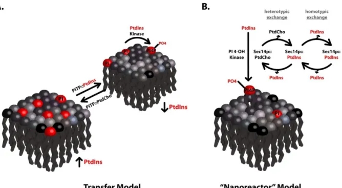
The slippery faces of lipid transfer activities
system to another (Figure 1a), such arguments are inherently circular. That is, PITPs are defined on the basis of an operational transfer assay of uncertain functional significance, and the transfer activity is subsequently featured as the central cellular activity executed by the PITP. Arguments that directly translate PITP in vitro transfer activities to facilitated mobilization of lipid between intracellular membranes in vivo are wrapped in important biological assumptions. One central assumption made in such transfer models is that lipid synthesis is restricted to a few intracellular compartments. As our understanding of cellular lipid biosynthetic capabilities grows, this assumption is coming under increasing fire.
The general acceptance of lipid transfer mechanisms notwithstanding, there is little direct evidence to support simple transfer models for any individual PITP. This evidentiary gap reflects the difficulties in experimentally testing transfer models in physiologically relevant settings. Are there other perspectives from which to view the PITP/lipid transfer problem? Insights culled from studies on PITPs, particularly PITPs of the
Sec14-superfamily; do indeed suggest new and detailed mechanistic possibilities. The available evidence is most consistent with Sec14, and other Sec14-like proteins, functioning as
‘primed’ lipid biosensors that couple binding of lipids other than PtdIns (sensor function) to a PtdIns-presentation activity (Figure 1b). The PtdIns-presentation function potentiates
PITP-like modules in multi-domain proteins. The Sec14 paradigm provides new ideas from which to view mechanisms of PITP function, and recent evidence suggests these new concepts might extend to Type 1 PITPs.
Figure 1. Transfer vs. nanoreactor models for PITP function
Sec14-like PITPs as molecules
Sec14 and integration of PtdCho metabolic signals
Sec14, the major yeast PITP, is required for membrane trafficking through the trans-Golgi network/endosomal system where it acts in a retrograde endosome to TGN trafficking capacity, and is essential for yeast cell viability (Phillips, Vincent et al. 2006; Mousley, Tyeryar et al. 2008; Bankaitis, Mousley et al. 2010). ‘Bypass Sec14’ mutations that permit yeast viability in the absence of the normally essential Sec14 provide unique avenues for diagnosing how Sec14 translates its PtdIns/PtdCho-transfer activities to biological function (Cleves, Novick et al. 1989; Cleves, McGee et al. 1991; Cleves, McGee et al. 1991; Fang, Kearns et al. 1996; Rivas, Kearns et al. 1999; Li, Rivas et al. 2002). The ‘bypass Sec14’ mutants reveal a remarkably intimate coupling between the cellular requirement for Sec14 function and activity of the CDP-choline pathway for PtdCho biosynthesis. That is, inactivation of the CDP-choline pathway obviates the cellular Sec14 requirement (Cleves, McGee et al. 1991; Cleves, McGee et al. 1991). These studies also show that yeast mutants deranged for phospholipid biosynthesis, such that PtdIns is the major membrane
essential for membrane trafficking through the TGN/endosomal system (Cleves, McGee et al. 1991; Cleves, McGee et al. 1991; Fang, Kearns et al. 1996; Rivas, Kearns et al. 1999; Li, Rivas et al. 2002; Phillips, Vincent et al. 2006; Schaaf, Ortlund et al. 2008; Bankaitis, Mousley et al. 2010).
The anatomy of phospholipid exchange by Sec14-like PITPs
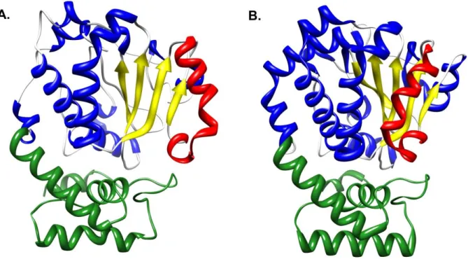
All ideas regarding mechanisms of PITP function assign an important role for the phospholipid-exchange activities of these proteins. These remarkable activities are sustained by thermal energy alone and require no additional co-factors. What are the mechanics of the phospholipid exchange reaction from the perspective of the PITP and from the perspective of phospholipid ligand? Crystallographic studies show the Sec14-domain (smart00516) to be a ca. 280 amino acid two-lobed globular structure that encases a large hydrophobic cavity which defines the phospholipid-binding pocket (Sha, Phillips et al. 1998; Phillips, Sha et al. 1999; Schaaf, Ortlund et al. 2008). Electron paramagnetic resonance measurements report that the hydrophobicity parameters of the pocket are such that this cavity, from the
Access to the binding pocket is gated by a helical substructure whose configuration is flipped open in apo-Sec14 conformers that occur when Sec14 is docked onto membrane surfaces (Figure 2a; Sha, Phillips et al. 1998; Ryan, Temple et al. 2007; Schaaf, Ortlund et al. 2008). The helical gate is closed in holo-Sec14 conformers (Figure 2b), and these represent solution configurations for Sec14::PtdIns and Sec14::PtdCho complexes. The transitions between the ‘open’ and ‘closed’ conformers that accompany phospholipid binding and release on membrane surfaces are dominated by an 18Å displacement of the helical gate (Figure 2). Helical gate dynamics are controlled by a compact ‘gating module’ that
The core engineering of a Sec14 nanoreactor
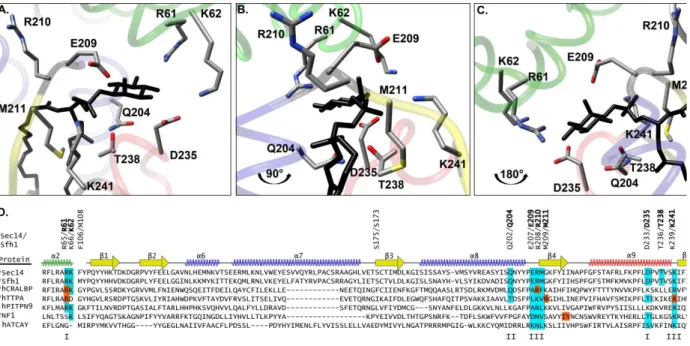
How does Sec14 use its PtdIns- and PtdCho-transfer activities to integrate PtdCho metabolism with phosphoinositide synthesis? The solution to this problem is encoded in the way Sec14 binds its phospholipid ligands. The most remarkable feature of Sec14 (and Sfh1) is the striking difference in the binding poses of PtdIns and PtdCho upon incorporation into the hydrophobic pocket (Figure 3). While the acyl chain regions of each phospholipid
Figure 2. The Sec14-fold.
occupy overlapping physical space, the respective headgroups are stabilized by distant regions of the hydrophobic pocket. This curious and unexpected engineering for how Sec14 binds distinct phospholipid headgroups is of functional significance as heterotypic
phospholipid exchange capability must be housed within individual protein molecules in order for Sec14 to potentiate PtdIns 4-OH kinase activities in vivo (Schaaf, Ortlund et al. 2008; Bankaitis, Mousley et al. 2010). In principle, Sec14 employs a coincidence-detection strategy that integrates PtdCho metabolic information with the action of PtdIns 4-OH
Primed PtdIns-presentation models versus lipid transfer models
The concept that Sec14 employs heterotypic exchange in a concerted phospholipid sensing/presentation cycle portrays the associated in vitro PtdIns-/PtdCho-transfer activities in a very different light than do lipid transfer models. An attractive aspect of
presentation/nanoreactor models is these frame specific and experimentally testable
hypotheses. Several are presented here for discussion. While the questions are framed in the context of Sec14, each of these ideas generates questions that generally pertain to functional interpretations of PITPs and other lipid transfer proteins.
Figure 3. Differential phospholipid binding strategies by Sec14-like PITPs.
Presentation models do not demand physical transfer of lipids from one intracellular destination to the other–it is the cycle that is the key. How many average exchange cycles does Sec14 complete during its membrane dwell time? Presentation models embrace the possibility that many such cycles are executed per membrane association window, while transfer models describe a scenario where there is one exchange cycle per transfer event.
Is physical disengagement of Sec14 from membranes required for execution of biological function? The simplest transfer models demand this be the case, although membrane ‘contact site’ models leave open the possibility that a membrane-bound Sec14 could still retain biological function. Space limitations prohibit detailed discussions of ‘contact-site’ models but the concept is reviewed elsewhere (Wu and Voelker 2002; Holthuis and Levine 2005), and readers are referred to these for further information. Presentation models easily accommodate scenarios where Sec14 (or other LTP domains) are biologically functional as membrane-bound multi-domain molecules.
of abortive PtdIns incorporation may occur per single PtdCho egress event–thereby providing a physical picture of priming. A formal corollary to this hypothesis is that an appropriate covalent adduct of PtdCho, or any other suitable steric obstacle, within the Sec14
hydrophobic pocket might still be compatible with the capability of Sec14 to stimulate PtdIns 4-OH kinase activity– even though this arrangement is physically incompatible with lipid exchange.
What trajectories do lipid molecules follow during entry/exit from the Sec14 hydrophobic pocket? Do PtdIns and PtdCho share similar trajectories (e.g. same entry and exit portals), or do these trace different paths? Reductionist questions of this sort are not particularly important for understanding inter-organelle lipid transfer mechanisms. However, solutions to these questions are central to an understanding of how presentation/nanoreactor mechanisms work because heterotypic phospholipid trajectories define the operative anatomy of the presentation process.
platform (protein or lipid) might contribute at some level. The PtdIns 4-OH kinase itself is an obvious candidate for such a landmark. However, the ability of the structurally unrelated vertebrate Type 1 PITPs to act as functional Sec14 surrogates in yeast argues against
privileged Sec14/PtdIns 4-OH kinase interactions (Skinner, Alb et al. 1993; Tanaka and Hosaka 1994; Ile, Kassen et al. 2010). As discussed in detail elsewhere (Bankaitis, Mousley et al. 2010), more stochastic arrangements can still be biologically productive (i.e. result in enhanced synthesis of phosphoinositide).
A PITP-centric strategy for linking lipid metabolism to phosphoinositide signaling The Sec14-fold is an evolutionarily ancient and versatile one conserved from single cell eukaryotes to man. Its expansion throughout the eukaryotic kingdom reflects an impressive diversification of the unit to bind a wide variety of lipids and lipophilic
Figure 4. The PtdIns-binding bar code in Sec14-like proteins.
Mammalian Sec14-domain protein disorders
Figure 5. Domain arrangements of Sec14-like proteins
Stand-alone Sec14-like proteins and disease
The stand-alone Sec14-like proteins regulate a variety of cellular events including the visual cycle, vitamin E homeostasis, apoptosis, and membrane trafficking. Individual
derangements in these stand-alone Sec14-like proteins primarily manifest themselves as neurological disorders. Some outstanding examples are summarized below. In some cases, the identities of the ligands which occupy the hydrophobic pocket are known. For others, no ligand that incorporates into the protein interior is known. Yet, many Sec14-like proteins exhibit a recognizable structurally defined PtdIns-binding bar code (Schaaf, Ortlund et al. 2008). Whether these proteins do indeed bind PtdIns, and whether these are capable of channeling PtdIns to phosphoinositide synthesis, raises interesting questions for study.
α-tocopherol transfer protein and vitamin E status
late-onset neurological deficits (Terasawa, Ladha et al. 2000; Yokota, Igarashi et al. 2001; Leonard, Terasawa et al. 2002).
At least 25 mutations have been described in the 278-amino acid, αTTP structural gene (TTPA). These fall into two clinical categories: those resulting in severe AVED with early onset and, and those characterized by milder AVED with late onset (Di Donato,
Bianchi et al. 2010). Several mutations in conserved residues (e.g. R59W, E141K, and R221W)
compromise α-TOH binding/transfer and result in severe AVED (Morley, Panagabko et al. 2004). By contrast, the R192H, H101Q, and A120T missense substitutions involve partially
conserved residues, do not strongly compromise α-TOH binding /transfer, and result in mild AVED (Qian, Atkinson et al. 2006; Morley, Cecchini et al. 2008).
It is generally accepted that αTTP is the master regulator of plasma vitamin E levels. In comparative studies, αTTP preferentially binds and transfers α-tocopherol (α-TOH) between membranes in vitro relative to other tocopherols (Akihiro, Makoto et al. 1997; Panagabko, Morley et al. 2003; Zhang, Frahm et al. 2009). It is thought that αTTP employs such a transfer activity to channel α-TOH to a secretory, rather than a degradatory, fate (Traber 2007; Clarke, Burnett et al. 2008). Some models suggest that αTTP does so by mediating direct transport of α-TOH from endosomes to the plasma membrane for
The Sec14 nanoreactor concept suggests an alternative model—αTTP may link heterotypic α-TOH/PtdIns binding/exchange to generation of a phosphoinositide pool dedicated to biogenesis of α-TOH-rich exocytic vesicles. This model predicts that compromise of the PtdIns-binding bar code in αTTP will inactivate the protein. In this regard, the R221W missense substitution, which results in severe AVED, directly alters the
PtdIns-binding bar code. This position corresponds to Sec14 residue K239. This residue
helps coordinate binding of the phosphate moiety through which the Ins headgroup is esterified to the glycerol backbone. Substitutions at this position specifically compromise PtdIns binding by Sec14 (Phillips, Sha et al. 1999; Schaaf, Ortlund et al. 2008). Similarly, the R192H AVED-associated missense substitution in αTTP corresponds to Sec14 amino acid
G210 – a residue positioned adjacent to core elements of the PtdIns-binding bar code.
Caytaxin and cerebellar ataxia
Ortlund et al. 2008). Analyses of Cayman-type ataxic individuals reveal two
polymorphisms. One disrupts an exon-intron boundary leading to truncation of much of the protein, and an S301R missense mutation (Bomar, Benke et al. 2003). Our analyses project
S301 to fall into the helical gate region of the caytaxin Sec14-fold, suggesting gate dynamics
that regulate transitions between open and closed caytaxin conformers might be
compromised. An interesting question for future address is whether compromise of the putative caytaxin PtdIns-binding bar code inactivates the protein.
Much of what is known about caytaxin is derived from the study of rodent models. Caytaxin derangements result in a spectrum of motor malfunctions/dystonia in the jittery (ji), hesitant (jihes), sidewinder (jiswd) and wobbly mice
[http://mutagenetix.scripps.edu/home.cfm], and the well-characterized dystonic (dt) rat (Xiao and LeDoux 2005). A battery of electrophysiological and biochemical studies define the olivocerebellar pathway, particularly in the response of Purkinje cells to climbing fiber projections, as the point of functional abnormality in the dt rat (LeDoux and Lorden 2002). The dt rat cerebellar cortex exhibits altered transcript levels for signaling pathway
Caytaxin physically interacts with number of proteins—including the E3 ubiquitin ligase CHIP (Grelle, Kostka et al. 2006), and peptidyl-prolyl isomerase during neuronal differentiation (Buschdorf, Chew et al. 2008). These associations suggest caytaxin function may be regulated by its binding to these proteins. Furthermore, the caytaxin Sec14-domain binds the kidney-type glutaminase – an enzyme which converts glutamine to the abundant neurotransmitter glutamate. Caytaxin over-expression results in the translocation of the enzyme from the cell body to neurite terminals, and reduces steady state glutamate levels by inhibiting glutaminase. On this basis, it is speculated that caytaxin deficiency-associated glutamate elevation underlies the clinical manifestations of cayman-type cerebellar ataxia (Grelle, Kostka et al. 2006). Moreover, overexpression of either caytaxin (or its Sec14-domain alone) result in the elongation of processes in MCF-7 cells, as is the case with overexpression of the caytaxin homologue BNIP-2 (Hayakawa, Itoh et al. 2007; Aoyama, Hata et al. 2009). Finally, caytaxin also scaffolds kinesin light chain 1 in cultured
hippocampal cells, thereby facilitating transport of vesicle cargo (Aoyama, Hata et al. 2009).
including: retinitis pigmentosa, fundus albipunctatus, Newfoundland rod/cone dystrophy, and Bothnia dystrophy. It is suggested that all of these disorders are manifestations of retinitis punctata albescens (a flecked retinal dystrophy characterized by early onset night blindness, uniform white-yellow spots across the fundus and the progression of macula and retina atrophy resulting in legal blindness) which is also a manifestation of CRALBP1
insufficiencies (Thompson and Gal 2003; Saari and Crabb 2005). Moreover, mice deficient in CRALBP1 manifest large reductions in rates of rhodopsin regeneration, 11-cis-RAL production, and dark adaptation after illumination. Unlike the case in humans, photoreceptor degeneration is not observed (Saari, Nawrot et al. 2001).
lipid exchange reaction), or whether acidic phospholipid interactions with the protein surface evoke conformational changes that eject 11-cis-retinal.
Several naturally-occurring mutations in CRALBP1 compromise retinoid binding (R151Q, M226K), or enhance its binding (e.g. R234W; Maw, Kennedy et al. 1997; Golovleva,
Bhattacharya et al. 2003). In this regard, the autosomal recessive, Bothnia dystrophy presents an interesting case. Pathologies include night blindness in early childhood, and progressive macular/peripheral retinal degeneration (Burstedt, Sandgren et al. 1999; Golovleva, Köhn et al. 2010). Bothnia dystrophy occurs in 1:3500 births worldwide with increased incidence in northern Sweden, primarily as a result of inheritance of the R234W and
M226K variants (Golovleva, Köhn et al. 2010). The crystal structures of 11-cis-RAL-bound
CRALBP1 and the R234W mutant were recently solved (He, Lobsiger et al. 2009). These
studies reveal that R234W further stabilizes bound 11-cis-RAL by increasing packing
interactions within the binding cavity. Additionally, R234 resides in a conserved basic cleft of
CRALBP1. R234 corresponds to Sec14 residue R208 which helps coordinate PtdIns binding
by Sec14 and is a component of the Sec14 structural bar code for PtdIns binding (Phillips, Sha et al. 1999; Schaaf, Ortlund et al. 2008).
Multi-domain Sec14-like proteins and disease
The Sec14 superfamily is too large to cover in one review. The distant members of the superfamily, the BNiP proteins which primarily function in apoptosis, are reviewed elsewhere (Zhang, Cheung et al. 2003; Curwin and McMaster 2008), and will not be emphasized here. Rather, we focus on a set of examples relevant to human disease.
Rho guanine nucleotide exchange proteins with Sec14 domains
The Dbl family of RhoGEFs is defined by a ca. 200-residue Dbl homology (DH) domain positioned adjacent to a C-terminal ca.100-residue plekstrin homology (PH) domain. Of the approximately 70 Dbl-family RhoGEFs, four (Dbl, Dbs/Ost, Duo/Kalirin and Trio) exhibit Sec14-domains (Rossman, Der et al. 2005; García-Mata and Burridge 2007; Curwin and McMaster 2008). All four RhoGEFs are expressed as multiple sliceoforms, not all of which harbor a Sec14-domain, thereby offering mechanisms for differentially regulating the functional properties and subcellular localization of individual isoforms (Johnson, Penzes et al. 2000; Ueda, Kataoka et al. 2004; Kostenko, Mahon et al. 2005; Portales-Casamar, Briançon-Marjollet et al. 2006).
DBL
Dbl, the founding member of the Dbl family of RhoGEFs, is represented by least four splice variants—three of which contain a Sec14-domain. This domain regulates Dbl
Kalirin /Duo
Kalirin/Duo is a neuronal RhoGEF represented by at least eleven forms, six of which contain a Sec14-domain (Kalirin-SOLO, 4, 7, 8, 9, and 12) (McPherson, Eipper et al. 2002; Rabiner, Mains et al. 2005; Schiller, Ferraro et al. 2008). Full-length kalirin (Kalirin-12) is a complex protein that exhibits a Sec14-like domain, nine spectrin-like repeats, two DH, two PH, two SH3, one Ig, one FnIII, and one Ser/Thr protein kinase-like domain (Figure 5) (Penzes, Johnson et al. 2001). The isolated Kalirin Sec14-domain is reported to bind phosphoinositides based on crude lipid blot assays (Schiller, Ferraro et al. 2008). Kalirin nullizygous mice show cognitive and working memory deficiencies associated with reduced neuronal spine densities and abnormal spine morphologies (Cahill, Xie et al. 2009). These neuronal morphology defects are also manifested in ex vivo culture (Xie, Cahill et al. 2010). Additionally, Kalirin is implicated as a genetic risk factor for ischemic stroke (Krug, Manso et al. 2010), coronary artery disease (Wang, Hauser et al. 2007), Alzheimer’s disease (Youn, Ji et al. 2007), and schizophrenia (Cahill, Xie et al. 2009; Hayashi-Takagi, Takaki et al. 2010).
The predominant Kalirin, Kalirin-7, is an important regulator of dendritic spine
development and functional plasticity (Penzes and Jones 2008; Saneyoshi, Fortin et al. 2010). NMDA receptor activation in pyramidal neurons induces a CaMkII-dependent
domain interfaces with other Kalirin-7 domains is not understood. As the Sec14-domain is implicated as a negative regulators of other Dbl family members (Kostenko, Mahon et al. 2005), and phosphorylation of the Sec14-domain promotes GEF activity, a negative regulatory role is a distinct possibility (Xie, Srivastava et al. 2007). Whether lipid binding is involved in such a circuit remains to be determined. Moreover, variants produced from an alternative translation start site truncate the Sec14 domain and the first four spectrin repeats (Δ-Kalirin-7) exhibit distinct properties with regard to regulation of endocytosis, solubility, oligomerization state, cytoskeleton binding and subcellular localization (Schiller, Ferraro et al. 2008).
TRIO
Purkinjie cells, localizes to endosomes where it activates Rho GTPases and promotes neurite elongation in developing Purkinjie cells. The Sec14-domain is suggested to contribute to endosomal localization of this isoform (Sun, Nishikawa et al. 2006). Human Trio also potentiates the nerve growth factor pathway for RhoG- and Rac1-dependent neurite
outgrowth in PC12 cells. The Sec14-domain is dispensable for the neurite promoting activity of Trio, however (Estrach, Schmidt et al. 2002).
DBS
RhoGAPS
The RhoGAP family is populous – counting in excess of 70 members. Of those, p50RhoGAP/Cdc42GAP and BPGAP1 are similar proteins that exhibit N-terminal Sec14-domains appended to RhoGAP Sec14-domains by proline-rich linker Sec14-domains (Figure 5;
Tcherkezian and Lamarche-vane 2007).
CDC42GAP/p50RhoGAP
Mice deficient for Cdc42GAP exhibit multiple premature aging defects including reduction in body mass, loss of subdermal adipose, muscular atrophy, osteoporosis and delayed wound healing (Wang, Yang et al. 2007), and present enhanced rates of JNK-mediated basal apoptosis (Wang, Yang et al. 2005). As may be expected, Cdc42GAP-deficient murine embryonic fibroblasts display elevated Cdc42 activity, and these cells are prone to spontaneous formation of filipodia with defects in directional migration (Yang, Wang et al. 2006). Cdc42GAP derangements are implicated in human disorders such as Waldenstrom Macroglobulinemia (Hatjiharissi, Ngo et al. 2007) and human chronic myeloid leukemia (Jin, Liu et al. 2009). CDC42GAP is also suggested to be a counter-regulator of tubule formation, forecasting a role in angiogenesis (Engelse, Laurens et al. 2008).
controlled in part by intermolecular interactions between amino acids 1-48 and 169-197 which reside in the Sec14-like domain. Interaction with the prenyl group of small GTPases promotes the release of autoinhibition (Moskwa, Paclet et al. 2005).
Neurofibromin RasGAPs
Neurofibromin NF-1 encodes for a 2818 residue RasGAP that is homologous to the yeast RasGAPs, Ira1 and Ira2 (Cichowski and Jacks 2001; D'Angelo, Welti et al. 2006). Defects in NF-1 result in the progressive, autosomal dominant disorder, neurofibromatosis type 1 affecting 1:3500 individuals world-wide. The disease manifests through multiple brown skin macules (café-au-lait spots), intertriginous freckling, iris hamartoma (Lisch nodules), and learning disabilities. NF1 patients are also at a higher risk for optical gliomas and neurofibromas (Friedman 1999; Ferner, Huson et al. 2007). NF-1 primarily regulates p21-Ras-GTP levels, thereby modulating downstream cascades including Ras/MAPK and Akt/mTOR pathways. Loss of NF-1 activity deregulates of these pro-proliferative pathways and inhibits apoptosis (Gottfried, Viskochil et al. 2010).
NF-1 has three discrete domains: the RasGAP catalytic module (Xu, O'Connell et al. 1990), the Sec14-like domain (Aravind, Neuwald et al. 1999), and a PH-domain (D'Angelo, Welti et al. 2006). Several other domains have recently been defined largely on the basis of bioinformatic analyses (Figure 5; Bonneau, Lenherr et al. 2009). A number of missense substitutions elicit NF1 loss-of-function phenotypes without destabilizing the protein
biological activity. A series of these disease-associated mutations affect residues that either comprise, or flank, the hinge domain of Sec14-like proteins. These substitutions likely interfere with conformational transitions of the helix which gates the hydrophobic pocket (Welti, Fraterman et al. 2007). Additionally, a tandem repeat mutation identified in a neurofibromatosis patient with Noonan’s disease duplicates a linker region between the NF1 Sec14- and PH-domains—indicating inter-domain communications between the Sec14-, PH-, and RasGAP-domains are required for proper NF1 activity (D'Angelo, Welti et al. 2006).
Sec14-like protein tyrosine phosphatase
A Sec14-module is incorporated into the 68kDa, cytoplasmic, protein-tyrosine phosphatase MEG2/PTPN9 (Huynh, Wang et al. 2003; Alonso, Sasin et al. 2004; Saito, Tautz et al. 2007). In vitro experiments suggest PTP-MEG2 binds PtdIns(3,5)P2,
PtdIns(4,5)P2, PtdIns(3,4,5)P3 and phosphatidylserine (Zhao, Fu et al. 2003). Thus,
these substitutions do inactivate the protein for stimulating vesicular fusion (Huynh, Wang et al. 2003; Saito, Williams et al. 2007). The available data suggest the MEG2 Sec14-domain executes functions in addition to membrane targeting.
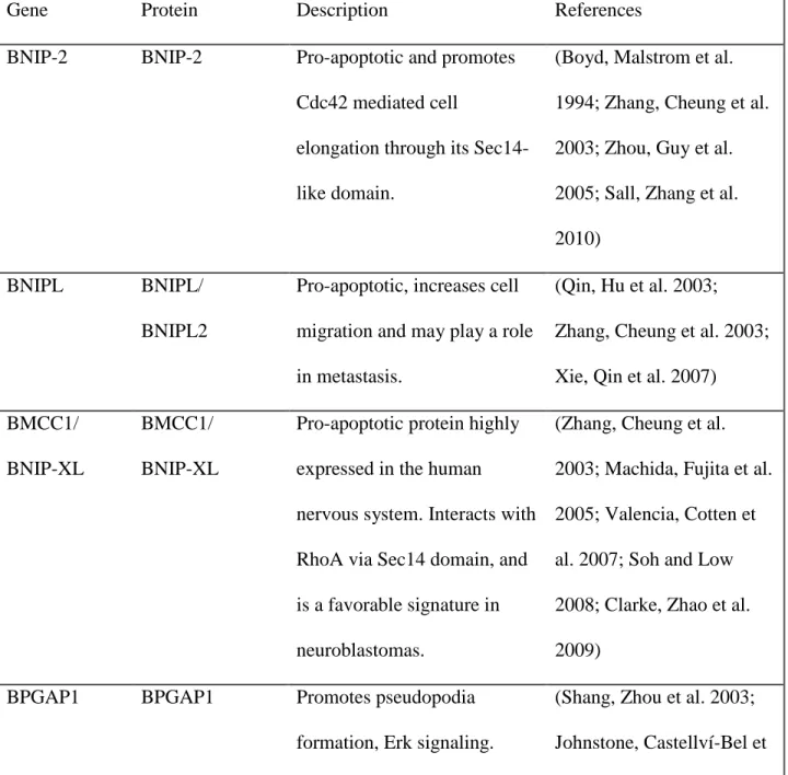
Table 1. Other selected Sec14-like proteins
Gene Protein Description References
BNIP-2 BNIP-2 Pro-apoptotic and promotes Cdc42 mediated cell
elongation through its Sec14-like domain.
(Boyd, Malstrom et al. 1994; Zhang, Cheung et al. 2003; Zhou, Guy et al. 2005; Sall, Zhang et al. 2010)
BNIPL BNIPL/
BNIPL2
Pro-apoptotic, increases cell migration and may play a role in metastasis.
(Qin, Hu et al. 2003; Zhang, Cheung et al. 2003; Xie, Qin et al. 2007) BMCC1/
BNIP-XL
BMCC1/ BNIP-XL
Pro-apoptotic protein highly expressed in the human nervous system. Interacts with RhoA via Sec14 domain, and is a favorable signature in neuroblastomas.
(Zhang, Cheung et al. 2003; Machida, Fujita et al. 2005; Valencia, Cotten et al. 2007; Soh and Low 2008; Clarke, Zhao et al. 2009)
BPGAP1 BPGAP1 Promotes pseudopodia
formation, Erk signaling.
Elevated levels associated with colorectal cancer and invasive cervical cancer.
al. 2004; Lua and Low 2004; Lua and Low 2005; Song, Lee et al. 2008) CRALBPL/
RLBP1L1
Clavesin1 Regulates late endosome/lysosome
morphology in neurons and is upregulated in hepatocellular carcinoma.
(Kong, Ye et al. 2006; Zhao, Xu et al. 2008; Katoh, Ritter et al. 2009)
Clavesin2 Regulates late endosome/lysosome morphology in neurons.
(Katoh, Ritter et al. 2009)
Sec14L1 SEC14L1 Regulates cholinergic transporters and synaptic vesicle formation.
(Ribeiro, Ferreira et al. 2007; Saito, Tautz et al. 2007)
Sec14L5 SEC14L5 Unknown (Saito, Tautz et al. 2007)
Sec14L2 SEC14L2/ TAP1/SPF
Role cholesterol synthesis during fasting. In humans a potential link to breast carcinogenesis and prostate cancer.
(Ni, Wen et al. 2005; Shibata, Jishage et al. 2006; Saito, Tautz et al. 2007; Wen, Li et al. 2007; Johnykutty, Tang et al. 2009; Wang, Ni et al. 2009)
TAP2/p45 induced lung adenocarcinoma in mice.
2003; Saito, Tautz et al. 2007; Bortner, Das et al. 2009)
Sec14L4 SEC14L4/TAP3 Unknown (Saito, Tautz et al. 2007)
SESTD1 Solo/
HIPLP
Elevated levels in the thalamus and brain with potential functions in cone guidance and smooth muscle contraction. It was shown to be embryonic lethal in mice and the Sec14 domain is involved in Rho activation.
(Bezzerides, Ramsey et al. 2004; Miehe, Bieberstein et al. 2010; Yang and
Cheyette 2013)
MOSPD2 MSPD2 Unknown function in humans
but it localized with zebrafish maternal expression in eggs
(Hong, Levin et al. 2010)
Type 1 START-like PITPs as molecules
mammalian START-like Type 1 PITPs (PITPα, RdgBβ, PITPβ). Essentially nothing is known about RdgBβ and we will ignore it for the remainder of this review. Rather, we focus on the homologous PITPα and PITPβ both of which are classical PITPs. These proteins are encoded by distinct genes, yet share 77% identity at the primary sequence level. PITPβ is expressed as two splice variants (termed canonical and alternative on a historical basis) which differ only in the extreme C-terminal primary sequence of the protein (Tanaka and Hosaka 1994; Morgan, Allen-baume et al. 2006; Phillips, Ile et al. 2006). Zebrafish (Danio rerio) also express a mammalian-like cohort of Type 1 PITPs with the addition of a unique PITPβ-like version designated PITPγ. The Type 1 PITP roster extends to interesting details of Type 1 PITP diversity— i.e. zebrafish execute precisely the same exon-skipping splicing event as do mammals in generating the canonical and alternative PITPβ splice variants (Ile, Kassen et al. 2010).
Crystal structures for both PITPα and PITPβ are available, and multiple structural models for PITPα have been solved (Figure 6). These include high resolution structures for PtdCho-bound and phospholipid-free forms (Yoder, Thomas et al. 2001; Schouten, Agianian et al. 2002), and a lower resolution structure for the PtdIns-bound form (Tilley, Skippen et al. 2004). As indicated above, Type 1 PITPs are structurally unrelated to Sec14-like PITPs and are characterized by a START structural fold that forms a single large lipid-binding cavity. Unlike the case for Sec14-like PITPs, PtdIns and PtdCho assume very similar poses within the Type 1 PITP lipid binding cavity (Figure 6). The Type 1 PITP strategy for phospholipid binding suggests these proteins may not operate in a Sec14-like
even though neither residue uniquely contacts PtdIns, or influences other residues that do so, in the holo-PITPα structure. The existence of such enigmatic ligand-specific binding/transfer mutants suggests that PtdCho and PtdIns trajectories during lipid exchange are different in Type 1 PITPs, but ultimately converge on similar poses in the closed conformer. Indeed, Type 1 PITPs rescue both cell viability and phosphoinositide production in yeast devoid of Sec14. Because yeast membranes are rich in PtdIns (20mol%), these data indicate Type 1 PITPs can function in a Sec14-like nanoreactor/PtdIns-presentation mode (Skinner, Alb et al. 1993; Tanaka and Hosaka 1994). Whether these do so in the PtdIns-poor mammalian cell (PtdIns represents 5mol% of bulk phospholipid) is difficult to demonstrate, yet, structural studies suggest that the apo-PITPα conformer displays an open channel which provides access to the headgroup binding region. This channel provides a path via which a lipid kinase could potentially access a PITP-bound PtdIns headgroup (Schouten, Agianian et al. 2002). Such a mechanism requires productive PITP-PtdIns kinase interactions to occur during the interfacial lipid exchange reaction. This concept is consistent with
nanoreactor/PtdIns-presentation modes of action.
The structural studies also suggest how Type 1 PITPs interact with membrane surfaces. Type 1 PITPs present a loop with adjacent Trp residues (Trp203 and Trp204 in
PITPα) and it is reported that compromise of this Trp-Trp motif inactivates the PITP --
that accompany lipid exchange reactions nor is it important for biological function in a vertebrate context (Phillips, Ile et al. 2006).
Figure 6. PITPα Structures.
Cellular functions
The similarity between PITPα and PITPβ notwithstanding, the proteins exhibit important differences including: (i) PITPα localizes to the cytosol/nucleus while PITPβ targets to the trans-Golgi complex, (ii) PITPβ is able to bind/transfer the ceramide-based PL sphingomyelin (SM), in addition to the glycerol-PLs PtdIns and PtdCho, while PITPα only binds/transfers PtdIns and PtdCho. From a functional perspective, PITPβ appears to execute important housekeeping function(s) in the face of robust PITPα expression (Alb, Phillips et al. 2002) , while PITPα is not essential for cell viability. As discussed in detail below, PITPα nullizygosity results in neonatal lethality–even though normal levels of PITPβ are expressed in the nullizygotes.
Remarkably little is known about the cellular functions of Type 1 PITPs. Data from permeabilized cell systems report PITPα stimulates Ca2+-activated secretory granule
exocytosis (Hay and Martin 1995), secretory vesicle and immature granule budding from hepatocyte and neuroendocrine trans-Golgi network (TGN; Ohashi, Jan de Vries et al. 1995), and plasma membrane receptor/G-protein-coupled phosphoinositide hydrolysis by
with murine models as primary focus. Both the congruence and the dissonance between cellular studies and animal studies of Type 1 PITPs are discussed.
Vertebrate models for type 1 PITP -associated disease
Precisely how Type 1 PITP biochemical properties translate to biological activity of the individual proteins remains to be determined. The RdgBβ remains uncharacterized, and only recently have insights in vertebrate PITPβ function been forthcoming (Carvou, Holic et al. 2010; Ile, Kassen et al. 2010). However, it is clear that PITPα, at least, is essential for the viability of vertebrate organisms–including mammals. Our understanding of the
physiological consequences that accompany impaired PITPα functionality derive from analyses of a series of mouse lines with graded reductions in PITPα activity. Hypomorphic lines include the vibrator homozygous mice (vb/vb) and vb/null heterozygous mice (Weimar, Lane et al. 1982; Hamilton, Smith et al. 1997; Alb, Phillips et al. 2007), which express 20% and 10% of wild-type levels of wild-type PITPα, respectively. The vb allele is the result of a serendipitous insertion of an IAP retro-transposon into an intronic region of the pitpa
structural gene--thereby reducing the efficiency with which the cognate pre-mRNA is processed (Weimar, Lane et al. 1982; Hamilton, Smith et al. 1997).
allele or the splice-trap insertion fail to produce detectable amounts of PITPα protein and exhibit indistinguishable phenotypes (Alb, Cortese et al. 2003). Most of the detailed characterizations executed to date involve mice homozygous for the deletion allele.
PITPα and neurological disease
PITPα is produced in most (if not all) cells, but it is particularly highly expressed in brain and cerebellum. In the adult rat, PITPα is produced most robustly in cerebellar Purkinje neurons and granule cells (Nyquist and Helmkamp 1989; Imai, Tanaka et al. 1997; Utsunomiya, Owada et al. 1997). Consistent with these expression data, murine model systems report an important role for PITPα in maintaining integrity of the spinocerebellar system. PITPα null (pitpα0/0) and hypomorphic mice exhibit striking neurological defects--the severities of which are proportional to defects--the level of PITPα expressed (Weimar, Lane et al. 1982; Alb, Cortese et al. 2003; Alb, Phillips et al. 2007).
By contrast, pitpα0/0 mice are born at the expected Mendelian frequencies but usually expire within several days of birth. In rare instances, the pitpα0/0 homozygotes can persist for 10-13 days after birth. The inability of the nullizygotes to thrive is hallmarked by obvious tremor and impaired motor capacity (Alb, Cortese et al. 2003). Unlike the case of vb/vb mice, the abbreviated lifespan of pitpα0/0 homozygotes is independent of genetic background, and is accompanied by two additional signature pathologies – hypoglycemia and intestinal chylomicron retention disease (CRD; Alb, Cortese et al. 2003; Alb, Phillips et al. 2007). These syndromes are addressed in subsequent sections and, as discussed below, contribute to the rate of onset of neurological disease in PITPα-deficient animals.
The neurodegenerative disease course of the vb mouse is classified into three phases. Phase I describes the “true vibrator” phenotype defined by fine, high-frequency postural tremors that become apparent ca. 15 days after the birth of pitpαvb/vb homozygotes. Phase I postural tremors are reminiscent of the enhanced physiological tremors encountered in clinical settings (Weimar, Lane et al. 1982; Elble 1996). While the etiology of enhanced physiological tremors in humans is not known, such tremors often present as a symptom of hyperthyroidism or metabolic dysfunction such as hypoglycemia and liver disease. In that regard, pitpα0/0 mice also display hepatic steatosis and severe hypoglycemia (see below; Alb, Cortese et al. 2003).
progressive, from Phase I symptoms. Furthermore, Phase II presents with clear anatomical signs of degeneration. Neurons in the lumbar and cervical spinal cord and in the cerebellum are vacuolated, aponecrotic, and display distended ER (see below). In genetic backgrounds where vb mice are reasonably long-lived (5-6 months), the disease progresses to a severe cerebellar atrophy (Weimar, Lane et al. 1982). For these reasons, Phase II defines the “degenerative” phase. In the inbred C57/B6 background, Phase II persists until hours before the animal perishes. The basic presentation of Phase II disease resembles the symptoms associated with clinical cases of stroke, inherited neurodegenerative disorders, and multiple sclerosis (Gauthier and Sniderman 1983; Schwab and McGeer 2008; Trapp and Nave 2008).
The terminal stages of PITPα-insufficiency define Phase III disease characterized by loss of consciousness, decreased motor tone and fasciculations, and a progressive ascending motor paralysis that ultimately leads to asphyxiation. For C57/B6 animals, Phase III signals imminent death (within hours) and appears ca. postnatal day 31-33 in the case of vb/vb homozygotes.
Anatomy of neurodegenerative disease in PITPα -deficient mice