UBIQUITIN AND UBIQUITIN-LIKE PROTEINS IN CELL CYCLE REGULATION
Christine A. Mills
A dissertation submitted to the faculty at the University of North Carolina at Chapel Hill in partial fulfillment of the requirements for the degree of Doctor of Philosophy
in the Department of Pharmacology in the School of Medicine.
Chapel Hill 2018
Approved by:
Michael J. Emanuele Lee Graves
ABSTRACT
Christine A. Mills: Ubiquitin and Ubiquitin-Like Proteins in Cell Cycle Regulation (Under the direction of Michael J Emanuele)
Cell cycle is a tightly regulated process; however, it is mis-regulated in many cancers, leading to increased proliferation. Our lab is interested in better
understanding cell cycle regulation, in particular, regulation by the ubiquitin
system, which controls targeted protein degradation. The following work focuses on the modular ubiquitin E3 ligase composed of SKP1/CUL1/F-box protein (SCF) with its substrate adapter Cyclin F. Cyclin F is a unique F-box protein in that it is highly cell cycle regulated, and has been revealed as a key regulator of cell cycle
progression despite few of its substrates having been identified. Our lab aims to identify novel Cyclin F substrates, and determine how these substrates regulate cell cycle processes.
The Cyclin F substrate, Nucleolar and Spindle Associated Protein 1 (NUSAP1), is a microtubule binding protein implicated in mitotic spindle stability and
Cell cycle and metabolic regulation are critical to the growth and proliferation of normal cells, and these systems can be rewired in cancer to promote
To my mentors, friends and parents, whose limitless support I have relied upon along the way. Special thanks to Charles Jervis, the teacher who ignited my love of
science, Dr. Tara Phelps-Durr, who encouraged me to go farther than I ever imagined, and finally, Dr. Michael J Emanuele, who has helped me grow into an independent scientist and given me confidence in my work. I could not have come
ACKNOWLEDGEMENTS
TABLE OF CONTENTS
LIST OF TABLES ... x
LIST OF FIGURES ... xi
LIST OF ABBREVIATIONS ... xiii
CHAPTER 1: INTRODUCTION ... 1
1.1Cell Cycle ... 1
Cell cycle Checkpoints ... 2
The Ubiquitin system ... 4
Ubiquitin E3 ligases ... 6
Ubiquitin-Like Modifications ... 8
1.2 Cyclin F in Cell Cycle Control ... 10
Identified Cyclin F substrates ... 10
1.3 Cell Cycle mis-regulation in Cancer ... 12
2.1 Introduction ... 17
2.2 Results ... 20
NUSAP1 localizes to dynamic spindle microtubules near chromatin ... 20
Identification of NUSAP1 interacting proteins using mass spectrometry ... 23
NUSAP1 does not control RanBP2 localization during mitosis ... 25
RanBP2 depletion impairs the response to taxol ... 27
2.3 Discussion ... 28
2.4 Materials and Methods ... 31
Mammalian cell culture... 31
Immunoblotting and immunoprecipitations ... 32
Immunological reagents ... 33
Mass Spectrometry analysis ... 34
Gel filtration chromatography ... 35
Chromatin Fractionation ... 36
Immunofluorescence Imaging ... 36
Flow Cytometry ... 38
CHAPTER 3: IDENTIFICATION OF A NOVEL SCFCYCLIN F TARGET, SIRTUIN 5, LINKING PROLIFERATION AND METABOLIC REGULATION ... 50
3.1 Introduction ... 50
Sirtuin 5 stability is increased in the absence of Cyclin F ... 54
Sirtuin 5 interacts with Cyclin F ... 55
SCFCyclin F can ubiquitinate Sirtuin 5 ... 56
Sirt5 protein levels influence G1 timing ... 57
Sirt5 protein levels increase with G0 arrest ... 58
3.3 Discussion ... 58
3.4 Materials and Methods ... 61
Mammalian cell culture... 61
Immunoblotting and immunoprecipitations ... 61
Immunological reagents ... 63
Flow Cytometry ... 63
CHAPTER 4: CONCLUSIONS AND FUTURE DIRECTIONS ... 73
LIST OF TABLES
Table 2.1. siRNA oligonucleotides used in Chapter 2. ... 49
Table 2.2. Antibodies used in Chapter 2. ... 49
Table 3.1. siRNA oligonucleotides used in Chapter 3. ... 72
LIST OF FIGURES
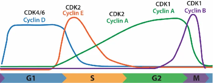
Figure 1.1. Cyclin levels through the cell cycle. ... 16
Figure 1.2. Cyclin F and its targets promote cell cycle progression. ... 16
Figure 2.1. NUSAP1 is a cell cycle regulated microtubule binding protein. ... 39
Figure 2.2. NUSAP1 is cell cycle regulated. ... 40
Figure 2.3. NUSAP1 mitotic localization. ... 41
Figure 2.4. NUSAP1 interacts with the RRU in a cell cycle dependent manner. ... 42
Figure 2.5. RanBP2 co-precipitates with NUSAP1 in both Nocodazole and Taxol arrested cells, and does not influence complex assembly... 43
Figure 2.6. NUSAP1 depletion does not affect mitotic localization of the RRU complex. ... 44
Figure 2.7. NUSAP1 depletion in U2OS cells does not alter RanBP2 localization. ... 45
Figure 2.7. NUSAP1 depletion in U2OS cells does not alter RanGAP1 localization. ... 46
Figure 2.9. NUSAP1 and RanBP2 interact in the cytosol of mitotic cells. ... 47
Figure 2.10. RanBP2 knockdown sensitizes cells to taxol treatment. ... 47
Figure 2.11. NUSAP1 contains a SAP domain in its N-terminus. ... 48
Figure 3.1. Schematic representing results of DRYGIN screen for potential Cyclin F substrates. ... 64
Figure 3.2. Sirt5 stability is increased in the absence of Cyclin F. ... 65
Figure 3.3. Sirt5 and Cyclin F co-immunoprecipitate. ... 66
Figure 3.5. Sirt5 is ubiquitinated in the presence of Cyclin F. ... 68
Figure 3.6. Sirt5 CRISPR KO cells exhibit redistribution of cell cycle phases. ... 68
Figure 3.7. Sirt5 protein expression influences G1 timing. ... 69
Figure 3.8. Sirt5 KO cells exhibit activated DNA damage response. ... 70
Figure 3.9. Sirt5 protein levels are increased in G0 cells. ... 70
LIST OF ABBREVIATIONS
AA amino acid
AEBSF 4-[2Aminoethyl] benzenesulfonyl fluoride
AKT RAC-alpha serine/threonine-protein kinase
APC/C Anaphase Promoting Complex/Cyclosome
apc5 Anaphase-promoting complex subunit 5
ATM Ataxia telangiectasia mutated
ATP Adenosine triphosphate
ATR Ataxia telangiectasia and Rad3-related protein
BSA Bovine serum albumin
C/EBP-β CCAAT/Enhancer-Binding Protein beta
Cdc20 Cell division cycle protein 20 homologue
Cdc25A M-Phase inducer phosphatase 1
Cdc4 Cell division control protein 4
Cdc53 Cell division control protein 53
Cdc6 Cell division control protein 6 homolog
Cdh1 Fizzy-related protein homolog Cdh1
CDK Cyclin Dependent Kinase
CENP-E Centromere-associated protein E
Chk1 Checkpoint Kinase-1
Chk2 Checkpoint Kinase-2
CP110 Centriolar coiled-coil protein o 110 kDa
CPC Chromosomal Passenger Complex
CPS1 Carbamoyl phosphate synthase 1
CRAPome Contaminant Repository for Affinity Purification Mass Spectrometry Data
CRISPR Clustered regularly interspaced short palindromic repeats
CSK Cytoskeletal buffer
CUL1 Cullin-1
CUL3 Cullin-3
DMEM Dulbecco's Modified Eagle Media
DMSO Dimethyl sulfoxide
DNA Deoxyribonucleic Acid
DRYGIN Data Repository of Yeast Genetic Interactions
DTT Dithiothreitol
DUB Deubiquitinating enzyme
EDTA Ethylenediaminetetraacetic acid
EdU 5-ethynyl-2'-deoxyuridine
EGTA Ethylene glycol-bis(β-aminoethyl ether)-N,N,N',N'-tetraacetic acid
Exo1 Exonuclease 1
FASP Filter-aided sample preparation
FBS Fetal bovine serum
FF Firefly luciferase
FoxM1 Forkhead box protein M1
HA Human influenza hemagglutinin epitope
HEC1 Kinetochore protein NDC80 homolog
HECT Homologous to E6-AP carboxy terminus
HEPES 4-(2-hydroxyethyl)-1-piperazineethanesulfonic acid
HIS Hexahistidine tag
HRP Horseradish peroxidase
hst3 NAD-dependent histone deacetylate hst3
IBR InBetweenRING domain
IF Immunofluorescence
INCENP Inner Centromere Protein
IP Immunoprecipitation
IPTG isopropyl β-D-1-thiogalactopyranoside
KCl Potassium chloride
KD Knock-down
kDa Kilodalton
KIF4 Chromosome-Associated kinesin KIF4A
KLH Keyhole Limpet Hemocyanin
KO Knockout
KOH Potassium hydroxide
LC Liquid chromotography
MCC Mitotic Checkpoint Complex
mcm3 DNA replication licensing factor MCM3
MDM2 E3 ubiquitin-protein ligase Mdm2
MEF Mouse embryonic fibroblasts
MgCl2 Magnesium chloride
mRNA messenger ribonucleic acid
MS Mass spectrometry
NAD Nicotinamide adenine dinucleotide
NaF Sodium fluoride
Nedd8 Neural precursor cell expressed developmentally down-regulated protein 8
NETN 20mM Tris-Cl, pH 8.0, 100mM NaCl, 0.5mM EDTA, 0.5% Nonidet P-40
Ni-NTA Nickel-nitrilotriacetic acid
NP-40 Nonidet P-40
NSLC Non small-cell lung cancer
NUSAP1 Nucleolar and Spindle Associated Protein 1
orc3 Origin recognition complex subunit 3
PBS Phosphate buffered saline
PBST Phosphate buffered saline, 0.05% tween-20
p-Chk1 phosphorylated Chk1
PFA Paraformaldehyde
PIAS Protein Inhibitor of Activated STAT2
PIPES piperazine-N-N'-bis(2-ethanesulfonic acid)
PLA Proximity ligation assay
PRC1 Protein Regulator of Cytokinesis 1
RanBP2 Ran Binding Protein 2
RanGAP1 Ran GTPase Activating Protein 1
RB Retinoblastoma protein
RING Really Interesting New Gene
RNA Ribonucleic acid
RNAi RNA interference
RNase A Ribonuclease A
RRM2 Ribonucleoside-diphosphate reductase subunit M2
SAC Spindle Assembly Checkpoint
SAF-A/B Scaffold Attachment Factor A/B
SAP SAF-A/B/Acinus/PIAS protein domain
SCF Skp1/Cul1/F-box
SDS Sodium dodecyl sulfate
SDS-PAGE Sodium dodecyl sulfate polyacrylamide gel electrophoresis
siRNA Small interfering RNA
Sirt5 NAD-dependent protein deacylase sirtuin-5, mitochondrial
Sirt7 NAD-dependent protein deacetylase sirtuin-7
Skp1 Supressor of Kinetochore Protein 1
sli15 Inner centromere protein-related protein SLI15
SOD1 Cu/Zn superoxide disumutase
SUMO Small ubiquitin-related modifier
swi5 Transcriptional factor SWI5
TOP2 DNA topoisomerase 2
TOP2A DNA topoisomerase 2-alpha
TOP2B DNA topoisomerase 2-beta
TSC Total spectral counts
UBC9 SUMO-conjugating ezyme
UBD Ubiquitin Binding Domain
UBL Ubiquitin-Like proteins
WCE Whole cell extract
CHAPTER 1: INTRODUCTION 1.1 Cell Cycle
Cell growth and division is a highly regulated process during which one cell becomes two daughter cells. This process, known as the cell cycle, is broken up into four phases; G1, S, G2 and Mitosis (M). During G1, cells monitor their surroundings and nutrient availability, assessing whether it is safe to proceed through the cell cycle. If the cell proceeds forward in the cell cycle, G1 acts as a preparatory phase for S, where DNA replication occurs. G1 cells contain only one copy of each
chromosome, but to divide, the DNA must be duplicated to ensure that each daughter cell receives the same DNA. To prepare for DNA replication, cells must make nucleotides and proteins, and license DNA replication origins. At the beginning of S, origins fire and DNA replication begins. Once DNA replication is complete and the cells are equipped with two copies of each chromosome, the cells proceed into G2. During G2, the cell ensures that the DNA has been properly
replicated and that it is of adequate size to proceed through mitosis. Once it is ready, the cell proceeds to mitosis, where it segregates sister chromatids equally, resulting in two genetically identical daughter cells. Mitosis is composed of six
stages; prophase, prometaphase, metaphase, anaphase, telophase and cytokinesis.
form active kinase complexes. There are a number of different CDKs and cyclins, which combine in specific pairs. While CDK protein levels remain constant
throughout the cell cycle, cyclin protein levels oscillate. When a particular cyclin is expressed, it binds its preferred CDK to promote signaling and cell cycle
progression (Figure 1.1). It is important to note, that while CDK/Cyclin pairs are considered the core of cell cycle regulation, abundance and activity of hundreds of proteins cycle throughout cell cycle and contribute to proper progression (1).
Cell cycle Checkpoints
There are many checkpoints during the cell cycle to prevent cells from prematurely beginning irreversible processes. These checkpoints are always “active” and monitoring the cellular state. For a cell to proceed past these
checkpoints it must satisfy them by fulfilling a number of requirements, with each checkpoint having a unique set of requirements. Cell cycle checkpoints include the restriction point, DNA damage checkpoint, and Spindle Assembly Checkpoint (SAC). Weakened checkpoints can allow the cell to proceed through the next phase of the cell cycle despite being unprepared, which can result in damage to the cell.
The DNA-damage checkpoint, which arrests cell so that damaged DNA can be repaired, is controlled by either of two large kinases. Ataxia telangiectasia mutated (ATM) or Ataxia telangiectasia and Rad3-related protein (ATR) signaling, depending on which stage of the cell cycle the cell it is in at the time of damage. Both ATM and ATR phosphorylate hundreds of target proteins. If DNA damage is sensed in G1, the ATM signaling pathway is activated, preventing cells from entering S-phase before the damage is repaired, ultimately preventing replication of damaged DNA. ATM does this, in part, by phosphorylating and activating Checkpoint Kinase 2 (Chk2), which inhibits M-Phase inducer phosphatase 1 (Cdc25A) (3–7). Normally, Cdc25A dephosphorylates and activates CDK2/Cyclin E to promote S-phase entry, and phosphorylation by ATM in turn prevents activation of CDK2/Cyclin E (8). ATM also phosphorylates p53, releasing it from its inhibitor E3 ubiquitin-protein ligase Mdm2 (MDM2), so it can induce DNA repair proteins as well as the CDK2/Cyclin E and CDK2/Cyclin A inhibitor p21 (9–18).
If DNA damage occurs during S or G2, signaling goes through a similar signaling pathway mediated by ATR, to arrest cells and give the cell time to repair the damage before proceeding through mitosis. At the core of ATR damage
response, ATR phosphorylates and activates Checkpoint Kinase 1 (Chk1), which, like Chk2, also inhibits Cdc25A (19–21). During S-phase however, Cdc25A
promotes CDK1/Cyclin B activity (22, 23). ATR signaling also activates the CDK1 inhibitor Wee1-like protein kinase (Wee1) (24, 25). If cells slip through the damage checkpoint and into mitosis, cells undergo mitotic catastrophe and die.
cell segregates mitotic chromosomes, until they have made the correct attachments to the mitotic spindles, meaning each sister of a chromosome pair is stably
attached to opposite spindle poles (26). This checkpoint is controlled by the Mitotic Checkpoint Complex (MCC) which targets the E3 ubiquitin ligase complex known as the Anaphase Promoting Complex/Cyclosome (APC/C) (27). The MCC sequesters the APC/C substrate adapter protein Cell division cycle protein 20 homologue (Cdc20), preventing the APC/C from being active (28). Once each pair of sister chromatids has been stably attached to opposite poles, the checkpoint is satisfied and APC/C is activated. Once active, APC/C targets Cyclin B for degradation, as well as Securin, the protein that sequesters Separase (29–33). Once Separase is
released, it can cleave Cohesin, the proteins holding sister chromatids together, and the cell can enter anaphase where sister chromatids are pulled to opposite poles of the mitotic cell (34, 35).
The Ubiquitin system
Targeted protein degradation is a major component of cell cycle regulation and allows the cell to degrade proteins within a few minutes of degradation onset, offering a quick switch-like mechanism for cells to release from, or even activate checkpoints. For example, once the SAC has been satisfied, cells trigger
degradation of Cyclin B via the Anaphase Promoting Complex/Cyclosome (APC/C) and within minutes, Cyclin B is degraded, allowing cells to progress into anaphase.
is added specifically to substrate lysines through an enzyme cascade composed of E1, E2 and E3 enzymes (37, 38). The first step to this cascade is ATP-dependent ubiquitin activation by the E1, or activating enzyme. This activation results in a thioester linkage between the C-terminus of the ubiquitin and a cysteine in the E1. The ubiquitin molecule is then passed to the catalytic cysteine of an E2, or
conjugating enzyme (39). Finally, the E2 interacts with an E3 ubiquitin ligase
(discussed in detail below), to ligate the ubiquitin molecule to a substrate lysine via an isopeptide bond with the C-terminal glycine of ubiquitin (39). Alternatively, ubiquitin can be conjugated to the amino-terminal of a target substrate (40).
Ubiquitin can be added to substrates a number of different ways. Mono-ubiquitination refers to a single ubiquitin molecule added to a substrate on a single lysine. Multi-mono-ubiquitination occurs when multiple lysines of the substrate have a single ubiquitin modification. Furthermore, because ubiquitin is itself a protein that contains lysines, it too can be modified by other PTMs or other ubiquitin
molecules, resulting in ubiquitin chains. There are many possible chain linkages due ubiquitin containing seven lysines, with different lysine linkages between ubiquitin molecules resulting in different chain topologies, which determine the outcome for the substrate (36, 41, 42).
number of proteolytic enzymes that hydrolyze peptide bonds, ultimately degrading proteins (46, 47). Deubiquitinating enzymes (DUBs) associated with the
proteasome cleave ubiquitin from target proteins, allowing it to be recycled (48, 49).
Apart from ubiquitin’s role in promoting degradation through the proteasome, ubiquitin also regulates cellular processes such as endocytosis, DNA repair and other signaling pathways (41, 50, 51). Ubiquitination of some proteins may
regulate binding partners through mechanisms such as steric hinderance, or even promote complex assembly (52). Ubiquitin can also be conjugated to histones or transcription factors to regulate transcription (52).
Ubiquitin E3 ligases
As described above, ubiquitin is added to substrates via an enzyme cascade, with the last member of the cascade is the E3 ubiquitin ligase. E3 ligases typically fall into one of three families; RING-type, HECT-type or RING-between-RING (RBR) type (53–55). The largest family of E3 ligases, RING-type ubiquitin ligases, are classified by the Really Interesting New Gene (or RING) domain or protein, which recruits an E2 to the enzyme complex (53). In RING-type ligases, the E3 binds a substrate protein and the E2 transfers ubiquitin directly to the substrate, with the E3 acting more as a scaffold and never contacting the ubiquitin directly (53). For HECT-type E3 ubiquitin ligases, which contain a Homologous to E6-AP carboxy terminus (HECT) domain, the E2 binds the E3 and transfers the ubiquitin to a
the ubiquitin is first transferred from the E2 to the E3, which then transfers it to a substrate lysine (55).
E3 ligase families can be further broken down into subfamilies, for example, the RING-type ligases include Cullin E3 ligases (56). These are modular E3 ubiquitin ligases based on a Cullin backbone, and were first discovered for their role in the cell cycle (example in Figure 1.2) (56, 57). There are up to nine Cullin proteins, and each uses a specific family of substrate adapter proteins to recruit substrates to the complex. In the following research, the Cullin 1 (CUL1) based E3 ubiquitin ligase, called the SCF (Skp1/CUL1/Fbox protein) is of particular interest. This E3 ligase was the first Cullin based E3 ligase to be identified, and is composed of a CUL1
backbone, which binds a RING protein on its C-terminus and Skp1 (Suppressor of Kinetochore Protein 1) on its N-terminus (58, 59). The Skp1 protein binds an F-box protein, which then recruits specific substrates to the E3 complex for ubiquitination while the RING protein recruits the E2 (58, 59). Humans possess ~70 different F-box proteins, each enabling the SCF unique target specificity.
E3 Ubiquitin ligases are antagonized by DUBs, which remove ubiquitin
Ubiquitin-Like Modifications
Ubiquitin is a member of the ubiquitin family of proteins, which primarily act as PTMs. Ubiquitin-Like proteins (UBLs) are classified into two types; type I have been identified as being conjugated to substrates, while type II have not, with ubiquitin being a type I UBLs. All UBLs undergo the same basic enzyme cascade for activation and conjugation to substrates, but so far, the best characterized of these type I UBLs, besides ubiquitin, include Neural precursor cell expressed
developmentally down-regulated protein 8 (Nedd8) and Small Ubiquitin-related Modifier (SUMO).
Nedd8 is the UBL most similar to ubiquitin (62). The role of neddylation in the cell is most closely tied to regulation of Cullin E3 ligases. Neddylation of Cullin backbones aids in recruitment of the ubiquitin loaded E2 and promotes a structural change that brings the E2 and substrate closer together (63–66). Furthermore, it helps stabilize the transition state, during which the E2 and substrate are
interacting, allowing chain elongation to occur (63).
the protein level), which can both be conjugated to substrates, however only SUMO2/3 can form SUMO chains (67). Currently, it is unclear what the differences between SUMO1 and SUMO2/3 modification of substrates are, although it has been suggested that SUMO2/3 conjugation plays more of a role in stress response (72, 73). It is also unclear if either paralog is preferred by particular SUMO E3 ligases, although research has suggested that SUMO1 modifications compose the majority of preferential SUMO modifications in vivo (74).
SUMOylation may influence many protein characteristics, including
localization, dimerization/interactions, activity, and stability. While a few SUMO substrates have been well characterized, there is still debate on the function of the SUMOylation. So far, studies aimed at identifying SUMOylation substrates have relied on stress induced SUMOylation or overexpression of SUMO or the
promiscuous E2, UBC9. Using overexpression methods in the identification of SUMO targets is difficult because the SUMO can be used by other UBL pathways, for
1.2 Cyclin F in Cell Cycle Control
Some members of the ubiquitin pathway are becoming more recognized for key roles in cell cycle progression. One such protein is Cyclin F (Figure 1.2). Cyclin F is the founding member of the F-box containing family of proteins, most of which are substrate adapters for the SCF E3 ubiquitin ligase complex (75–77). Cyclin F is a non-traditional cyclin, as it does not bind to and activate a CDK (78). However, like cyclins, Cyclin F protein levels are highly cell cycle regulated, and it is the most cell cycle regulated F-box protein (75, 78). Cyclin F was first identified as a protein that could rescue the yeast Cdc4mutant phenotype, which causes a G1 arrest and subsequent death (75). While many of the known Cyclin F substrates alluded to its importance for cell cycle progression, more recent studies have shown that
feedback between the APC/CCdh1 and SCFCyclin F is important for promoting the G1-S transition (79, 80). During early G1, when the APC/C substrate adapter Cdh1 is around, the APC/CCdh1 targets Cyclin F for ubiquitination and degradation (79). However, as Cyclin F protein levels slowly accumulate, a switch occurs and the SCFCyclin F targets Cdh1 for ubiquitination and degradation, promoting the transition into S-phase (79). This switch has been shown to be controlled by growth factor signaling through the RAC-alpha serine/threonine-protein kinase (AKT) pathway, which promotes the recruitment of Cyclin F into the SCF complex, resulting in the degradation of Cdh1 (80).
Identified Cyclin F substrates
Identified Cyclin F substrates to date include Centriolar coiled-coil protein of 110 kDa (CP110), Nucleolar and Spindle Associated Protein 1 (NUSAP1),
Stem-Loop Binding Protein (SLBP), Cell division control protein 6 homolog (Cdc6), and Fizzy-related protein homolog Cdh1 (79, 81–85). All of these substrates have roles in cell cycle progression and are involved in highly regulated processes.
RRM2, Cdc6 and Exo1 are all important in G1-S transition and DNA
replication. RRM2 is a subunit of ribonucleoside-diphosphate reductase complex, which is important for nucleotide synthesis needed for DNA replication. Cdc6 is important for pre-replication complex loading, an event critical to proper DNA replication (86, 87). Degradation of Cdc6 by Cyclin F is important for preventing re-replication (85). Exo1 is required for mismatch repair during DNA re-replication and strand resection during homologous recombination (88). Exo1 is targeted for degradation by Cyclin F in response to UV-induced DNA damage in G2, likely to prevent excessive strand resection (84).
CP110, NUSAP1 and Cdc6 are all important for G2-M transition and mitotic progression. Along with its role in DNA-replication initiation, Cdc6 prevents
premature mitotic entry when un-replicated DNA is present by regulating
phosphorylation of Chk1, promoting arrest until DNA replication is completed (86, 87, 89). CP110 controls centrosome duplication, an event critical for the
results in microtubule bundling which inhibits normal mitotic progression (93). SLBP is also targeted by Cyclin F for degradation during G2, preventing the translation and accumulation of H2A.X and γH2A.X (94). Upon high genotoxic stress, SLBP
accumulates and promote the induction of H2A.X and γH2A.X (94).
Finally, Cdh1 plays important roles in a number of cell cycle transitions. As one of the substrate adapter for the APC/C, it is especially important for mitotic exit, where it targets Cyclin B for degradation (30). As mentioned in the previous section, Cyclin F mediated degradation of Cdh1 is also important for the G1-S transition (79, 80).
Despite only a handful of Cyclin F substrates having been identified to date, the evidence is clear that Cyclin F plays key roles in regulating cell cycle
progression. Identification of more Cyclin F substrates is needed, and will help further clarify our understanding of cell cycle regulation.
1.3 Cell Cycle mis-regulation in Cancer
Cancer is a disease consisting of over proliferative cells, indicating disruptions in normal cell cycle regulation. Over the years, many cell cycle components have been shown to be mis-regulated in cancers, including CDK4/6/Cyclin D, p53 and mitotic machinery.
target; Retinoblastoma protein (RB). All of these changes result in an increase in E2F-mediated transcription of S-phase promoting genes, ultimately promoting proliferation. Several drugs have been developed to target this pathway, including the CDK4/6 inhibitors palbociclib, abemaciclib and ribociclib (96–98).
Cancers commonly have misregulated or weakened cell cycle checkpoints, and will not arrest properly in response to stress such as DNA damage. One clear misregulation of the DNA damage response pathway includes the mutation or loss of p53. p53 is a key player in promoting cell cycle arrest in response to DNA
damage, which is the most commonly mutated gene among cancers. For example, p53 is mutated in >90% of triple negative breast cancer (99). Drugs currently in clinical trials include inhibitors of the ubiquitin E3 ligase MDM2, such as idasanutlin, that allow p53 accumulation and activation, which induces cell cycle arrest and/or apoptosis (100, 101) (clinical trials.gov, Dec 2017). The Wee1 inhibitor MK1775 is also currently in clinical trials for treatment of p53 deficient cancers, in which it is shown to improve efficacy of DNA damaging agents (102) (clinicaltrials.gov, Dec 2017). Finally, ATR and Chk1 inhibitors have been developed and are being tested in combination with DNA damaging agents, preventing DNA damage checkpoint-induced cell cycle arrest (103–105) (clinicaltrials.gov, Dec 2017).
aneuploidy, which in some cases is thought to promote survival (106–108). In recent years, it has been shown that low levels of aneuploidy may promote
tumorigenesis (108). In an attempt to cause even higher rates of mis-segregation in cancer cells, with the goal of inducing cell death, mitotic spindle assembly has been targeted by traditional chemotherapeutic agents for years, using spindle poisons. Spindle poisons fall into two categories; 1) microtubule depolymerizing agents or 2) microtubule stabilizing agents (103, 109, 110). While these drugs are commonly used, cells can become resistant to spindle poisons via different
mechanisms (111, 112). Cells have a number of checks in place to ensure proper chromosome segregation, including the SAC, and maintenance of spindle stability. Much is known about establishment and maintenance of spindle stability, and it’s known that these are processes that are regulated by hundreds of proteins, further research is needed to understand the precise functions of many of those proteins, or how they may promote resistance, or sensitivity to spindle poisons.
The ubiquitin system has also been a target of cancer therapeutics.
Bortezomib (PS-341), the reversible proteasome inhibitor, has been approved for treatment of multiple cancers, having been shown to increase the cytotoxic effects of both radiation and chemotherapy (113–118). The NEDD8-activating enzyme inhibitor MLN4924 (or pevonedistat), which inhibits function of CRLs, is currently under clinical trials for use in treatment in a number of cancer types (119)
(clinicaltrials.gov, Dec 2017). More specific E3 ligase targeted drugs have also been developed, including CC-220, which specifically targets cerablon (120)
E3 ubiquitin ligase complex, and treatment with CC-220 promotes E3 ligase activity, leading to increased degradation of certain substrates (120).
Figure 1.1. Cyclin oscillations through the cell cycle. Each cyclin has precise cell cycle regulated expression, and bind to specific CDKs, forming active kinase complexes that promote cell cycle progression.
CHAPTER 2: NUCLEOLAR AND SPINDLE ASSOCIATE PROTEIN 1 (NUSAP1) INTERACTS WITH A SUMO E3 LIGASE COMPLEX DURING CHROMOSOME
SEGREGATION1 2.1 Introduction
The accurate partitioning of chromosomes during cell division is essential for cell survival and preventing chromosome instability. The movement of
chromosomes during mitosis requires the assembly and organization of a bipolar array of microtubules termed the mitotic spindle. Spindle dynamics are controlled by numerous microtubule associated proteins, and the molecular function of many of these remains to be characterized.
Nucleolar and Spindle Associated Protein 1 (NUSAP1) is a mitotic
phosphoprotein that binds microtubules and which has been implicated in cell division (91–93, 121–123). NUSAP1 is highly conserved among higher eukaryotes and genetic knockout in mice is embryonic lethal due to chromosome segregation defects (92). NUSAP1 is overexpressed in numerous malignancies, and high levels correlate with poor prognosis in aggressive triple-negative breast cancer (124). A central domain in NUSAP1 directly interacts with microtubules in vitro and in vivo, and its association with the mitotic spindle is controlled by phosphorylation (91,
1 This chapter previously appeared as an article in the Journal of Biological Chemistry. The original citation is as follows: Mills, C. A., Suzuki, A., Arceci, A., Mo, J.Y., Duncan, A., Salmon, E. D., and Emanuele, M.J. (2017) “Nucleolar and Spindle Associated Protein 1
(NUSAP1) interacts with a SUMO E3 ligase complex during chromosome segregation.” JBC
121, 123). NUSAP1 and has been implicated in mitotic progression, spindle formation and stability (91, 93, 121). In addition, NUSAP1 depletion sensitized a variety of cell types to the chemotherapeutic agent taxol, consistent with its role in spindle formation and stability (82, 125). Furthermore, studies in frog egg extracts have suggested a potential role for NUSAP1 in tethering microtubules to chromatin in a kinetochore independent manner (121).
Mass spectrometry based analysis of spindle associated factors demonstrated that NUSAP1 is among a small group of proteins, that includes PRC1/Ase1 and KIF4, whose binding to microtubules increases after anaphase compared to earlier stages of mitosis (126). Consistent with this observation, NUSAP1 phosphorylation by CDK1/Cyclin B, which is active in early mitosis, displaces it from microtubules (123). Together, these studies point to a crucial role for NUSAP1 is regulating both early and late mitotic events. Importantly, they strongly suggest that there exists a pool of microtubule-free NUSAP1 in early mitosis that could contribute to its
function during cell division.
We previously identified NUSAP1 as a substrate for a cell cycle regulated, SCF-type E3 ubiquitin ligase during S/G2 phase (82). NUSAP1 is also targeted for degradation during late mitosis and in early G1 by a second E3 ligase, the
interacting proteins. This analysis identified a cell cycle regulated interaction between NUSAP1 and a SUMO (small ubiquitin like modifier) E3 ligase complex.
SUMO is an ubiquitin related protein that is post-translationally appended to substrates, contributing to various aspects of signaling. SUMOylation has been linked transcriptional activation, protein stability, and regulating protein-protein interactions (130, 131). The first described SUMO E3 ligase is composed of three proteins; Ran Binding Protein 2 (RanBP2), Ran GTPase Activating Protein 1 (RanGAP1) and the SUMO E2 conjugating enzyme, UBC9 (69, 132, 133). During interphase, this complex is part of the nuclear pore where it functions in Ran mediated nuclear import and export (134, 135). However, following nuclear
envelope breakdown at mitotic entry, the RanBP2 SUMO E3 ligase dissociates from the nuclear pore complex and SUMOylates proteins important for chromosome segregation (132, 136–138). The DNA decatenating enzyme TOP2A is SUMOylated at the metaphase to anaphase transition by the RanBP2 E3; SUMOylation directs TOP2A localization to centromeres, where it functions in sister chromatid disjunction (137). Failure to SUMOylate TOP2A during mitosis has been linked to severe
The most well characterized SUMO ligases described to date are the PIAS family of SUMO E3s. PIAS proteins are a family of conserved SUMO ligases involved in various aspects of cellular physiology, including cell cycle control. PIAS proteins contain an amino-terminal SAP (SAF-A/B, Acinus and PIAS) domain that has been implicated in both enzyme activation and substrate targeting (143). Despite the significant size of the RanBP2-RanGAP1-UBC9 complex (RanBP2 has a predicted molecular weight of 358kDa), it lacks a recognizable SAP domain. Notably, NUSAP1 has an obvious SAP domain in its amino terminus and we propose that NUSAP1 could facilitate RanBP2 ligase function through the amino-terminal SAP domain.
2.2 Results
NUSAP1 localizes to dynamic spindle microtubules near chromatin
NUSAP1 is a cell cycle regulated, microtubule binding protein whose
expression has been shown previously, by us and others, to oscillate during the cell cycle (82, 91, 144). However, experiments performed to date were done on
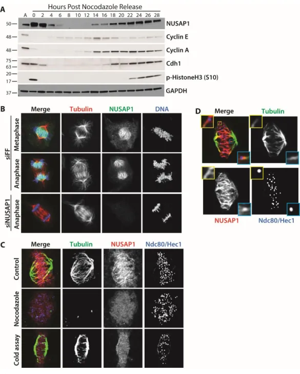
relatively short time scales after synchronization and release, making it difficult to know if its dynamics were due to the effects of chemical synchronization. To analyze NUSAP1 protein dynamics throughout an entire cell cycle we performed immunoblots on U2OS cells synchronized using nocodazole, isolated by shake-off, and followed for 28 hours after re-plating (Figure 2.1A). NUSAP1 levels are elevated in mitotic cells compared to asynchronous populations, concomitant with an
expression of Cyclin A, which marks the beginning of S-phase. NUSAP1 is also targeted by another E3 ligase, the SCFCyclin F, during S/G2 (82). Interestingly, abundance of the APC/C co-activator Cdc20, Cyclin F, and NUSAP1 are all abruptly diminished at mitotic exit, consistent with their coordinated degradation by APC/C and it other co-activator, Cdh1 (Figure 2.1A and Figure 2.2) (79).
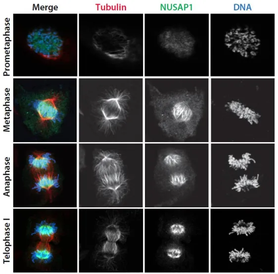
We used high-resolution immunofluorescent (IF) imaging to interrogate the localization of NUSAP1 during mitosis, when its protein levels are at their highest. The specificity of the NUSAP1 antibody was confirmed by comparing anti-NUSAP1 stained cells treated with either control siRNA targeting firefly luciferase (FF) or oligonucleotides targeting NUSAP1. RNAi depletion of NUSAP1 completely eliminated staining, confirming antibody specificity for IF. In prometaphase,
NUSAP1 staining was diffuse and localization to specific mitotic structures was not apparent (Figure 2.3). Later in mitosis NUSAP1 did not localize to the whole of the mitotic spindle, like the majority of known microtubule binding proteins in mitosis (Figure 2.1B). Instead, it localizes to the central spindle with the most concentrated area of NUSAP1 being near the chromatin (Figure 2.1B). Highly concentrated
This suggests that NUSAP1 represents a unique class of microtubule binding protein that localize in the vicinity of inter-digitated microtubules and that tracks chromatin localization in both early and late mitosis.
The localization of a pool of NUSAP1 on spindle microtubules near chromatin prompted us to determine if NUSAP1 localization is microtubule dependent. Prior to fixation, cells were treated with either DMSO (control) or the microtubule
depolymerizing drug nocodazole. NUSAP1 localization is lost when the spindle is depolymerized by nocodazole treatment, confirming that its localization it
microtubule dependent (Figure 2.1C). To determine which population of
microtubules NUSAP1 localizes to, we depolymerized dynamic spindle microtubules prior to fixation (Figure 2.1C). Cells were cold treated prior to fixation, which leads to the destabilization of microtubules that are not stably attached to kinetochores (k-fibers). NUSAP1 localization to the spindle was lost when non-kinetochore microtubules were depolymerized, suggesting that NUSAP1 localizes to dynamic microtubules during mitosis (Figure 2.1C). This observation, and the diffuse
NUSAP1 staining in prometaphase cells, is consistent with the notion that NUSAP1 binds to overlapping spindle microtubules. Finally, we analyzed single focal planes of NUSAP1 and tubulin staining by confocal microscopy. We observed NUSAP1 localization along microtubules, but not at the centromere, centrosome or
kinetochore (Figure 2.1D). Together, these data confirm that NUSAP1 is cell cycle regulated, and demonstrate its chromatin-centric localization to dynamic
Identification of NUSAP1 interacting proteins using mass spectrometry
NUSAP1 has a unique mitotic localization pattern compared to known microtubule binding proteins (Figure 2.1). Since NUSAP1 has been implicated in spindle stability and chromosome segregation we were interested in the mechanism by which NUSAP1 contributes to mitotic progression. To address this question, we analyzed protein interaction partners that bind NUSAP1 using endogenous NUSAP1 immunoprecipitation (IP) followed by protein identification using mass spectrometry (MS/MS). We performed IP experiments using control IgG and endogenous NUSAP1 antibodies in multiple cell lines (HeLa and HEK-293T). In addition, since NUSAP1 levels peak during mitosis (Figure 2.1A) we also performed IPs from both
asynchronous and mitotic HEK-293T cells arrested using nocodazole. By performing endogenous IPs in multiple cell lines and physiological conditions we sought to identify the strongest interactors that are most likely to be physiologically relevant in controlling mitotic progression.
We filtered out non-specific interactions identified in control IgG IPs, which were performed in parallel with each experiment, and removed known
contaminants based on the CRAPome dataset (148). We then overlapped the remaining interactions between the three IPs to identify the highest-confidence set of NUSAP1 interacting proteins (Figure 2.4A). This resulting list of 14 proteins included the known NUSAP1 interacting protein Importin-β (93).
IPs were saturating in that we detected a similar number of NUSAP1 TSCs between asynchronous and mitotic 293T samples. This allowed us to compare the relative number of RanBP2, RanGAP1 and Ubc9 TSCs between asynchronous and mitotic experiments. Our data show an enrichment of all three proteins in the mitotic sample relative to asynchronous cells, indicating that their interaction is cell cycle regulated (Figure 2.4B). Further supporting an interaction between NUSAP1 and RanBP2, their binding was detected in a recent, large scale interactome study using a tagged version of NUSAP1 (93).
To confirm our IP-MS/MS findings we tested whether RanBP2 co-IPed with endogenous NUSAP1 in multiple cell lines. Importantly, isolated endogenous
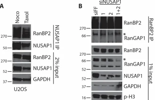
NUSAP1 precipitated from nocodazole arrested U2OS, HeLa, HEK-293T and HCT116 cell lines co-precipitated endogenous RanBP2 (Figure 2.4C). Similarly, when we precipitated endogenous RanBP2 from nocodazole arrested HEK-293T cells we co-precipitated endogenous NUSAP1, as well as its known interactor RanGAP1 (Figure 2.4D). This interaction was also detected in Taxol arrested cells, which prevents microtubule depolymerization, indicating that their interaction is not due to gross changes in microtubule dynamics (Figure 2.5).
RanBP2 and RanGAP1 (Figure 2.4F). Together this data strongly supports an interaction between a pool of available NUSAP1 and the RanBP2 SUMO E3 ligase complex.
Interestingly, only a subset of SUMOylated RanGAP1 co-migrated with RanBP2 based on the size exclusion chromatographic analysis. The majority of SUMOylated and unSUMOylated RanGAP1 eluted in fraction of ~500 kDa (Figure 2.4E). This demonstrates that there are RanBP2 bound and unbound pools of RanGAP1 in mitotic 293T cells and contrasts with a recent study suggesting that all of RanBP2 and RanGAP1 are complexed together in HeLa cells (136). The reason for this discrepancy is unknown, but could be cell line dependent. The peak elution of NUSAP1 partially overlapped with the peak elution of RanGAP1 that lacked RanBP2 and IPs from these fractions demonstrate that RanGAP1 and NUSAP1 interact in those fractions (lanes 7-10; Figure 2.4E and F). The full composition of these different NUSAP1 complexes remains unknown.
NUSAP1 does not control RanBP2 localization during mitosis
The RanBP2 complex regulates the SUMOylation of TOP2A and Borealin, both of which have distinct mitotic localization patterns (132, 136–138). In addition, RanBP2 localizes at the kinetochore and on the spindle (149). We hypothesized that NUSAP1 could recruit the RanBP2 SUMO E3 ligase to the spindle. We performed IF; probing for RanBP2 and RanGAP1 localization in control (FF) and NUSAP1 depleted cells. We observed the previously reported RanBP2 and RanGAP1 localization
SUMOylation of TOP2 regulates its centromeric localization, we also analyzed the localization of TOP2A and TOP2B on chromatin in control and NUSAP1 depleted cells using biochemical fractionation. Similarly, we observed no change in the localization of TOP2 on chromatin in control and NUSAP1 depleted cells (Figure 2.6D). We
conclude that NUSAP1 is not involved in the localization of RanBP2 and RanGAP1, nor that of the RanBP2-RanGAP1-UBC9 SUMO substrate TOP2.
Since our IF staining was unable to distinguish clear co-localization of
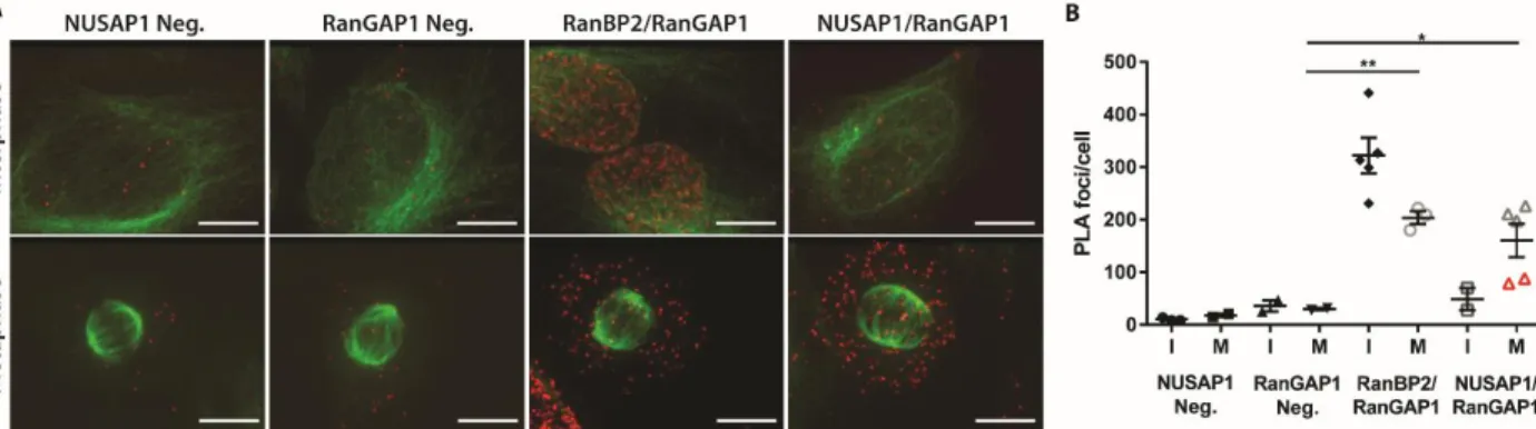
NUSAP1 with RanBP2 or RanGAP1 and there are soluble pools of NUSAP1, RanBP2 and RanGAP1 during mitosis, we determined where these proteins interact using a proximity ligation assay (PLA; Figure 2.9). PLA relies on the proximity of
co-localizing antibodies during immune staining of fixed cells, which allows for the rolling circle amplification of a DNA probe that is detected using fluorescence
hybridization. The result is a fluorescent foci at each site of interaction between the target proteins (151). Performing PLA in asynchronous cells with either NUSAP1 or RanGAP1 antibody alone produced a low background (Figure 2.9A), quantified in Figure 4B. Co-staining RanBP2 and RanGAP1 served as a positive control since they interact in both interphase and mitotic cells. Co-staining with NUSAP1 and RanGAP1 antibodies showed a strong increase in the number of foci in the cytosol of mitotic cells (Figure 2.9A and B). Intriguingly, the mitotic cells with the lowest number of foci in the NUSAP1 and RanGAP1 stained samples were in the late stages of mitosis (telophase and after; identified by red triangles). This suggests that the interaction between NUSAP1 and the RanBP2 E3 ligase complex decreases in late mitosis as the cells begin to rebuild their nuclear membranes/pores. Consistent with
between NUSAP1 and RanBP2-RanGAP1, the PLA signal was unchanged between single-antibody stained controls (NUSAP1 and RanGAP1 only) and dual-antibody (combined NUSAP1/RanGAP1) stained interphase cells. This supports the
observation that NUSAP1 interacts with RanBP2-RanGAP1 in a cell cycle dependent manner, and suggest that NUSAP1 binds RanBP2-RanGAP1 independent of the mitotic spindle, consistent with the binding observed in nocodazole treated cells.
RanBP2 depletion impairs the response to taxol
Previous reports have shown that NUSAP1 depletion sensitizes cells to
spindle poisons, such as taxol or nocodazole (82). To determine if RanBP2 depletion would show a consistent phenotype, we depleted cells of RanBP2 using siRNA and treated them with increasing doses of taxol overnight. RanBP2 was effectively depleted by siRNA based on immunoblot analysis (Figure 2.10B). Propidium iodide staining for DNA content in control depleted cells shows a progressive increase in G2/M phase cells in response to taxol, indicating an increased number of cells arresting in response to spindle checkpoint activation (Figure 2.10A). RanBP2
depleted cells had substantially reduced numbers of cells in G2/M phase at all doses of taxol tested, consistent with a defect in maintaining their mitotic arrest in
response to checkpoint activation. Consistent with a slippage through mitosis, there was also a reduction in Cyclin B levels in RanBP2 depleted cells compared to
2.3 Discussion
NUSAP1 is an important regulator of mitotic progression and chromosome segregation. NUSAP1 is essential for mouse development, and its inactivation by RNAi leads to defects in chromosome segregation (92). The NUSAP1 protein is tightly controlled post-translationally during the cell cycle. Its stability is controlled by at least two E3 ubiquitin ligases: SCFCyclin F during S/G2 phase and by APC/C in G1 (82, 127). Furthermore, NUSAP1 phosphorylation is upregulated during cell cycle progression on upwards of 20 different residues (128, 129). Nevertheless, little is known about where NUSAP1 fits mechanistically in the mitotic spindle apparatus.
We used confocal imaging to determine the precise localization of NUSAP1 on the mitotic spindle, providing a high-resolution snap-shot of NUSAP1 localization at each stage of mitosis. Interestingly, NUSAP1 exhibits a prominent, chromatin-centric localization pattern during metaphase and anaphase that is unique among microtubule binding proteins. We demonstrate here that NUSAP1 is localized on microtubules, and that its localization is dependent on dynamic spindle
PRC1 and KIF4 all showed increased microtubule binding after anaphase, suggesting a potential relationship between these factors in controlling spindle integrity (126).
To further define the role of NUSAP1 we examined endogenous binding partners using mass spectrometry. Through this analysis we identified and validated a cell cycle regulated interaction between NUSAP1 and the RanBP2-RanGAP1-UBC9 SUMO E3 ligase. Their interaction was identified first using
endogenous NUSAP1 pulldown followed by mass spectrometry and was validated by co-IP of both endogenous proteins in multiple cell lines. We were surprised not to identify a larger set of overlapping conditions between datasets, and predict that inter-cell lines differences could be explained by variances in the oncogenic
repertoire of the different cell types. An interaction between NUSAP1 and RanBP2 was also detected in a large scale study that globally mapped protein-protein interaction networks, providing further validation for their interaction (152).
RanBP2-RanGAP1-UBC9 is a critical SUMO ligase involved in cell division. However, little is known about which substrates it targets, how those substrates are
recognized, how its activity is regulated, and how its localization is controlled.
contain an identifiable SAP domain. NUSAP1, however, has a well conserved SAP domain at its N-terminus, with nearly all of the key, conserved residues found in the PIAS protein SAP domains (Figure 2.11). Like the PIAS proteins and other SAP domain containing proteins, the NUSAP1 SAP domain has been shown to be
important for its interactions with DNA, however, this may not be its only function (153). It is unknown how RanBP2 SUMO ligase is activated and how it specifies substrates for SUMOylation. We speculate that NUSAP1 could be a regulatory subunit for the complex, mediating substrate interactions and/or complex
activation, similar to the role of substrate adapters in cullin E3 ligases. Importantly, depletion of NUSAP1 using multiple siRNA reagents does not interfere with RanBP2-RanGAP1 complex assembly (Figure 2.5B). It is noteworthy that despite being the first discovered SUMO E3, little is known about the enzymology of the intact complex, due in large part to the size of RanBP2 (136, 154).
Despite the prominent localization of NUSAP1 during metaphase and anaphase to microtubules in the vicinity of chromatin, its binding to RanBP2-RanGAP1 is cytoplasmic. Thus, NUSAP1 could contribute to mitotic progression through multiple mechanisms: at the site overlapping microtubules on the mitotic spindle and through interactions with RanBP2 in the cytoplasm.
Recent large-scale studies have sought to identify targets of SUMOylation and have even examined cell cycle dependent changes in SUMOylation. However, NUSAP1 has not been identified in any of these large-scale SUMO substrate
as a SUMO substrate, we currently lack evidence supporting it as target of SUMOylation and were unable to detect NUSAP1 in SUMO pulldowns.
Little is known about how SUMO E3 ligases interact with, and subsequently SUMOylate their targets and how these interactions are regulated. If NUSAP1 did mediate enzymatic activity of the RanBP2 SUMO E3 ligase, this would provide important insight into the functions of not only the RanBP2 complex, but possibly how other SUMO E3 ligases are regulated as well. Further study of the interaction between NUSAP1 and the RanBP2 SUMO E3 ligase, and possibly the SUMO
pathway, could elucidate the mechanisms involved in the regulation of other SUMO E3 ligases.
2.4 Materials and Methods
Mammalian cell culture
HEK-293T, U2OS, HCT116 and HeLa cells were grown in Dulbecco's Modified Eagle's Medium (DMEM; Gibco) supplemented with 10% FBS (Atlanta Biologicals) and Pen/Strep (Gibco). For live cell imaging, cells were imaged in Fluorobrite DMEM (Gibco) + 10% FBS. Nocodazole (Sigma 487928) was used at 150 ng/mL for U2OS and 200 ng/mL for 293T. All siRNA transfections were performed using
Immunoblotting and immunoprecipitations
Samples analyzed by immunoblot were lysed in NETN (20mM Tris-Cl, pH 8.0, 100mM NaCl, 0.5mM EDTA, 0.5% Nonidet P-40 (NP-40)) supplemented with
1ug/mL apoprotinin, 1ug/mL pepstatin, 10ug/mL leupeptin, 1mM Na3VO4, 1mM NaF and 1mM AEBSF (4-[2Aminoethyl] benzenesulfonyl fluoride). Protein
concentration was estimated using the Bradford assay (Bio-Rad). Laemmli buffer was added to samples, which were then separated by SDS-PAGE gel electrophoresis using home-made or commercially available gels (Bio-Rad). Gels were transferred to nitrocellulose membranes and blotted using standard immunoblotting
procedures.
NUSAP1 interacting proteins were identified using endogenous immunoprecipitation followed by tandem mass spectrometry. The mass spectrometry analysis was carried out by the UNC Hooker Proteomics Facility (described below). As a source of starting material, we used asynchronous HEK-293T and HeLa cells, or HEK-HEK-293T cells that were arrested in mitosis by overnight incubation in nocodazole. Whole cell extracts (WCE) were prepared on ice in the aforementioned NETN lysis buffer. Protein A/G agarose beads were covalently
coupled to control IgG or anti-NUSAP1 antibodies using dimethyl pimelimidate (74). WCE was clarified by centrifugation at 14,000 rpm for 10 minutes at 4˚C in a
benchtop centrifuge. Clarified lysates were mixed with antibody coated beads on a rotary mixer for 4 hours at 4˚C. Samples were quickly washed three times with lysis buffer, eluted using 100mM Glycine, pH 2.5 and neutralized with Tris buffer (pH7.5). Elutions were then digested with trypsin and analyzed by mass
For the co-IP experiments in Figure 2, cells were lysed in hypotonic lysis buffer (10mM HEPES, pH 7.9, 10mM KCl, 1.5mM MgCl2, 0.5mM DTT),
supplemented with 1ug/mL apoprotinin, 1ug/mL pepstatin, 10ug/mL leupeptin, 1mM Na3VO4, 1mM NaF and 1mM AEBSF (4-[2Aminoethyl] benzenesulfonyl fluoride). Protein A/G DynaBeads (Thermo) were bound to control rabbit IgG, NUSAP1 or RanBP2 antibodies overnight at 4°C. Samples were incubated with beads for 4 hours at 4°C, which were subsequently washed three times in lysis buffer and eluted with 2X Laemmli sample buffer at 95°C for 10 minutes.
Immunological reagents
Commercially available antibodies used in this study, including their use (immunoblotting, immunofluorescence, etc.), catalog numbers and specific dilutions are included in Table 2.2.
An antibody against RanBP2 was generated in-house for these studies. The DNA sequence encoding amino acids 1000-1200 was cloned into the pET28A using traditional PCR amplification to generate an amino-terminally tagged hexahistidine tagged version of the fragment. The cloning was verified by Sanger sequencing and resulting plasmid DNA was introduced into BL21 (DE3) E.coli for recombinant
incubated in batch with Ni-NTA agarose (Thermo) on a rotary mixer for 90 minutes at 4ºC. Beads washed extensively with 20mM Tris pH7.5, 500mM NaCl, 0.5% NP-40, 30mM Imidizole and then eluted in 20mM Tris pH7.5, 200mM NaCl, 300mM Imidizole. Eluted samples were analyzed by Coomassie blue staining, combined, and tested by Bradford. 6HIS-RanBP21000-1200 was conjugated to KLH and injected into rabbits for antiserum production by Pocono Rabbit Farm & Laboratory (PRF&L, Canadensis, PA). The serum was affinity purified over a column of
recombinant protein using described protocols and dialyzed into PBS (159).
For immunoblotting, antibodies were diluted in a solution of 5% nonfat dry milk in phosphate buffered saline, 0.05% tween 20 (PBST). Antibodies were either incubated at room temperature for 2 hours or overnight at 4°C. Detection was performed using HRP conjugated secondary antibodies (Jackson ImmunoResearch Laboratories, Inc; 1:10000), ECL reagent (Pierce), and exposure to film.
Mass Spectrometry analysis
Samples provided in solution were digested using the FASP (Filter assisted sample preparation) protocol. This includes reduction, alkylation, and digested with trypsin. The peptides were extracted, lyophilized, and resuspended in 2%
acetonitrile/98% (0.1% formic acid). The peptides were loaded onto a 2 cm long X 360 µm o.d. × 100 µm i.d. microcapillary fused silica precolumn packed with Magic 5 µm C18AQ resin (Michrom Biosciences, Inc.). After sample loading, the
rate of 250 nL/min by increasing the percentage of solvent B to 40% with a Nano-Acquity HPLC solvent delivery system (Waters Corp.). The LC system was directly connected through an electrospray ionization source interfaced to an LTQ Orbitrap Velos ion trap mass spectrometer (Thermo Fisher Scientific). The mass
spectrometer was controlled by Xcalibur software and operated in the data-dependent mode in which the initial MS scan recorded the mass to charge (m/z) ratios of ions over the range 400–2000. The 10 most abundant ions were
automatically selected for subsequent collision-activated dissociation. All files were searched using MASCOT (Matrix Science, Ver. 2.3.02) via Proteome Discoverer (Thermo., Ver. 1.3.0.339) against a recently downloaded human FASTA database. The search parameters included peptide mass tolerance of 10 ppm, fragment ion tolerance of 0.6 mass unit. The search allowed variable modifications for
methionine oxidation and carbamidomethylation of Cys.
Gel filtration chromatography
Mitotically arrested 293T cells were analyzed by gel filtration
chromatography. Cells were arrested overnight in nocodazole and lysed in
hypotonic buffer as described above. The cell extract was clarified via centrifugation followed by filtration through a 0.22 µm syringe filter. Protein complexes in the clarified lysate were then separated using a size exclusion column (Superose 6 10/30, G.E. Healthcare) that had been pre-quilibrated in hypotonic lysis buffer. During separation, 0.4 mL fractions were collected and later analyzed by
Chromatin Fractionation
Cells were lysed in CSK buffer (10mM PIPES, pH 7.0, 300mM sucrose, 100mM NaCl, 3mM MgCl2, 0.1% triton X-100) supplemented with 1ug/mL
apoprotinin, 1ug/mL pepstatin, 10ug/mL leupeptin, 1mM Na3VO4, 1mM NaF and 1mM AEBSF (4-[2Aminoethyl] benzenesulfonyl fluoride). Protein concentration was determined using Bradford and a portion of the lysate was taken for WCE samples. Samples were then pelleted at 3,000rpm for 5 minutes at 4°C. Supernatant was saved as the soluble fraction (S). Each pellet was washed with CSK buffer on ice and pelleted. The supernatant was removed and the pellet was resuspended in Laemmli buffer diluted in CSK and boiled for 5 minutes before the DNA was sheared using a needle to produce the insoluble fraction (I).
Immunofluorescence Imaging
Cells were plated on poly-L-Lysine coated coverslips approximately one day before fixation. Cells were fixed in PHEM buffer (60mM PIPES, 25mM HEPES, 10mM EGTA, 2mM MgCl2, adjusted to pH 7.0 using KOH) + 3% PFA for 13 minutes at 37°C. Cells were washed with PHEM buffer and permeabilized using PHEM + 0.5% NP-40 for 15 minutes at room temperature. Cells were washed in PBS before blocking in PBS + 5% BSA. All antibodies were subsequently diluted in PBST + 5% BSA. Primary antibodies and their dilutions used: α-NUSAP1 (1:500), α- RanGAP1
(1:100), α-RanBP2 (1:100), α-tubulin (1:200), mouse anti-HEC1 (abcam ab3613;
1ug/mL Hoechst 33342 for 5 minutes at room temperature. All samples were mounted onto glass slides in ProlongGold media.
The cold stability assay was conducted as detailed in Suzuki et al. Nat Comm 2015 (160). Briefly, cells were treated with ice cold media for 10 minutes before fixation and staining. Proximity Ligation Assay (PLA) was performed using the Sigma Duolink In Situ Red Starter Kit Mouse/Rabbit (DUO92101 Sigma). Cells were plated and fixed as described above. Staining was performed following the DuoLink kit protocol, with primary antibodies against NUSAP1, RanBP2 and RanGAP1 being used at the concentrations described above. Tubulin counterstaining was performed using AlexaFluor488 conjugated α-tubulin at a dilution of 1:100 for 40min at 37°C.
For image acquisition, 3D stacked images were obtained sequentially at 200 nm steps along the z -axis through the cell using MetaMorph 7.8 software
Flow Cytometry
Figure 2.1. NUSAP1 is a cell cycle regulated microtubule binding protein. A) U2OS clls were synchronized by overnight treatment with nocodazole and released by mitotic shake-off. Samples were analyzed by immunoblot as cells progress through the cell cycle. B) NUSAP1 localization to the mitotic spindle analyzing by immunofluorescent imaging of mitosis in U2OS cells. (Scale bars = 10μM.) C) NUSAP1 localization was analyzed in in nocodazole treated cells and following incubation with ice-cold buffer to destabilize non-kinetochore microtubules. (Scale bars indicate 5μM.) D) Single plane confocal imaging of NUSAP1 localization on the spindle during metaphase. Insets highlight two kinetochore-microtubule
Figure 2.4 NUSAP1 interacts with the RRU in a cell cycle dependent manner. A) Venn diagram showing overlap of IP-MS/MS experiment results. B) Total Spectral Counts (TSC) for each of the RRU complex members determined by mass spectrometry. C) Endogenous NUSAP1 IPs were performed in four different nocodazole arrested cells and analyzed for RanBP2. D) Endogenous RanBP2 IP performed in nocodazole arrested 293T cells. E) Size exclusion chromatography was performed on extracts from nocodazole arrested 293T cells. Extracts were analyzed on a Superose 6 column. Previously tested size markers migrated in the indicated fractions. F) Endogenous NUSAP1 IPs were performed using each of the gel
Figure 2.9 NUSAP1 and RanBP2 interact in the cytosol of mitotic cells. A) PLA in U2OS cells using endogenous against NUSAP1, RanGAP1, RanBP2, or control IgG. Tubulin is shown in green with PLA signal in red. (Scale bars indicate 10μM) B) Average number of foci/cell for each PLA condition shown in A. Foci were counted using ImageJ.
Figure 2.11. NUSAP1 contains a SAP domain in its N-terminus. A)
Target siRNA sequence, 5'-3' Firefly luciferase CGUACGCGGAAUACUUCGA NUSAP1 #1 GAUAAUGAGCAUAAGCGUU NUSAP1 #2 CCACUUUAGUCACGAGAUC NUSAP1 #3 CAGCCAACGACGCUCGCAA RanBP2 #1 CGAAACAGCUGUCAAGAAA RanBP2 #2 GAAAGAAGGUCACUGGGAU RanBP2 #3 GAAAGGACAUGUAUCACUG RanBP2 #4 GAAUAACUAUCACAGAAUG
Table 2.1. siRNA oligonucleotides used in Chapter 2.
Target Company
Catalog
number Dilution
NUSAP1 Proteintech 12024 IB 1:10000; IF 1:500 RanGAP1 abcam ab119092 IB 1:500; IF 1:100
Ubc9 Santa Cruz 10759 1:1000
Tubulin Santa Cruz sc32293 IB 1:1000; IF 1:200
CENP-C MBL IF 1:1000
Ndc80/Hec1 abcam ab3613 IF 1:500 GAPDH Santa Cruz sc25778 1:10000
Cyclin E CST 4129 1:1000
Cyclin A Santa Cruz sc751 1:5000 Cdh1 (Fzr1) abcam ab3242 1:1000
TopoisomeraseIIα BDBiosciences 611327 1:2000
TopoisomeraseIIβ BDBiosciences 611492 1:2000
Cyclin B1 abcam ab32053 1:10000
Cdc20 Bethyl A301-107A 1:1000
Cyclin F Santa Cruz sc-952 1:2000
CHAPTER 3: IDENTIFICATION OF A NOVEL SCFCYCLIN F TARGET, SIRTUIN 5, LINKING PROLIFERATION AND METABOLIC REGULATION
3.1 Introduction
Precise cell cycle progression is important for maintaining genomic integrity in dividing cells. Cell cycle is highly regulated, in part, by the ubiquitin-proteasome system, which targets proteins for degradation. One Cullin E3 ubiquitin ligase, a modular complex composed of Skp1, CUL1 and F-box protein (SCF) with the
substrate adapter Cyclin F, has been identified as an important cell cycle regulator. Most recently, it has been recognized as having an important role in G1-S transition via feedback with the Anaphase Promoting Complex/Cyclosome with its substrate adapter Cdh1 (APC/CCdh1) during late G1 (79, 80).
While a number of Cyclin F substrates have been characterized, using traditional protein-protein interaction, mass spectrometry (MS) based approaches to identify novel Cyclin F substrates has proven challenging. This is, in part, due to the transient nature of F-box-substrate interactions, a feature common to E3 ubiquitin ligase and substrate interactions. Furthermore, the substrate is typically being targeted for degradation, making it difficult to capture. Finally, these
experiments are often performed in asynchronous cells, which is problematic because Cyclin F, and many of its substrates, are cell cycle regulated, making it difficult to detect in asynchronous cell extracts. In this report, based on genetic yeast interactions and conservation in the ubiquitin system, we examined whether a human sirtuin could be regulated by Cyclin F.
Here we identify Sirtuin 5 (Sirt5) as a novel SCFCyclin F target important for the G1-S transition. Sirtuins are a class of deacylating enzymes involved in regulating a variety of processes including epigenetic regulation, DNA damage response and metabolism (162, 163). This is the first described role for Sirt5, a mitochondrial sirtuin, in cell cycle control. Sirt5 has specific deacylating activity towards succinyl, malonyl, glutaryl and acetyl post-translational modificiations (PTMs) (164–167). Sirt5 is one of three mitochondrial sirtuins, (Sirtuins 3-5), which are classified by their ability to be imported into the mitochondria, but are not restricted to the mitochondria. Sirt5 also localizes to the cytoplasm and nucleus, however its roles outside of the mitochondria are less clear. Sirt5 is best known for its role in regulating metabolic enzymes, such as carbamoyl phosphate synthase 1 (CPS1), which catalyzes the production of carbamoyl phosphate from ammonia and
to desuccinylate and activate Cu/Zn superoxide disumutase (SOD1), which
functions in reactive oxygen species response (171, 172). Sirt5 has been implicated in a number of other metabolic processes as well, including glycolysis, purine
metabolism, fatty acid oxidation, as well as the citrate cycle (173). While it is known that Sirt5 is localized to the mitochondria as well as the nucleus and
cytoplasm, it has yet to be determined whether it has different activities based on localization.
Here we describe the interaction between Cyclin F and Sirt5, and its impact on the G1-S transition. These data provide a link between cell cycle ubiquitin machinery, and metabolic regulation, a connection of which little has been described.
3.2 Results
Cyclin F was originally identified in a gain-of-function cDNA screen searching for human genes that could rescue the yeast Cdc4 temperature sensitive mutant, which caused G1 arrest and subsequent death (75). Cyclin F was identified as a gene that rescued the G1 arrest phenotype, and based on the fact that it cycled and contained a cyclin homology domain, Cyclin F was classified as a traditional Cyclin (75). However, subsequent studies identified Cyclin F as a substrate adapter for the SCF E3 ubiquitin ligase, and Cdc4 is a yeast F-box substrate adapter (174).
(DRYGIN), a database containing data from global synthetic genetic interactions in yeast, for mutants or deletions that also rescue the Cdc4 mutant phenotype as a tool to identify new potential Cyclin F targets (175). To narrow our list down we also compared this to a list of deletions that rescue the Cdc53 mutant G1 arrest phenotype as well (Figure 3.1). Cdc53 is a yeast cullin, which complexes with the F-box protein, Cdc4, to form an E3 ubiquitin ligase. A list of ~40 genes that rescued the G1 arrest and lethality of both the Cdc4 and Cdc53 mutations was identified, which we narrowed down by first eliminating all the genes that had no connection to cell cycle, either directly or through genetic interactions. We then eliminated apc5, orc3 and mcm3 due to their incorporation into large protein complexes with low turnover rates. The yeast specific transcription factor, swi5, was eliminated because no human homologue has been identified. Finally, we chose not to
interrogate sli15 because it is a component of the Aurora B complex, which is only active in mitosis. This eliminated all but one gene; hst3, a yeast sirtuin family deacetylase. When we looked at genes with similar genetic interactions to hst3, what we identified was a list of genes important for DNA replication and DNA damage response, indicating a potential role in S-phase entry and progression. Interestingly, hst3 is ubiquitinated by the yeast SCFCdc4 ligase.
Sirtuin 5 stability is increased in the absence of Cyclin F
Since our screen identified a yeast sirtuin as a potential Cyclin F target, we analyzed human sirtuin levels in Cyclin F CRISPR KO HeLa cells, with the