Efficacy of Gene Therapy Is Dependent
on Disease Progression in Dystrophic Mice
with Mutations in the FKRP Gene
Charles Harvey Vannoy,
1Will Xiao,
1Peijuan Lu,
1Xiao Xiao,
2and Qi Long Lu
11McColl-Lockwood Laboratory for Muscular Dystrophy Research, Cannon Research Center, Carolinas Medical Center, Carolinas Healthcare System, Charlotte, NC 28203,
USA;2Division of Molecular Pharmaceutics, Eshelman School of Pharmacy, University of North Carolina at Chapel Hill, Chapel Hill, NC 27599, USA
Loss-of-function mutations in the Fukutin-related protein (FKRP) gene cause limb-girdle muscular dystrophy type 2I (LGMD2I) and other forms of congenital muscular dystro-phy-dystroglycanopathy that are associated with glycosylation defects in thea-dystroglycan (a-DG) protein. Systemic admin-istration of a single dose of recombinant adeno-associated virus serotype 9 (AAV9) vector expressing humanFKRPto a mouse model of LGMD2I at various stages of disease progression was evaluated. The results demonstrate rescue of functional glyco-sylation of a-DG and muscle function, along with improve-ments in muscle structure at all disease stages versus age-matched untreated cohorts. Nevertheless, mice treated in the latter stages of disease progression revealed a decrease in bene-ficial effects of the treatment. The results provide a proof of concept for future clinical trials in patients withFKRP-related muscular dystrophy and demonstrate that AAV-mediated gene therapy can potentially benefit patients at all stages of disease progression, but earlier intervention would be highly preferred.
INTRODUCTION
Dystroglycanopathies are a subgroup of muscular dystrophy that result from aberrant glycosylation of a-dystroglycan (a-DG) and are characterized pathologically by muscle fiber degeneration and clinically by progressive muscle weakness.1 These disorders are caused by mutations in a multitude of genes, including the gene en-coding Fukutin-related protein (FKRP; OMIM 606596). TheFKRP gene, along with fukutin, is part of a family of genes that possess the putative catalytic DXD motif—a conserved motif found in many families of glycosyltransferases—and is predicted to function as a ribitol 5-phosphate transferase, which is fundamentally required for the post-translational modification ofa-DG.2–5In normal tissues, the mucin-like domain ofa-DG is modified with numerous oligosac-charides that are essential for function—anchor the structural frame-work inside each cell (cytoskeleton) to the lattice of extracellular matrix proteins.6–8Missense mutations in theFKRP gene lead to FKRP protein deficiencies that impair the glycosylation pathway of a-DG.9,10 As a consequence of reduced FKRP protein activity,
a-DG is hypoglycosylated and has a reduced binding capacity to the extracellular matrix proteins, effectively weakening the bridge
between the dystrophin-glycoprotein complex and the extracellular matrix and disrupting the skeletal muscle basal lamina.11
Mutations in the FKRP gene exhibit a wide spectrum of clinical severity, ranging from severe congenital muscular dystrophies to limb-girdle muscular dystrophy type 2I (LGMD2I).9,10,12LGMD2I can present as mild or severe depending on the age of onset. Early childhood onset of LGMD2I usually indicates a severe clinical course with affected individuals becoming non-ambulatory as early as their teens. The late- or adult-onset form of LGMD2I is a slowly progres-sive, milder form of the disorder. Respiratory and cardiac involve-ment is prominent in all disease severities.13The variable phenotypic severity has been partly attributed to the differences in location of point mutations within the coding sequence, affecting protein trans-portation and its glycosyltransferase activity differentially.14–16 Therefore, it is likely that any type of therapeutic intervention could potentially have different efficacies for individuals at different stages of disease progression.
Currently, recombinant adeno-associated virus (AAV) gene therapy is one of the most promising strategies for replacing a gene with loss-of-function mutations. AAV is a small (25 nm in diameter) hu-man parvovirus that packages a linear single-stranded DNA genome and is replication defective. The lack of pathogenicity of the virus and its ability to persist stably in transduced cells, especially in post-pro-liferative muscle tissues, make it a desirable and effective delivery vehicle for gene therapy applications to muscular dystrophies.17,18 As a result, AAV gene therapy has been tested in various animal models of many limb-girdle muscular dystrophies,19–24along with ongoing/completed clinical trials for dysferlinopathy (ClinicalTrials.
Received 30 December 2016; accepted 20 February 2017; http://dx.doi.org/10.1016/j.omtm.2017.02.002.
Correspondence: Charles Harvey Vannoy, McColl-Lockwood Laboratory for Muscular Dystrophy Research, Cannon Research Center, Carolinas Medical Cen-ter, Carolinas Healthcare System, Charlotte, NC 28203, USA.
E-mail:[email protected]
Correspondence:Qi Long Lu, McColl-Lockwood Laboratory for Muscular Dys-trophy Research, Cannon Research Center, Carolinas Medical Center, Carolinas Healthcare System, Charlotte, NC 28203, USA.
gov identifier NCT02710500), LGMD2C (g-sarcoglycan) (ClinicalTrials.govidentifier NCT01344798), and LGMD2D (a -sar-coglycan) (ClinicalTrials.govidentifier NCT00494195).
In this study, an AAV serotype 9 vector containing a full-length hu-manFKRPgene (AAV9-FKRP) under control of a muscle creatine kinase-based promoter was administered by a single intravenous tail-vein injection at a dose of 2.51013vector genomes per kilogram
(vg/kg) at onset or later stages of disease progression in our dystro-phic mouse model containing a missense mutation in the FKRP gene. Our objective is to systematically evaluate the therapeutic po-tential of AAV-mediatedFKRPgene delivery and assess the efficacy of the gene replacement therapy for individuals with LGMD2I exhib-iting various degrees of disease pathology before a viable treatment option is available.
RESULTS
SystemicFKRPDelivery Restores Functionala-DG in Dystrophic Mice
For these experiments, we used a mouse model containing a homozy-gous missense mutation (c.1343C > T, p.Pro448Leu) in the FKRP gene (FKRPP448Lmutant), as previously described.25,26Onset of the dystrophic attributes can be observed as early as 3 weeks, with pro-gressive pathological changes as the mouse ages. Accordingly, we formulated four age groups of FKRPP448Lmutant mice (5, 13, 26,
and 39 weeks of age) representing disease progression from an early stage, when muscle degeneration has just become clearly identifiable, to a late stage characterized by severe dystrophy andfibrosis in skel-etal muscles and noticeable defects in cardiac muscle function. To correct for the FKRP deficiency, we utilized a skeletal/cardiac muscle-tropic AAV serotype 9 vector expressing a full-length human
FKRP coding sequence under control of a muscle-specific creatine kinase-based (CK7) promoter (AAV9-CK7-HuFKRP, abbreviated as AAV9-FKRP) (Figure S1). A single tail-vein injection of AAV9-FKRPat a dose of 2.51013vg/kg was administered to all age groups. Mice were monitored and functionally assessed for a 13-week period and then were subsequently euthanized for analysis, giving rise to cohorts T18, T26, T39, and T52, respectively ( Fig-ure 1A). Age-matched untreated FKRPP448Lmutant mice were used as negative controls. All mice remained healthy in appearance, activ-ity, and body weight over the 13-week observation period.
Analysis of glycosylation on a-DG by immunohistochemistry at euthanasia demonstrates that intravenous delivery of AAV9-FKRP reconstituted functional glycosylation ofa-DG in skeletal muscles
throughout the body, including the heart (Figure 1B). Age-dependent variation in levels of functional glycosylation ofa-DG was visualized by immunohistochemistry with a monoclonal anti-a-DG antibody (IIH6C4) specific to glycosylated epitopes ona-DG,27also known as functionala-DG. The IIH6C4 signals were localized to the sarco-lemma and relatively homogeneous in a large portion of musclefibers and were generally higher in the diaphragm and heart compared to the skeletal muscles, which is likely due to the higher tropism of AAV9 to the cardiac muscle. Positive signals were relatively stronger in the muscles of the early-stage treatment cohorts in comparison to the late-stage cohorts, especially in the skeletal muscles. In stark contrast, a positive immunofluorescence signal for functionally glyco-sylateda-DG was undetectable in all muscles of the age-matched un-treated cohorts, except for a few revertantfibers in the tibialis anterior muscles. Western blot analysis confirmed that the AAV-treated mus-cle produced normal glycosylated forms ofa-DG (150–250 kDa; car-bohydrate composition differs depending on tissue type) similar to that detected in the same muscle type of wild-type tissue (Figure 1C). Similar to the immunofluorescence staining, the levels of functional glycosylation were evidently different in all tissues between the early- and late-stage treatment cohorts, with the highest levels in the T18 cohort and lowest levels in the T52 cohort. Only 12%, 34%, and 53% of normal levels of functionally glycosylateda-DG were de-tected in the tibialis anterior, diaphragm, and heart muscles of the oldest T52 age cohort, respectively, whereas approximately 87%, 88%, and 70% of normal levels were detected in the corresponding three muscles from the youngest T18 cohort. Interestingly, an age-related decrease in levels of functionally glycosylateda-DG was also muscle-type dependent, predominantly in the tibialis anterior muscle. As expected, there was little or no immunoreactivity detectable in the age-matched untreated FKRPP448Lmutant samples. IIH6C4 expres-sion in the tibialis anterior was representative of all skeletal muscles tested (quadriceps, gastrocnemius, bicep, and triceps; data not shown).
Western blot (Figure 1D) and quantitative real-time PCR analysis (Figure 1E) demonstrated similar levels of FKRP transgene expression in the AAV-treated cohorts. Expression of vector-driven FKRP pro-tein was analyzed on whole-muscle lysates by western blotting with an antibody raised against a peptide mapping near the C terminus of FKRP (human origin). FKRP protein was clearly detected in the ti-bialis anterior muscle of all AAV-treated cohorts, with a distinct signal band at approximately 110 kDa that corresponds to a dimeric FKRP. The intensity of the FKRP signal was similar for all AAV-treated cohorts, whereas it was undetectable in all age-matched
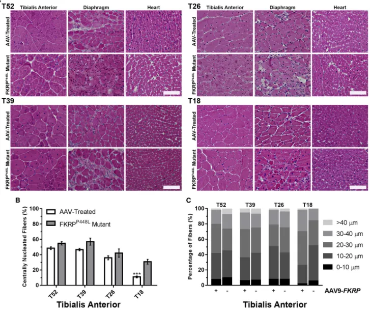
Figure 1. Study Design and Rescue ofa-DG Glycosylation in Dystrophic Mice
(A) Experimental design of FKRPP448L
mutant mice injected with AAV9-FKRPat 5 (T18, n = 4), 13 (T26, n = 4), 26 (T39, n = 4), and 39 weeks of age (T52, n = 4) in a 13-week treatment study. (B) Immunofluorescence staining of glycosylateda-DG in tibialis anterior, diaphragm, and heart tissue from indicated mice. Scale bars, 200mm. (C) Western blot analysis of glycosylateda-DG expression in tibialis anterior, diaphragm, and heart tissue from FKRPP448L
mutant mice injected with AAV9-FKRP (+) or untreated (). Glycosylateda-DG was detected with the IIH6C4 antibody at a dilution of 1:200 and 1:1,000 for immunofluorescence staining and western blot, respectively. Anti-actin antibody was used as the protein loading control. (D) Western blot analysis of exogenous FKRP protein in tibialis anterior tissues from indicated mice. FKRP was detected with an FKRP (C-terminal) antibody at a dilution of 1:400. Anti-GAPDH antibody was used as the protein loading control. (E) FKRP transgene expression in tibialis anterior tissues was analyzed by quantitative real-time PCR methods. All levels are relative to those in untreated FKRPP448L
untreated FKRPP448L mutant samples. We also validated the expression level of the FKRP transgene by quantitative real-time PCR analysis in all AAV-treated and age-matched untreated FKRPP448Lmutant cohorts. These results show that a single injection of AAV9-FKRPis efficacious in rescuing functional glycosylation of a-DG in FKRPP448Lmutant mice at all stages of disease pathology.
AAV9-FKRPGene Therapy Improves Muscle Pathology
Similar to the clinical severity observed in patients with LGMD2I, FKRPP448Lmutant mice exhibit a mild-to-moderate phenotype of muscular dystrophy represented as myofiber
regeneration/degenera-tion, significantfiber size variability, mononuclear cell infiltration, and pronouncedfibrosis in the later stages of disease progression. Ex-amination of the muscle morphology by H&E staining indicates that degeneration/regeneration in the limb skeletal muscles of the un-treated FKRPP448Lmutant begins to plateau around 26 weeks of age, as demonstrated by the presence of large areas of necroticfibers and a considerable number of centrally nucleatedfibers (Figure 2A). An increase infibrosis becomes clearly recognizable in the diaphragm and functional defects in skeletal and cardiorespiratory systems become detectable from the age of 26 weeks onward. Conversely, sys-temicFKRPgene delivery ameliorated pathological phenotypes of the
Figure 2.FKRPGene Therapy Improves the Internal Structure and Hyper-/Hypotrophy of Skeletal and Cardiac Muscle Fibers
(A) Cross-sections from tibialis anterior, diaphragm, and heart tissue of AAV-treated and FKRPP448L
mutant mice stained with H&E. Scale bars, 100mm. (B) Quantification of centrally nucleated fibers in AAV-treated and untreated FKRPP448L
mutant tibialis anterior muscles (n = 4). ***p%0.001 (Student’s t test, each condition versus age-matched FKRPP448L
mutant mice). (C) The equivalent radius of individual myofibers in tibialis anterior muscles from FKRPP448Lmutant mice injected with AAV9-FKRP
dystrophic skeletal muscles of all mice from both early- and late-treated cohorts, which included a normalization offiber size distribu-tion, a reduction in centralized nuclei, and less pronouncedfibrosis.
Quantitative analysis of the tibialis anterior muscles from each cohort of untreated FKRPP448Lmutant mice showed that the number of cen-trally nucleatedfibers increased from younger to older cohorts, with 30.8%±3.1%, 42.2%±5.4%, 57.0%±4.6%, and 54.9%±2.1%, from T18 to T52, respectively, reaching a plateau around 39 weeks of age (Figure 2B). In contrast, the T18 cohort from the AAV-treated mice contained a significantly smaller percentage (11.0% ± 1.2%, p % 0.001) of centrally nucleated fibers. This percentage also decreased in the T26 (36.0% ± 2.6%, p = 0.3399), T39 (46.6% ± 1.5%, p = 0.0730), and T52 (48.5% ± 1.8%, p = 0.0633) cohorts from the AAV-treated mice, but it lacked significance compared to the age-matched untreated controls. The similar percentage of cen-trally nucleated fibers in the two older (T39 and T52) cohorts is consistent with the notion that muscle degeneration and regeneration reaches a plateau around 26 weeks of age in FKRPP448Lmutant mice and that the time required for peripheralization of nuclei could take several months or longer to complete. Quantitative analysis of the col-lective myofiber radius revealed a decreased proportion offibers with large and small diameters representing hypertrophy and regenerating fibers, respectively, in all cohorts of the AAV-treated muscles when compared to untreated FKRPP448Lmice (Figure 2C). The population of largefibers (>40mm) was very small in the T18 cohort, and the dif-ference between treated and untreated FKRPP448Lmutant mice was not clear. Together, these results suggest a normalization of fiber size distribution, representing a clear deceleration or halting of pro-longed cycles of regeneration/degeneration.
Another histological hallmark of the FKRPP448Lmutant mouse is pro-nounced and progressivefibrosis in the diaphragm as the mouse ages. To evaluate fibrotic changes after treatment, Masson’s trichrome staining was performed on tissue sections of the diaphragm from each cohort (Figure 3A). The results show a significant reduction in
the amount of collagen present within AAV-treated cohorts
compared with the untreated cohorts. Quantitative data confirmed that deposition of connective tissues follows a linear increase with age in both AAV-treated and untreated FKRPP448Lmutant mice ( Fig-ure 3B). More importantly, areas of connective tissue were
signifi-cantly more prominent in untreated FKRPP448L mutant mice compared to AAV-treated mice within each age cohort (T52: 56.2%±2.4% versus 45.7%±0.7%, p = 0.0060; T39: 44.2%±1.7% versus 35.5% ± 0.9%, p = 0.0051; T26: 32.0% ± 0.7% versus 20.5% ±1.3%, p = 0.0002; and T18: 22.8% ±1.3% versus 9.5%± 0.9%, p = 0.0002). Quantitative comparisons additionally reveal that AAV-mediated delivery ofFKRPto FKRPP448Lmutant mice re-duces the level offibrosis of each treatment cohort to a similar level of a younger untreated cohort corresponding to the treatment interven-tion time point (Table S1). Collectively, these results demonstrate that timely administration of an intervention that restores functionally glycosylateda-DG can slow or halt the development offibrosis in the diaphragm before irreversible damage can occur.
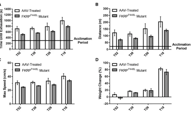
Gene Therapy with AAV9-FKRPImproves Muscle Function To assess the effect of rescuing functionally glycosylateda-DG on physical function, we conducted treadmill exhaustion tests 11 weeks post-injection. Initially, the mice were allowed to undergo an acclima-tion period, after which the test was started and mice were run to the point of exhaustion. The untreated FKRPP448Lmutant mice showed a
reduced tolerance to exercise compared with AAV-treated mice ( Fig-ures 4A–4C). On average, mice from each AAV-treated cohort ran approximately 20%–28% longer at higher speeds than age-matched untreated FKRPP448L mutant mice. Also, AAV-treated mice were able to run longer distances than age-matched untreated FKRPP448L
mutant mice, although a gradual decline was observed for both as age increased (untreated FKRPP448L mutant versus AAV-treated mice, respectively: T18: 140.3 ± 9.6 versus 203.3 ± 38.4 m, p = 0.1623; T26: 96.3 ± 9.5 versus 152.5 ± 37.5 m, p = 0.1962; T39: 81.8±5.5 versus 113.5±15.9 m, p = 0.1075; and T52: 71.0±7.2 versus 122.3± 22.0 m, p = 0.0690). Together, these data indicate that muscle impairment can be ameliorated at all stages of disease progression.
Figure 3. Gene Therapy with AAV9-FKRPSlows the Progression of Fibrosis in the Diaphragm
(A) Masson’s trichrome staining of diaphragm tissue of AAV-treated and FKRPP448L
mutant mice in each cohort. Scale bar, 200mm. (B) Quantification of the fibrotic area of the diaphragm in AAV-treated and FKRPP448L
mutant mice for each cohort (n = 4). **p%0.01; ***p%0.001 (Student’s t test, each condition versus age-matched FKRPP448L
The assessment of body weight (measured every 6–7 weeks) was re-ported as a percent change relative to baseline and revealed that all study groups gained body mass over the 13-week observation period, except for untreated FKRPP448Lmutant mice in the T52 cohort ( Fig-ures 4D andS2). No significant difference was observed between
the AAV-treated and untreated FKRPP448Lmutant mice in the T18, T26, or T39 cohorts.
Effect of AAV9-FKRPTreatment on Respiratory and Cardiac Parameters
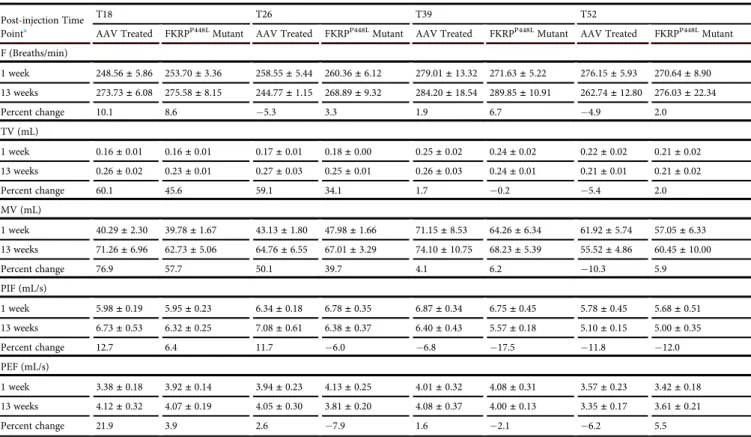
To evaluate respiratory function, unrestrained and conscious AAV-treated and unAAV-treated FKRPP448Lmutant mice were monitored by whole-body plethysmography. This non-invasive technique provides a comprehensive pulmonary analysis by measuring sensitive respira-tory parameters that include breathing frequency (F; the number of breaths per minute), tidal volume (TV; amount of air inhaled and exhaled normally at rest), minute volume (MV; amount of air inhaled and exhaled per minute), peak inspiratoryflow (PIF; maximalflow rate achieved during the inspiratory cycle), and peak expiratory flow (PEF; maximalflow rate achieved during the maximally forced expiration initiated at full inspiration) (Table 1). Analysis of the re-sults indicates an improvement trend in a majority of the respiratory parameters of the early-stage AAV-treated cohorts (T18 and T26) when compared to age-matched untreated FKRPP448Lmutant mice.
However, no statistical significance was reached with the small size of the cohorts. Consistent with more progressivefibrosis in the dia-phragm as observed from H&E and Masson’s trichrome staining,
minimal or no improvement at all is observed with the late-stage AAV-treated cohorts (T39 and T52). Assessment of cardiac morphology and function was conducted via echocardiography and summarized (Table S2). The results indicate that there was no sig-nificant difference in cardiac morphology or function between the AAV-treated and untreated FKRPP448L mutant mice cohorts at each time point.
DISCUSSION
Great advancements have been made in transforming gene replace-ment therapy into an efficient, sustainable method for treating dystro-phic disorders. However, many questions remain to be answered before the therapeutic approach can be effective in a clinical study focused on a majority of patients with muscular dystrophy. Pre-clin-ical studies related to muscular dystrophy have proven that AAV-mediated delivery can result in transgenic expression at therapeutic levels in body-wide skeletal muscles and organs of interest. Recently, our research group and collaborators reported that AAV-mediated delivery ofFKRPwas able to effectively express sufficient levels of FKRP protein, triggering the restoration of functional glycosylation ofa-DG in mouse models of LGMD2I containing point mutations of proline to leucine at position 448 (FKRPP448L) and leucine to isoleucine at position 276 (FKRPL276I).22,23Administration at an early stage of the disease ameliorated disease progression, with significant improvement in dystrophic pathology, serum creatine kinase levels, and muscle function void of any deleterious evidence. The results showed that transgenic FKRP expression, levels of functionally
Figure 4. Improvement of Muscle Performance and Body Weight
(A–C) Treadmill exhaustion test assessing the running time until exhaustion (A), distance covered (B), and maximum running speed (C) of AAV-treated and FKRPP448L mutant mice in each cohort. (D) Percent change in body weight observed from initial treatment to euthanasia. *p%0.05 (Student’s t test, each condition versus age-matched FKRPP448L
glycosylateda-DG, and other therapeutic effects persisted long after treatment. However, disease progression of muscular dystrophy is the result of muscle wasting and accumulation offibrotic tissue, both of which are likely to have a profound effect on the efficacy of gene therapy. Consequently, knowing the degree of efficacy that can be achieved in individuals with different severities or at different stages of disease progression is critical.
To our knowledge, we are thefirst to demonstrate the phenotypic correction ofFKRP-related muscular dystrophies at multiple stages of disease progression in a mouse model by a single intravenous administration of an AAV vector. The results of this study address the important issue as to whether gene replacement will have similar efficacies at different stages of disease progression. This is especially important because FKRP-related muscular dystrophies exhibit a wide spectrum of clinical severity, variability in age at onset, and vary-ing degrees of myogenic atrophy. These variables significantly complicate the clinical study process, including population selection, dose determination, and comprehensive evaluation of the results. However, these challenges can be overcome by understanding the ef-fect of the underlying variables to the therapy in animal models rele-vant to clinical manifestations. We propose that several aspects of the disease can be conjectured to affect the efficiency of transgene
expres-sion and efficacy of the therapy. Specifically, pathological changes with aging and an increasing amount offibrosis with disease progres-sion are two important factors that can determine the efficiency of transgene expression and its functional impact on diseased muscles. Our FKRPP448Lmutant mouse model serves this purpose well by pre-senting dystrophic phenotypes that closely resemble those demon-strated in patients with LGMD2I. This model exhibits severely reduced expression of functionally glycosylateda-DG in all skeletal and cardiac muscles, along with progressive degeneration as the mouse ages, which is associated with a gradual loss of muscle mass and an increase infibrosis, especially in the diaphragm.
Our results indicate that administration of AAV9-FKRPat a dose of 2.51013vg/kg has the greatest therapeutic effect in the youngest T18 cohort. This includes the highest levels of restoration of func-tional glycosylation ofa-DG, maximum improvement in both skel-etal and respiratory functions, and prevention of dystrophic pathol-ogy. Treatment at later stages of disease progression also improves pathology and functions, but at reduced rates. More importantly, the therapeutic effect could only reach a level similar to the point at which the treatment was initiated. For example, AAV-treated mice in the T39 cohort (treated at 26 weeks of age) showed a reduced per-centage of centrally nucleatedfibers as well as reduced areas offibrotic
Table 1. Assessment of Respiratory Function
Post-injection Time Pointa
T18 T26 T39 T52
AAV Treated FKRPP448LMutant AAV Treated FKRPP448LMutant AAV Treated FKRPP448LMutant AAV Treated FKRPP448LMutant
F (Breaths/min)
1 week 248.56±5.86 253.70±3.36 258.55±5.44 260.36±6.12 279.01±13.32 271.63±5.22 276.15±5.93 270.64±8.90
13 weeks 273.73±6.08 275.58±8.15 244.77±1.15 268.89±9.32 284.20±18.54 289.85±10.91 262.74±12.80 276.03±22.34
Percent change 10.1 8.6 5.3 3.3 1.9 6.7 4.9 2.0
TV (mL)
1 week 0.16±0.01 0.16±0.01 0.17±0.01 0.18±0.00 0.25±0.02 0.24±0.02 0.22±0.02 0.21±0.02
13 weeks 0.26±0.02 0.23±0.01 0.27±0.03 0.25±0.01 0.26±0.03 0.24±0.01 0.21±0.01 0.21±0.02
Percent change 60.1 45.6 59.1 34.1 1.7 0.2 5.4 2.0
MV (mL)
1 week 40.29±2.30 39.78±1.67 43.13±1.80 47.98±1.66 71.15±8.53 64.26±6.34 61.92±5.74 57.05±6.33
13 weeks 71.26±6.96 62.73±5.06 64.76±6.55 67.01±3.29 74.10±10.75 68.23±5.39 55.52±4.86 60.45±10.00
Percent change 76.9 57.7 50.1 39.7 4.1 6.2 10.3 5.9
PIF (mL/s)
1 week 5.98±0.19 5.95±0.23 6.34±0.18 6.78±0.35 6.87±0.34 6.75±0.45 5.78±0.45 5.68±0.51
13 weeks 6.73±0.53 6.32±0.25 7.08±0.61 6.38±0.37 6.40±0.43 5.57±0.18 5.10±0.15 5.00±0.35
Percent change 12.7 6.4 11.7 6.0 6.8 17.5 11.8 12.0
PEF (mL/s)
1 week 3.38±0.18 3.92±0.14 3.94±0.23 4.13±0.25 4.01±0.32 4.08±0.31 3.57±0.23 3.42±0.18
13 weeks 4.12±0.32 4.07±0.19 4.05±0.30 3.81±0.20 4.08±0.37 4.00±0.13 3.35±0.17 3.61±0.21
Percent change 21.9 3.9 2.6 7.9 1.6 2.1 6.2 5.5
Respiratory function parameters in AAV-treated and FKRPP448Lmutant mice for T18, T26, T39, and T52 cohorts at post-injection time point intervals of 1 and 13 weeks. F, breathing
frequency; MV, minute volume; PEF, peak expiratoryflow; PIF, peak inspiratoryflow; TV, tidal volume.
tissue, but both percentages remained higher than those of the un-treated FKRPP448Lmutant T26 cohort. This could be due, in part, to the fact that even though AAV9 demonstrates early expression (within 7 days post-injection), maximum vector expression may not occur until 100 days post-injection.28A similar trend is evidenced in the functional indices assessed by the treadmill exhaustion test. Therefore, the results support the notion that it may be difficult for an AAV gene therapy to reverse existing pathological changes such as fibrosis and fragmented fibers that can hinder the function of diseased muscles. It remains to be investigated as to whether a higher dose and prolonged period of treatment could further improve func-tional outcomes of skeletal, respiratory, and cardiac muscles, reaching statistical significance in all cohorts.
In general, the correction of dystrophic muscle pathology is highly dependent on the restoration of functional glycosylation ofa-DG. We observed that the rescue of functional glycosylation of a-DG was variable between tissues in the treated mice and the amount was highly dependent on the age at which the mouse was treated. The reason behind the lower levels of functionally glycosylated a-DG in muscles, especially skeletal muscles, of the late-stage treated cohorts is not clearly understood. One possible explanation is that there is a lower level of infectivity of AAV in the muscles of older mice, which have experienced progressive accumulation of the extra-cellular matrix, thereby reducing the accessibility of the virus to dystrophicfibers. Another possible explanation is that the persistent degeneration is expected to lead to a loss of viral vectors, especially before transgene expression is able to achieve its protective effect. This theory is consistent in tissues where degeneration andfibrosis are minimal and progress slowly, such as in the case of the cardiac muscles, which showed relatively similar levels of functionally glyco-sylateda-DG in all AAV-treated cohorts. However, such hypotheses cannot be fully validated until we are able to understand the amount of virus and the localization of viral vectors within each individual muscle. The effort to quantify the total number of viral vector copy numbers has been met with difficulty for interpretation, as localiza-tion of the virus, whether in connective tissue cells or musclefibers, could not be determined by whole-tissue analysis. The expression levels of the FKRP transgene are currently the only data we have, which appear to show similar levels in the same muscle tissue of all AAV-treated cohorts. Therefore, muscle-specific and age-related variation in the efficiency of glycosylation of a-DG cannot be excluded. However, it should be noted that the levels of FKRP protein in the tibialis anterior muscles are very low and only detectable by western blot analysis utilizing an FKRP (C-terminal) antibody. Perhaps also relevant is that the FKRP protein is detected as a dimer, judged by the molecular weight, as opposed to the monomer form of FKRP, which is undetectable. It is possible that the detectable dimer form of FKRP might not represent the actual amount or even active form of FKRP expressed by the AAV vector.
Despite the difficulty in determining the mechanism(s) involved, there is a clear indication that levels of functional glycosylation of a-DG can be considerably lower in severely affected muscles and at
later stages of disease progression. This has significant clinical implications, considering the fact that most patients, even those with relatively mild LGMD2I, are likely to have more severe muscle degeneration and extensive extracellular matrix accumulation compared to the muscles of a dystrophic mouse model, in which sig-nificantfibrosis is largely limited to the diaphragm. The efficiency of AAV viral infection, transgene expression, and/or glycosylation of a-DG in patients is likely to be considerably lower and even more var-iable than that observed in the skeletal muscles of the late-stage treated cohorts. Our results strongly support the notion that early treatment is critical for achieving high efficacy. Encouragingly, AAV gene therapy at all stages of disease progression has the ability to maintain existing muscle mass and improve, albeit limited, muscle function as shown in our study. One possible alternative to compen-sate for the diminishing efficiency in transgene expression and glyco-sylation ofa-DG in advanced diseases could be the use of a higher viral dosage, which brings with it more risks that include non-target tissue expression, loss of efficacy, and the potential to trigger anti-capsid cytotoxic T lymphocyte responses.29Studies related to this issue are currently being investigated by our research group.
more sensitive and consistent detection method for FKRP can be developed.
In conclusion, this study provides direct evidence that a single-dose therapy can result in viable FKRP expression that is able to success-fully generate functional glycosylation ofa-DG and ameliorate the dystrophic phenotypes at multiple stages of disease progression. Our results are particularly important with regard to the clinical translation ofFKRPgene therapy, suggesting that early therapeutic interventions are critical for achieving high efficacy and are likely to have the best chance to counteract the deleterious effects of LGMD2I. Conversely, substantial correction of the disease pathology is unlikely to occur in the advanced stages of disease progression, where muscle tissue is progressively replaced by adipose andfibrotic tissue. Never-theless, limited benefits are achievable for more advanced stages of the disease.
MATERIALS AND METHODS
Study Design
Rationale and Design of Study
This was a proof-of-concept study designed to search for possible differences among experimental treatment groups. Animals were assigned to treatment groups on the basis of availability of gene replacement vector.
Randomization and Blinding
This was an open-label and non-randomized study.
Replication
Repeated functional measures were conducted over time in animals as indicated. Technical replicates were performed in immunohisto-chemical, western blot, quantitative real-time PCR, and histological analyses.
Ethics Statement
All animal studies were approved by the Institutional Animal Care and Use Committee (IACUC) of Carolinas Medical Center. All mice were housed in the vivarium of Carolinas Medical Center ac-cording to animal care guidelines of the institute. Food and water were available ad libitum during all phases of the study.
Mouse Model
FKRPP448L mutant mice were generated by the
McColl-Lock-wood Laboratory for Muscular Dystrophy Research.25,26 These mice contain a homozygous missense mutation (c.1343C > T, p.Pro448Leu) in theFKRPgene with thefloxed neomycin resistant (Neor) cassette removed from the insertion site.
Construction of AAV Vector and Administration
The recombinant AAV vector construction was performed as described previously.32Full-length humanFKRPcDNA was synthe-sized for high expression in mice and was subsequently subcloned into a single-stranded AAV9 vector under control of a muscle-specific CK7 promoter. Recombinant AAV vector stocks were produced
ac-cording to the three-plasmid cotransfection method reported previously.33The viral particles were purified twice by cesium chlo-ride density gradient ultracentrifugation using the previously published protocol.34Vector titers were determined by both dot-blot and real-time PCR methods using previously published protocols.35,36The concentration of viral vectors was kept in the range of 51012vg/mL and stored at80C until future use.
FKRPP448Lmutant mice were administered with AAV9-FKRPat a
dose of 2.51013vg/kg via a single tail-vein injection at four different age points: 5, 13, 26, and 39 weeks (n = 4 for each cohort; 2 male and 2 female). Mice were euthanized 13 weeks after injection. All un-treated FKRPP448Lmutant mice (n = 4 for each cohort; 2 male and 2 female) were euthanized at the same age point as the AAV-treated mice. FKRPP448Lmutant mice (n = 4 for each cohort; 2 male and 2 female) aged 5 and 13 weeks were used as controls for fibrotic area comparisons. C57BL/6 mice (n = 2; 1 male and 1 female) aged 14 weeks were used as controls for western blot normalization.
Immunohistochemical and Western Blot Analysis
Tissues were dissected and snap-frozen in dry-ice-chilled 2-methyl-butane. Tissues were cryosectioned (6-mm thick), positioned on glass microscope slides, and then stored at80C until future use. Anti-bodies used in this study were obtained as follows: mouse monoclonal a-DG (clone IIH6C4) was from EMD Millipore, rabbit polyclonal actin and laminin was from Sigma-Aldrich, rabbit polyclonal glycer-aldehyde 3-phosphate dehydrogenase (GAPDH) was from Thermo Fisher Scientific, and an affinity-purified rabbit polyclonal FKRP (C-terminal, sequence: NPEYPNPALLSLTGG) was produced in our laboratory.
For immunohistochemical detection, frozen tissue sections were incubated with 1 Tris-buffered saline (TBS) for 5 min and then immediately blocked with 10% normal donkey serum diluted in 1 TBS for 30 min. Sections were then incubated overnight at 4C with primary antibodies IIH6C4 (1:200) or laminin (1:200) diluted in 1 TBS. Sections were washed two times with 1 TBS and appropriate secondary antibodies were incubated at room tem-perature for 1 hr. Sections were washed three times with 1 TBS and mounted with fluorescence mounting medium (Dako) con-taining 1 DAPI. Images were visualized using an Olympus BX51/BX52 fluorescence microscope (Opelco) and were captured using the Olympus DP70 digital camera system (Opelco).
Fisher Scientific) or 3% milk in 1TBS with Tween 20 (0.05%) for 1 hr at room temperature and then incubated with the following pri-mary antibodies overnight at 4C: IIH6C4 (1:1,000), FKRP (C-termi-nal) (1:400), actin (1:8,000), and GAPDH (1:2,000). Appropriate horseradish peroxidase (HRP)-conjugated secondary antibodies were applied to the membranes for 1 hr. All blots were developed by electrochemiluminescence immunodetection (PerkinElmer), exposed to BioMax Light Film (Sigma-Aldrich), and processed by a Mini-Medical imaging system (AFP Imaging) or by manualfilm pro-cessing. The detection of actin and GAPDH confirmed that a similar amount of protein was loaded for each sample.
Quantitative Reverse Transcriptase PCR Assays
Total RNA was extracted from tibialis anterior tissues using TRIzol reagent (Life Technologies), and cDNA was synthesized using a High-Capacity RNA-to-cDNA kit (Applied Biosystems). PCR anal-ysis was performed using PowerUp SYBR Green Master Mix (Life Technologies), and amplified PCR products were quantified and normalized using GAPDH as a control. Cycling conditions for FKRP transgene expression were as follows: initial uracil-DNA glyco-sylase (UDG) activation (50C/2 min), Dual-Lock DNA polymerase (95C/2 min), 40 cycles (95C/15 s; 55C/15 s; and 72C/1 min). This was followed by melting curve analysis starting at 65C and increasing to 95C at a programmed rate of 0.1C/s. The primer pair used is 50-CCACAGCACAGACAGACACT-30 (forward) and 50-CTCCGGGGCATCCTTAGAAA-30 (reverse) (Integrated DNA Technologies). All reactions were performed in triplicate.
Histopathological and Morphometric Analysis
Frozen tissue sections were processed for H&E staining using stan-dard procedures. For H&E images, one representative40 magnifi-cation image from each cohort was used. For morphometric analysis, tibialis anterior muscles were subject to immunostaining as described above with rabbit anti-laminin primary antibody (Sigma-Aldrich) and an anti-rabbit IgG (H+L) secondary antibody, Alexa Fluor 594 conjugate (Life Technologies). Muscle cross-sectional fiber radii and the percentage of myofibers with centrally located nuclei were quantified manually from photographs taken at10 magnification (average ofR450fibers). Images were visualized using an Olympus BX51/BX52 fluorescence microscope and were captured using the Olympus DP70 digital camera system. All measurements and calcu-lations were conducted using MetaMorph Microscopy Automation and Image Analysis Software (version 7.7.0.0; Molecular Devices).
Masson’s Trichrome Staining
Tissue slides were thawed andfixed in 10% neutral buffered formalin overnight at room temperature, rinsed with water, and then baked at 56C in Bouin’sfixative for 1 hr. Slides were rinsed with water and then dipped in Weigert’s iron hematoxylin for 10 min. Slides were washed twice in deionized water and then incubated in Biebrich scarlet-acid fuchsin solution for 30 min. After a brief washing in de-ionized water, slides were put in phosphotungstic phosphomolybdic acid for 12 min and stained with aniline blue for 20 min. Slides were washed in deionized water and subsequently placed in acidified
ethanol containing 1% glacial acetic acid for 2 min. Slides were then dipped three times each in 95% ethanol, 100% ethanol, and xylene (Sigma-Aldrich). All reagents were acquired from Poly Scientific R&D unless otherwise stated. Masson’s trichrome was used to deter-mine the amount offibrosis within the diaphragm, which was calcu-lated from the entire muscle cross-sectional area and quantified using ImageJ software (Java version 1.6.0_20; NIH). The color threshold of the images was adjusted in ImageJ using the hue/saturation/bright-ness filter sets dividing the image into two-color ranges. Particles on the created masks were subsequently analyzed and the total areas were used to create the blue-to-total ratio indicating the percentage of fibrous tissue. One representative20 magnification image for each diaphragm per animal was used.
Treadmill Exhaustion Test
All mice were subjected to a treadmill exhaustion test. Mice were placed on the belt of afive-lane motorized treadmill (Columbus In-struments) supplied with shock grids mounted at the back of the treadmill, which delivered a 0.2-mA current to provide motivation for exercise. Initially, the mice were subjected to an acclimation period (time, 5 min; speed, 8 cm/s; distance, 24 m; and incline, 0). Immediately after the acclimation period, the test commenced at a speed of 8 cm/s and was subsequently increased 2 cm/s every min-ute until exhaustion. The test was stopped and the time to exhaustion was determined when the mouse remained on the shock grid at the back of the treadmill for 5 s without attempting to re-engage the treadmill or if the mouse repeatedly failed to make it at least halfway up the treadmill lane. Stop times were rounded to the nearest 15-s interval.
Whole-Body Plethysmography
Respiratory functional analysis in conscious, freely moving mice was measured using a whole-body plethysmography technique. The plethysmograph apparatus (emka Technologies) was connected to a ventilation pump for the purpose of maintaining constant airflow, a differential pressure transducer, a USBamp signal amplifier, and a personal computer running iox2 software (emka Technologies) with the respiratoryflow analyzer module, which was used to detect pressure changes due to breathing and recording the transducer signal. 20 mL of air was injected and withdrawn via a 20-mL syringe into the chamber for the purpose of calibration. Mice were placed in-side the plethysmograph chamber and allowed to acclimate for 5 min in order to minimize any effects of stress-related changes in ventila-tion. Resting ventilation was measured for a duration of 15 min after the acclimation period. Body temperatures of all mice were assumed to be 37C and to remain constant during the ventilation protocol.
Transthoracic Echocardiography
left ventricle were obtained and these images were analyzed for left ventricular function parameters in triplicate. The heart was also imaged under B-mode with the B-mode placement imaging the heart at the level of the aortic sphincter. Cardiac examination time was approximately 15 min.
Statistical Analysis
All data are expressed as means±SEM unless stated otherwise. Sta-tistical analyses were performed with GraphPad Prism software (version 7.01 for Windows; GraphPad Software). Individual means were compared using multiple Student’s t tests. Differences were considered to be statistically significant at p% 0.05, p%0.01, or p%0.001.
SUPPLEMENTAL INFORMATION
Supplemental Information includes twofigures and two tables and can be found with this article online athttp://dx.doi.org/10.1016/j. omtm.2017.02.002.
AUTHOR CONTRIBUTIONS
Conceptualization, C.H.V. and Q.L.L.; Methodology, C.H.V. and Q.L.L.; Validation, C.H.V.; Formal Analysis, C.H.V. and W.X.; Inves-tigation, C.H.V., W.X., and P.L.; Resources, Q.L.L. and X.X.; Data Curation, C.H.V. and W.X.; Writing – Original Draft, C.H.V.; Writing – Review & Editing, C.H.V. and Q.L.L.; Visualization, C.H.V.; Supervision, Q.L.L.; Project Administration, C.H.V. and Q.L.L.; Funding Acquisition, X.X. and Q.L.L.
CONFLICTS OF INTEREST
The authors declare no conflicts of interest.
ACKNOWLEDGMENTS
This work was supported by NIH grant 4R01NS082536-04 and the Carolinas Muscular Dystrophy Research Endowment through the Carolinas HealthCare Foundation. The authors thank Anthony Blaeser (McColl-Lockwood Laboratory, Carolinas Medical Center [CMC]) for analytical assistance and helpful discussions, Kim Mi-halko and all of the vivarium staff (Animal Facility, CMC) for caring for the animals, and Mallory Korman and Brooke King (Histology Core, CMC) for assistance with cryosectioning of tissues and histo-logical staining.
REFERENCES
1.Godfrey, C., Foley, A.R., Clement, E., and Muntoni, F. (2011). Dystroglycanopathies: coming into focus. Curr. Opin. Genet. Dev.21, 278–285.
2.Aravind, L., and Koonin, E.V. (1999). The fukutin protein family--predicted enzymes modifying cell-surface molecules. Curr. Biol.9, R836–R837.
3.Esapa, C.T., Benson, M.A., Schröder, J.E., Martin-Rendon, E., Brockington, M., Brown, S.C., Muntoni, F., Kröger, S., and Blake, D.J. (2002). Functional requirements for fukutin-related protein in the Golgi apparatus. Hum. Mol. Genet.11, 3319–3331. 4.Kuchta, K., Knizewski, L., Wyrwicz, L.S., Rychlewski, L., and Ginalski, K. (2009). Comprehensive classification of nucleotidyltransferase fold proteins: identification of novel families and their representatives in human. Nucleic Acids Res.37, 7701– 7714.
5.Kanagawa, M., Kobayashi, K., Tajiri, M., Manya, H., Kuga, A., Yamaguchi, Y., Akasaka-Manya, K., Furukawa, J., Mizuno, M., Kawakami, H., et al. (2016). Identification of a post-translational modification with ribitol-phosphate and its defect in muscular dystrophy. Cell Rep.14, 2209–2223.
6.Ibraghimov-Beskrovnaya, O., Ervasti, J.M., Leveille, C.J., Slaughter, C.A., Sernett, S.W., and Campbell, K.P. (1992). Primary structure of dystrophin-associated glyco-proteins linking dystrophin to the extracellular matrix. Nature355, 696–702. 7.Nilsson, J., Nilsson, J., Larson, G., and Grahn, A. (2010). Characterization of
site-spe-cific O-glycan structures within the mucin-like domain ofa-dystroglycan from hu-man skeletal muscle. Glycobiology20, 1160–1169.
8.Yoshida-Moriguchi, T., Yu, L., Stalnaker, S.H., Davis, S., Kunz, S., Madson, M., Oldstone, M.B., Schachter, H., Wells, L., and Campbell, K.P. (2010). O-mannosyl phosphorylation of alpha-dystroglycan is required for laminin binding. Science 327, 88–92.
9.Brockington, M., Blake, D.J., Prandini, P., Brown, S.C., Torelli, S., Benson, M.A., Ponting, C.P., Estournet, B., Romero, N.B., Mercuri, E., et al. (2001). Mutations in the fukutin-related protein gene (FKRP) cause a form of congenital muscular dystro-phy with secondary laminina2 deficiency and abnormal glycosylation of a-dystro-glycan. Am. J. Hum. Genet.69, 1198–1209.
10.Beltran-Valero de Bernabé, D., Voit, T., Longman, C., Steinbrecher, A., Straub, V., Yuva, Y., Herrmann, R., Sperner, J., Korenke, C., Diesen, C., et al. (2004). Mutations in the FKRP gene can cause muscle-eye-brain disease and Walker-Warburg syndrome. J. Med. Genet.41, e61.
11.Michele, D.E., Barresi, R., Kanagawa, M., Saito, F., Cohn, R.D., Satz, J.S., Dollar, J., Nishino, I., Kelley, R.I., Somer, H., et al. (2002). Post-translational disruption of dys-troglycan-ligand interactions in congenital muscular dystrophies. Nature 418, 417–422.
12.Brown, S.C., Torelli, S., Brockington, M., Yuva, Y., Jimenez, C., Feng, L., Anderson, L., Ugo, I., Kroger, S., Bushby, K., et al. (2004). Abnormalities ina-dystroglycan expres-sion in MDC1C and LGMD2I muscular dystrophies. Am. J. Pathol.164, 727–737. 13.Poppe, M., Cree, L., Bourke, J., Eagle, M., Anderson, L.V.B., Birchall, D., Brockington,
M., Buddles, M., Busby, M., Muntoni, F., et al. (2003). The phenotype of limb-girdle muscular dystrophy type 2I. Neurology60, 1246–1251.
14.Martin, P.T. (2007). Congenital muscular dystrophies involving the O-mannose pathway. Curr. Mol. Med.7, 417–425.
15.Roscioli, T., Kamsteeg, E.-J., Buysse, K., Maystadt, I., van Reeuwijk, J., van den Elzen, C., van Beusekom, E., Riemersma, M., Pfundt, R., Vissers, L.E., et al. (2012). Mutations in ISPD cause Walker-Warburg syndrome and defective glycosylation ofa-dystroglycan. Nat. Genet.44, 581–585.
16.Willer, T., Lee, H., Lommel, M., Yoshida-Moriguchi, T., de Bernabe, D.B., Venzke, D., Cirak, S., Schachter, H., Vajsar, J., Voit, T., et al. (2012). ISPD loss-of-function mu-tations disrupt dystroglycan O-mannosylation and cause Walker-Warburg syn-drome. Nat. Genet.44, 575–580.
17.Fisher, K.J., Jooss, K., Alston, J., Yang, Y., Haecker, S.E., High, K., Pathak, R., Raper, S.E., and Wilson, J.M. (1997). Recombinant adeno-associated virus for muscle directed gene therapy. Nat. Med.3, 306–312.
18.Daya, S., and Berns, K.I. (2008). Gene therapy using adeno-associated virus vectors. Clin. Microbiol. Rev.21, 583–593.
19.Qiao, C., Li, J., Zhu, T., Draviam, R., Watkins, S., Ye, X., Chen, C., Li, J., and Xiao, X. (2005). Amelioration of laminin-a2-deficient congenital muscular dystrophy by so-matic gene transfer of miniagrin. Proc. Natl. Acad. Sci. USA102, 11999–12004. 20.Bartoli, M., Roudaut, C., Martin, S., Fougerousse, F., Suel, L., Poupiot, J., Gicquel, E.,
Noulet, F., Danos, O., and Richard, I. (2006). Safety and efficacy of AAV-mediated calpain 3 gene transfer in a mouse model of limb-girdle muscular dystrophy type 2A. Mol. Ther.13, 250–259.
21.Mendell, J.R., Rodino-Klapac, L.R., Rosales-Quintero, X., Kota, J., Coley, B.D., Galloway, G., Craenen, J.M., Lewis, S., Malik, V., Shilling, C., et al. (2009). Limb-girdle muscular dystrophy type 2D gene therapy restoresa-sarcoglycan and associated pro-teins. Ann. Neurol.66, 290–297.
23.Qiao, C., Wang, C.-H., Zhao, C., Lu, P., Awano, H., Xiao, B., Li, J., Yuan, Z., Dai, Y., Martin, C.B., et al. (2014). Muscle and heart function restoration in a limb girdle muscular dystrophy 2I (LGMD2I) mouse model by systemic FKRP gene delivery. Mol. Ther.22, 1890–1899.
24.Vannoy, C.H., Xu, L., Keramaris, E., Lu, P., Xiao, X., and Lu, Q.L. (2014). Adeno-asso-ciated virus-mediated overexpression of LARGE rescuesa-dystroglycan function in dystrophic mice with mutations in the fukutin-related protein. Hum. Gene Ther. Methods25, 187–196.
25.Chan, Y.M., Keramaris-Vrantsis, E., Lidov, H.G., Norton, J.H., Zinchenko, N., Gruber, H.E., Thresher, R., Blake, D.J., Ashar, J., Rosenfeld, J., and Lu, Q.L. (2010). Fukutin-related protein is essential for mouse muscle, brain and eye development and mutation recapitulates the wide clinical spectrums of dystroglycanopathies. Hum. Mol. Genet.19, 3995–4006.
26.Blaeser, A., Keramaris, E., Chan, Y.M., Sparks, S., Cowley, D., Xiao, X., and Lu, Q.L. (2013). Mouse models of fukutin-related protein mutations show a wide range of dis-ease phenotypes. Hum. Genet.132, 923–934.
27.Ervasti, J.M., and Campbell, K.P. (1991). Membrane organization of the dystrophin-glycoprotein complex. Cell66, 1121–1131.
28.Zincarelli, C., Soltys, S., Rengo, G., and Rabinowitz, J.E. (2008). Analysis of AAV se-rotypes 1-9 mediated gene expression and tropism in mice after systemic injection. Mol. Ther.16, 1073–1080.
29.Mingozzi, F., and High, K.A. (2013). Immune responses to AAV vectors: overcoming barriers to successful gene therapy. Blood122, 23–36.
30.Salva, M.Z., Himeda, C.L., Tai, P.W.L., Nishiuchi, E., Gregorevic, P., Allen, J.M., Finn, E.E., Nguyen, Q.G., Blankinship, M.J., Meuse, L., et al. (2007). Design of tissue-spe-cific regulatory cassettes for high-level rAAV-mediated expression in skeletal and car-diac muscle. Mol. Ther.15, 320–329.
31.Beedle, A.M., Nienaber, P.M., and Campbell, K.P. (2007). Fukutin-related protein as-sociates with the sarcolemmal dystrophin-glycoprotein complex. J. Biol. Chem.282, 16713–16717.
32.Li, J., Dressman, D., Tsao, Y.P., Sakamoto, A., Hoffman, E.P., and Xiao, X. (1999). rAAV vector-mediated sarcogylcan gene transfer in a hamster model for limb girdle muscular dystrophy. Gene Ther.6, 74–82.
33.Xiao, X., Li, J., and Samulski, R.J. (1998). Production of high-titer recombinant ad-eno-associated virus vectors in the absence of helper adenovirus. J. Virol.72, 2224– 2232.
34.Snyder, R.O., Xiao, X., and Samulski, R.J. (1996). Production of recombinant adeno-associated viral vectors. In Current Protocols in Human Genetics, N. Dracopoli, ed. (John Wiley & Sons), pp. 12.1.1–12.1.20.