Hepatocytic expression of human
sodium-taurocholate cotransporting polypeptide enables
hepatitis B virus infection of macaques
Benjamin J. Burwitz
1
, Jochen M. Wettengel
2
, Martin A. Mück-Häusl
2
, Marc Ringelhan
2,3
, Chunkyu Ko
2
,
Marvin M. Festag
2
, Katherine B. Hammond
1
, Mina Northrup
1
, Benjamin N. Bimber
4
, Thomas Jacob
5
,
Jason S. Reed
1
, Reed Norris
4
, Byung Park
6
, Sven Moller-Tank
7,8,9
, Knud Esser
2
, Justin M. Greene
1
, Helen L. Wu
1
,
Shaheed Abdulhaqq
1
, Gabriela Webb
1
, William F. Sutton
4
, Alex Klug
4
, Tonya Swanson
4
, Alfred W. Legasse
4
,
Tania Q. Vu
5,10
, Aravind Asokan
7,8,9
, Nancy L. Haigwood
4
, Ulrike Protzer
2,11
& Jonah B. Sacha
1,4
Hepatitis B virus (HBV) is a major global health concern, and the development of curative
therapeutics is urgently needed. Such efforts are impeded by the lack of a physiologically
relevant, pre-clinical animal model of HBV infection. Here, we report that expression of the
HBV entry receptor, human sodium-taurocholate cotransporting polypeptide (hNTCP), on
macaque primary hepatocytes facilitates HBV infection in vitro, where all replicative
inter-mediates including covalently closed circular DNA (cccDNA) are present. Furthermore, viral
vector-mediated expression of hNTCP on hepatocytes in vivo renders rhesus macaques
permissive to HBV infection. These in vivo macaque HBV infections are characterized by
longitudinal HBV DNA in serum, and detection of HBV DNA, RNA, and HBV core antigen
(HBcAg) in hepatocytes. Together, these results show that expressing hNTCP on macaque
hepatocytes renders them susceptible to HBV infection, thereby establishing a physiologically
relevant model of HBV infection to study immune clearance and test therapeutic and curative
approaches.
DOI: 10.1038/s41467-017-01953-y
OPEN
1Vaccine and Gene Therapy Institute, Oregon Health and Science University, Beaverton, OR 97006, USA.2Institute of Virology, Technical University of Munich/Helmholtz Zentrum Munich, Munich 81675, Germany.3Department of Internal Medicine II, Klinikum rechts der Isar Technical University of Munich, Munich 81675, Germany.4Oregon National Primate Research Center, Oregon Health and Science University, Beaverton, OR 97006, USA.5Department of Biomedical Engineering, Oregon Health and Science University, Portland, OR 97239, USA.6Public Health and Preventative Medicine, Oregon Health and Science University, Portland, OR 97239, USA.7Gene Therapy Center, The University of North Carolina at Chapel Hill, Chapel Hill, NC 27599, USA. 8Department of Genetics, The University of North Carolina at Chapel Hill, Chapel Hill, NC 27514, USA.9Department of Biochemistry and Biophysics, The University of North Carolina at Chapel Hill, Chapel Hill, NC 27599, USA.10Center for Spatial Systems Bioscience, Oregon Health and Science University, Portland, OR 97201, USA.11German Center for Infection Research, Munich partner site, Munich 81675, Germany. Benjamin J. Burwitz, Jochen M. Wettengel, and Martin A. Mück-Häusl contributed equally to this work. Ulrike Protzer and Jonah B. Sacha jointly supervised this work. Correspondence and requests for materials should be addressed to B.J.B. (email:[email protected])
123456789
H
BV continues to infect humans worldwide despite the
availability of an effective prophylactic vaccine since 1981,
and it is estimated that one third of the world
’
s
popula-tion has been infected
1. While the majority of adults clear HBV
infection, exposure of young children or individuals with immune
deficiencies mainly results in chronic infection. Currently, more
than 260 million chronic HBV carriers are at high risk of
developing serious liver pathology including cirrhosis and
hepa-tocellular carcinoma. Hepahepa-tocellular carcinoma is the second
leading cause of death from cancer worldwide and approximately
half of these diagnoses are due to chronic hepatitis B virus
infections
2. Anti-viral drug therapies that employ reverse
tran-scriptase inhibitors can suppress HBV replication, normalize
inflammation, and slow progression of liver
fibrosis
3. However,
these treatments are not curative due to persistence of cccDNA in
the nuclei of infected hepatocytes, and do not eliminate cancer
risk. While recombinant interferon-
α
can cure about 15% of
patients treated, it is rarely used due to its severe side effects.
Pre S 1
Pre S2 S
P
X
Pre C
C
P68S
S90T T139AL144P P297S N319S
N459H D466N
C591S
M704V
F749Y
G785R R208S
P238S
P248LV270D
G34E Q87R
I88M N149TQ2H
S210P
R10K
HBV
HBV P68S mcHBV HBV
HBV P68S mcHBV
Cynomolgus PH HepG2-hNTCP
HBsAg (O.D.)
0.0 0.1 0.2 0.3
0 100 200 300
Alanine transaminase (IU/L)
Day post-HBV challenge
–10 0 10 20 30 40 50 60
HBV DNA (IU/ml)
HBV genotype D serotype ayw*
0.0 0.2 0.4 0.6 0.8 1.0
HBsAg (O.D.)
33451 33453 (CD3-IT) HBV P68S
34079 (CD3-IT) 34084 mcHBV
Day 4
Day 7 Day 10
0 2000 4000 6000
CD3+ T cell count /
μ
l blood
CD3-IT HBV DNA positive
(≥10 IU/ml) (below OD450 cutoff)Anti-HBc positive
5000
3000
1000
101
102
103
ONPRC Mauritius
(feral) Serum 0/27 0 0/27 0
Breeding colony
a
b
c
d
e
f
g
Origin Tissue N % N %
Fig. 1Mauritian-origin HBV (mcHBV) does not infect MCM in vitro or in vivo.aTwenty-seven MCM caught on the island of Mauritius and housed at the ONPRC were screened for HBV DNA and anti-HBc.bSchematic of HBV proteome (black) showing amino acid differences for the recently described mcHBV. *=HBV sequence described by Pasek et al.18.cCynomolgus PH and HepG2-hNTCP cells were infected with HBV, HBV P68S, and mcHBV (MOI
Thus, there remains an urgent need to better understand HBV
clearance, and to identify and test innovative curative HBV
therapies.
Multiple animal models of HBV infection have been utilized
over the past four decades including the Peking duck
4,5,
wood-chuck
6–8, mouse
9, 10, and chimpanzee
11–13. These models have
contributed significantly to our understanding of virus-host
interactions, but each have drawbacks that limit their utility for
drug development. First, ducks and woodchucks are infected with
species-specific hepatitis viruses that are related to HBV, but
differ in genomic sequence, protein composition, and virion
structure
14. In addition, both animals are outbred, ill-defined
genetically, and scarcely used outside the HBV
field, which
con-sequentially limits the availability of important reagents, such as
major histocompatibility complex:peptide tetramers and
immu-nophenotyping antibodies. Second, because HBV does not infect
macNTCP
K IR G
S LI VPV L
P IL I TC L VI G
P 157
Y
0.00% 7.40% 56.96%
Rhesus PH Baboon PH HepG2-hNTCP
M E A H N A S A P F N F T L P P N K F G K R P T D -NH2 K M S M L AL L IV L MF V
I FF S LM
L TA P
G IM A QY V
I AL L A G F VL G
E F S K K I T CG L
A HL W P K A E I N K L R F V
L IA L G CV G PS C
L NG V NS A LS F
K G D M N
LS Y
K C M PL M
A LG T F S
T TC V M I L LY I IG K
Y
S IV VLV L
P IL I TC L VI G S
R G
IY D G D
L K D K V P S K R P
Q Y M RY V
L SA V
A VT C SV L
I IL M I G I NV G
I K
G S
S IM FA
M T
F I LL P
S TA P ML S
G IF L LF L VY G
A L
F C LN G R Q LC V
C QN E TG M
T VS R R C S TI L
N V A F
P P EV
I G
P L
P FF F Y LL Q FI M
E GL L LG I AI L
W C Y E K F K T P K D K T K M I Y T A A T T E E T I P G A L G N G T Y K G E D C S P C T A K HOOC-hNTCP Day 4 Day 7 Day 10
0 2 4 6 8 10
100 101 102 103 104 105 106 107 108 HDAd-hNTCP AAV8-hNTCP HDAd-hNTCP AAV8-hNTCP HepG2-hNTCP Rhesus PH Baboon PH
HBV DNA (gc/ml)
Rhesus PH (HBV MOI 100)
HepG2-hNTCP
HBV cccDNA (gc)
Day post-HBV challenge Human Chimp Rhesus Cyno Baboon 157 165 0 20 40 60 HBsAg (IU/ml) HDAd-hNTCPAAV8-hNTCP (–) Control HDAd-hNTCPAAV8-hNTCP
(–) Control (+) Control
0 20 40 60
HBeAg (IU/ml)
AAV8-hNTCP (MOI 1 x 10⁴)
AAV8-hNTCP (MOI 5 x 10²)
(–) control
0 20 40 60
0.0 0.5 1.0 1.5
Day post-HBV challenge
HBsAg (O.D.)
0 –103 103 104
104
103
102
101
100 105–1030103 104 105–103 0103 104 105 0 20 K 40 K 60 K 80 K 100 K SSC-A MyrB-atto488
No treatment AAV8-hNTCP HDAd-hNTCP
AAV+AAV– HBV+HBV–
+
– T5 exo
PF-rcDNA PF-dlDNA cccDNA SM Rhesus PH 3.2 kb 2.2 kb 1.9 kb 1.4 kb rcDNA remnants + – HepG2-hNTCP
cccDNA (%) 100 95 100 91
a
b
c
d
e
f
g
mouse hepatocytes, current mouse models rely on artificial
expression of HBV, either as a germline transgene or as a linear
genome transferred by viral vectors
15, or on humanized livers that
require significant immunomodulation to maintain. Additionally,
murine hepatocytes do not support the formation of cccDNA,
restricting their use in cure research. The relatively short lifespan
of mice further detracts from the model, as liver dysfunction and
fibrosis are generally gradual processes that can take years to
manifest in humans. Finally, the NIH recently suspended all
funding for chimpanzees in biomedical research, effectively
ending the only physiologically relevant HBV animal model.
Thus, development of a new non-human primate model where
immune responses and cccDNA clearance can be studied using
readily available reagents would mark a milestone in HBV
research.
Dupinay et al.
16recently described endogenous HBV
circu-lating in a geographically isolated cynomolgus macaque
popula-tion on the island of Mauritius (mcHBV). Mauritian cynomolgus
macaques (MCM) descended from a founder population left on
the island ~500 years ago, and due to the absence of endogenous
HBV in non-Mauritian cynomolgus macaques, Dupinay et al.
16conjectured that mcHBV was transmitted from humans to MCM
since the time of their population bottleneck. Indeed, the mcHBV
sequence is 99% identical to clinical isolates of HBV genotype D
serotype ayw3.
Here we show that MCM housed at the Oregon National
Primate Research Center (ONPRC) have no evidence of current
or prior HBV infection in the blood, as measured by HBV DNA
qPCR and anti-HBc ELISA, respectively. Subsequent high-dose
challenge of MCM with recombinant mcHBV did not result in
productive infection. Therefore, in a parallel attempt to overcome
the HBV species-barrier, we transduced rhesus macaque
hepa-tocytes with hNTCP, followed by infection with a recombinant,
clinical isolate of HBV. We show that non-human primate
hepatocytes can support HBV replication in vitro following
transduction with viral vectors encoding hNTCP. Remarkably,
when we administered these viral vectors to rhesus macaques and
challenged with HBV, we detected increasing amounts of HBV
DNA in the serum over time, indicating active HBV replication
in vivo. Further analysis established replication in the liver, where
we detected HBV DNA, RNA, and HBV core antigen (HBcAg) in
hepatocytes. These
findings pave the way for a physiologically
relevant, rhesus macaque HBV infection model that can be
uti-lized to better understand HBV clearance and for the testing of
curative HBV treatments.
Results
mcHBV challenge of MCM
. Dupinay et al.
16recently published
the discovery of mcHBV, a naturally occurring HBV in MCM,
whereby 25.8% of MCM serum samples tested positive for HBV
DNA (range: 10
1–
10
6copies per ml). In order to confirm this
frequency of HBV infection, and to identify MCM with the
highest viral loads for serum transfer experiments, we collected
serum samples from 27 wild-caught MCM imported from
Mauritius to ONPRC and performed a previously published,
clinically validated HBV DNA qPCR assay
17. In addition, we
screened these serum samples for anti-HBc seroconversion using
a clinical, competitive ELISA assay to identify any MCM with
previously cleared HBV infections. We did not detect HBV DNA
or anti-HBc seroconversion in any of the MCM serum samples
tested at ONPRC (Fig.
1
a, Supplementary Table
1
). Given that the
probability of discovering zero infected MCM out of 27 MCM is
only 0.0317% using an exact binomial distribution based on the
prevalence reported by Dupinay et al.
16, these results indicate that
mcHBV is likely not endogenous in MCM on the island of
Mauritius.
The published sequence of mcHBV (GenBank accession#:
HE815465.1) is 99% identical to clinical isolates of HBV genotype
D serotype ayw3. There are a total of 23 amino acid differences
between a commonly used HBV genotype D serotype ayw strain
first described by Pasek et al.
18and mcHBV, distributed
throughout all four HBV open reading frames (Fig.
1
b). To test
the hypothesis that a single mutation within the Pre-S1 region,
P68S, enables mcHBV binding to macaque NTCP and facilitates
mcHBV entry and replication in MCM hepatocytes
16, we
generated recombinant, high-titer stocks of mcHBV and HBV
P68S (containing only the P68S mutation in Pre-S1) based on the
HBV backbone described by Pasek et al.
18. We confirmed the
genomic sequences of these stocks by deep sequencing and
infected cynomolgus macaque primary hepatocytes (PH) or
HepG2 cells expressing hNTCP (HepG2-hNTCP) at a
multi-plicity of infection (MOI) of 100 virions (Supplementary Fig.
1
).
Another commonly used HBV genotype D serotype ayw clinical
isolate, purified from HepAD38 cells and further referred to as
HBV
19, was used as a positive control virus in the assay. We
observed replication of all 3 viruses in HepG2-NTCP, but did not
detect HBV replication in cynomolgus macaque PH nor secretion
of HBV surface antigen (HBsAg) by ELISA (Fig.
1
c).
Next, we challenged MCM in vivo with mcHBV and HBV
P68S. We challenged four MCM intravenously with 1 × 10
9virions (Dane particles) of either mcHBV (
N
=
2) or HBV P68S
(
N
=
2) and monitored for signs of infection over the course of
8 weeks. In addition, we depleted CD3+ T cells in one MCM from
each group by administration of anti-CD3-immunotoxin
20to
remove early T-cell immunity and provide the highest chance of
seeding an acute HBV infection (Fig.
1
d). We found elevated ALT
activity in serum at single-time points from two MCM during the
first few weeks post-challenge, and animal 33451 exhibited
increases in absolute CD3+ T cell counts over the
first 4 weeks
post-infection (Fig.
1
d, e). However, these changes were not
associated with markers of productive HBV infection including
serum HBsAg ELISA, HBV DNA qPCR, and anti-HBc ELISA
(Fig.
1
f, g, Supplementary Table
2
). Cumulatively, these results
cast doubt on the use of mcHBV in MCM as a robust and
reproducible pre-clinical model of HBV, and suggest that
alternate approaches are necessary to generate a non-human
primate model of HBV.
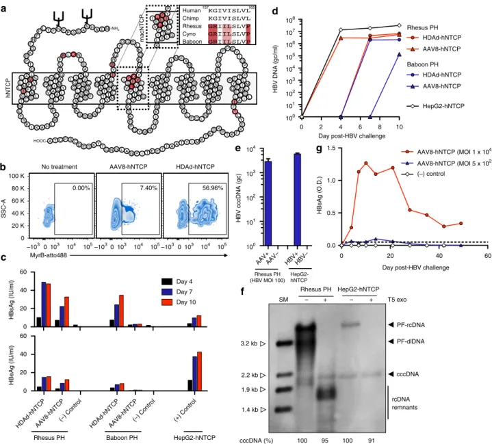
HBV replicates in non-human primate PH expressing hNTCP
.
HBV requires hNTCP to enter hepatocytes, and the HBV binding
region of hNTCP has been identified (amino acids 157
–
165)
21.
Macaque NTCP harbors 5 amino acid differences within this
region, precluding its binding to the pre-S1 region of the large
HBV envelop protein (Fig.
2
a)
21–23. We hypothesized that HBV
would replicate efficiently in non-human primate PH following
binding and entry. To test this hypothesis, we generated
helper-dependent adenovirus (HDAd, serotype 5) and adeno-associated
virus (AAV, serotype 8) vectors expressing hNTCP and
trans-duced freshly isolated rhesus macaque and baboon PH in vitro.
Three days after HDAd-hNTCP or AAV8-hNTCP transduction
we detected expression of hNTCP on the surface of PH by surface
staining with the hNTCP binding, pre-S1-derived peptide
Myr-cludex B (Fig.
2
b). Following confirmation of hNTCP surface
expression, we challenged parallel transduced PH with HBV at an
MOI of 100 virions per cell.
revealed packaging of HBV DNA, indicating the formation and
release of Dane particles (Fig.
2
d). Indeed, we detected both Dane
particles (~40 nm diameter) and spherical bodies (~15 nm
diameter) in supernatants collected from HBV-infected rhesus
macaque and baboon PH (Supplementary Fig.
2
). These spherical
bodies represent subviral HBV particles and were found at a
much higher frequency than Dane particles within the
super-natant. Finally, and most importantly, we detected the formation
of T5 exonuclease-resistant HBV cccDNA in rhesus macaque PH
by qPCR and Southern blotting (Fig.
2
e, f). T5 exonuclease readily
digested relaxed-circular (rcDNA) and duplex-linear DNA
(dlDNA), leading to visualization of DNA remnants, a
0 4 8
–4 Study weeks
N = 2 NN = 2 = 2 = 2
AAV8-hNTCP 5 x 10¹²GC/kg
HDAd-hNTCP 3.5 x 10¹¹GC/kg
HBV 1 x 10⁹ Dane particles
–1
101
102 103
33381 33506
33980 34193
AAV8-hNTCP HDAd-hNTCP
HBV DNA (GE/ml)
33381 33506 33980 34193 104
103
102
101
100
DNA
RNA
Rhesus macaque
HBV copies / 10
6 hepatocytes
Week post-HBV challenge
Alanine transaminase (IU/L)
33381 33506 33980 34193 0
100 200 300 400
C
P
Pre-S/S
X
IFN-γ
SFC / 10
6 cells
Rhesus macaque L R
+ + +
+ + +
C
L R C
L R C
L R C
Anti-HBc serconversion
*
0 2 4 6 8 10
0 50 100 150 200
33381
Left lobe
34193
Caudate lobe
HBV naive rhesus macaque Human chronic HBV infection
Right lobe
% HBcAg-positive nuclei
0.0 0.2 0.4 0.6 0.8 1.0
Naive CHB 34193 33381
L R C
L R C
+ –
– – –
–
a
b
c
d
e
f
phenomenon previously described
24. Thus, rhesus macaque and
baboon hepatocytes expressing hNTCP support the entire HBV
replication cycle.
To further confirm these
findings and to assess the durability of
HBV infection on rhesus macaque PH, we transduced rhesus
macaque PH with a high (1 × 10
4) or low (5 × 10
2genome copies)
dose of AAV8-hNTCP and monitored HBV infection
long-itudinally (Fig.
2
f). We found that HBsAg levels in the
supernatant were directly related to the transducing
AAV8-hNTCP dose and that HBV infection could be sustained on
rhesus macaque PH in vitro for as long as 49 days (Fig.
2
g). Thus,
expression of hNTCP on rhesus macaque PH renders these cells
permissive to HBV infection for prolonged periods of time,
indicating that chronic HBV infection should be possible in
rhesus macaques.
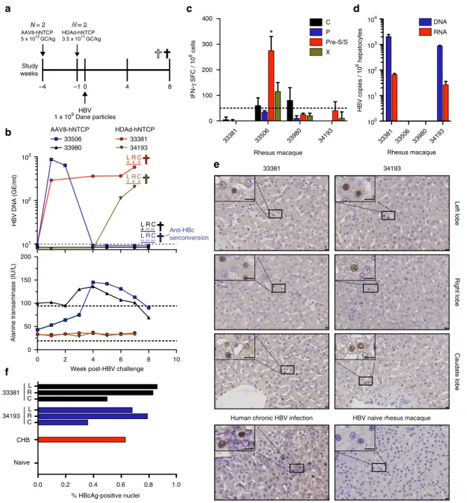
HBV infects rhesus macaques expressing hNTCP in vivo
. Given
our in vitro results demonstrating that hNTCP-expressing
macaque PH can support HBV infection for extended periods
of time, we next tested whether hNTCP-expression on
hepato-cytes in the liver could facilitate HBV infection in vivo. We
injected four rhesus macaques with either HDAd-hNTCP (
N
=
2)
or AAV8-hNTCP (
N
=
2) and subsequently challenged these
animals with HBV (Fig.
3
a). Importantly, we confirmed liver
transduction and hNTCP expression by qPCR and qRT-PCR,
respectively, showing hNTCP expression in all tested lobes
(Supplementary Fig.
3
). We also observed no cellular or humoral
immunity against hNTCP in these animals (Supplementary
Fig.
4
). We sent blinded serum samples to the Oregon Health and
Science University clinical diagnostic laboratory for detection of
HBV DNA and detected HBV viral loads in several consecutive
blood samples from three animals, showing that HBV is capable
of infecting and replicating in macaque livers in vivo following
transduction with hNTCP expressing vectors (Fig.
3
b
–
top panel).
Importantly, as we
first detected HBV DNA in the serum of
animal 34193 at week 6 post-challenge, we can exclude the
pos-sibility that we are simply detecting input virus. In further
sup-port of this, we were unable to detect HBV P68S and mcHBV in
MCM after inoculation with identical virus doses. We also
monitored changes in serum ALT levels following HBV challenge
(Fig.
3
b
–
lower panel). Rhesus macaque 33506 showed elevated
levels of serum ALT, concomitant with a drop in HBV viral load
to undetectable levels. This temporal relationship between
ele-vated serum ALT and clearance of circulating HBV DNA raised
the potential of immune-mediated clearance of HBV infection.
Consistent with HBV DNA and ALT results obtained from
ani-mal 33506, IFN-
γ
ELISpot analysis using liver-resident
lympho-cytes from this animal showed a statistically significant T cell
response against a preS/S peptide pool (Fig.
3
c). Less pronounced
responses against HBV C and X were also detected (Fig.
3
c). We
also assessed anti-HBc seroconversion in all four rhesus
maca-ques at the
final time point and found anti-HBc seroconversion in
33506, but in none of the other animals (Fig.
3
b
–
top panel,
Supplementary Table
3
). Finally, we measured commonly
asses-sed serological markers of HBV infection, including anti-HBs,
anti-HBe, and HBsAg at the
final time point, and found none of
these markers, suggesting that increased hNTCP expression
across macaque hepatocytes will be necessary to observe these
parameters (Supplementary Table
4
).
Next, we examined the liver for the presence of HBV
replication. We extracted total DNA from liver tissue taken from
the left, right, and caudate lobes and detected HBV DNA in three
HBV challenged animals by nested PCR, showing that HBV
replication is occurring in liver tissue (Fig.
3
b
–
top panel and
Supplementary Fig.
5
).
Because it was possible that the HBV DNA detected could be
from blood perfusing the liver sections, we also obtained
highly-purified hepatocytes from the right liver lobes of all four rhesus
macaques by collagenase perfusion. We performed qPCR and
qRT-PCR for the detection of HBV DNA or RNA, respectively, in
this population of purified hepatocytes. Supporting our nested
PCR data, we found both HBV DNA and RNA in hepatocytes of
animals 33381 and 34193, but not in hepatocytes from 33506 and
33980 (Fig.
3
d). Thus, analysis of HBV DNA and RNA levels in
the liver of 33506 further support our assertion of
immune-mediated HBV clearance in this animal.
Finally, we performed IHC on liver sections collected from the
left, right, and caudate lobes of rhesus macaques 33381 and 34193
to determine the level of HBcAg expression. We compared these
results to the livers of a chronically HBV-infected human and
rhesus macaque 34200, which was treated with AAV8-hNTCP
but never HBV challenged. Importantly, we found HBcAg
staining in all sections analyzed from HBV-infected rhesus
macaques (Fig.
3
e). HBcAg staining was localized to small
pockets of expression with only 0.5
–
1.0% of nuclei in
HBV-infected rhesus macaque livers staining HBcAg-positive (includes
parenchymal and non-parenchymal cells), supporting the
low-level HBV replication observed in the serum (Fig.
3
f). This
frequency of HBcAg-positive nuclei was comparable to the liver
section acquired from a chronically HBV-infected human
(Fig.
3
f). Taken together, our data show for the
first time that
HBV infects rhesus macaques expressing hNTCP and that viral
replication can be sustained for at least 6 weeks in vivo, leading to
the induction of HBV-specific T cell responses.
Discussion
Our study presents evidence that experimental HBV infection of
rhesus macaques is possible, paving the way for testing of novel
HBV therapies in a well-characterized non-human primate
infectious disease model. We show that after introducing hNTCP,
HBV enters and replicates in the livers of rhesus macaques. This
replication was associated with HBV-specific cellular and
humoral immunity in the animal with the highest HBV viral load,
indicating that higher HBV replication may be required to
con-sistently induce these immune responses. This is an important
finding, as one advantage of the rhesus macaque model over other
current HBV models is the closely related immune systems
shared between humans and macaques
25–27. Indeed, many of the
emerging HBV therapies rely on modulating anti-HBV
immu-nity, rather than targeting the virus directly
9, 28, 29. Thus, we
believe that further characterization of this model will provide an
important resource to study immune control of HBV and for
testing HBV therapies targeting host immunity.
We were unable to recapitulate the
finding that a particular
strain of HBV reported earlier infects MCM using both in vitro
and in vivo studies
16. The reasons for this remain unclear, but our
findings indicate that the zoonotic transmission of HBV from
human to MCM may have occurred within the particular colony
studied and not on the island of Mauritius. Alternatively, there
may be hurdles to overcome with using recombinantly produced,
high-titer HBV stocks rather than the simple transfer of serum
between HBV-infected animals. Indeed, the majority of animal
HBV research relies on transmission of HBV through serum
transfer. We were, however, also unable to detect any HBV
related virus in any of the captured animals tested. Regardless of
our inability to repeat the results of Dupinay et al.
16, we show
here that expressing hNTCP on the surface of macaque
hepato-cytes is sufficient for HBV entry and replication.
radiolabeled peptides corresponding to the NTCP binding region
of pre-S1 are retained in the livers of mice, rats, and dogs, but not
cynomolgus macaques
21, 23. However, the binding of pre-S1
peptides (and by extension HBV particles) to the livers of mice,
rats, and dogs does not render these species susceptible to
pro-ductive HBV infection, indicating a post-entry host-restriction
preventing productive HBV infection. Importantly, we show here
that no post-entry restriction of HBV infection exists in rhesus
macaques, as replication of HBV in rhesus macaque PH was
abundant following infection. Thus, expression of hNTCP is the
only requirement for infection of macaque hepatocytes in vitro
and in vivo.
We also found that cccDNA is formed in HBV-infected rhesus
macaque PH. This is an important difference to current mouse
models in which cccDNA is not formed following HBV infection,
precluding their use in HBV cure research
30, 31. In addition,
because all steps of the HBV life cycle are supported by rhesus
macaque PH, in vitro
findings can be rapidly transitioned within
the same species to an in vivo model. This stands in contrast to
in vitro
findings using HBV-infected human PH, which require
immunomodulated humanized mice to transition to the in vivo
setting. While we were unable to detect cccDNA in hepatocytes
in vivo, this may be due to the low level of HBV replication.
Indeed, our in vitro work with rhesus macaque PH shows that
cccDNA is formed, indicating that further assessment of cccDNA
formation in rhesus macaques in vivo is necessary.
One hurdle currently facing this macaque HBV model is the
requirement to transduce high numbers of hepatocytes in vivo
with hNTCP expressing viral vectors. In our study, we utilized
HDAd and AAV vectors to deliver hNTCP to the liver. To
achieve efficient hepatocyte targeting, we used an AAV serotype 8
capsid, and hNTCP was expressed under control of a ubiquitous
chicken beta-actin promoter. In animals transduced with
AAV8-hNTCP, HBV infection was variable, with only one animal
exhibiting HBV DNA in serum following challenge. Our HDAd
vector expressed hNTCP under the control of a hepatocyte
spe-cific transthyretin promotor, and both animals exhibited active
viral replication as assessed by serum HBV DNA quantification.
Unfortunately, we were unable to determine the number of
macaque hepatocytes expressing hNTCP due to cross-reactivity
of all available antibodies with macaque NTCP, and loss of the
Myrcludex B epitope on hNTCP during formalin
fixation.
However, the limited number of hNTCP DNA copies per diploid
genome detected in our qPCR assay indicates that hepatocyte
transduction was low. Indeed, if compared to previous AAV
transduction studies in mouse and rhesus macaque livers, the
number of hNTCP DNA copies per diploid genome we detected
corresponds to
<
5% of total hepatocytes transduced
32, 33.
Superior tools for the delivery of hNTCP to a greater number of
hepatocytes, or experiments looking at persistent transduction or
integration of hNTCP into neonate hepatocytes may yield better
hNTCP expression and thus higher infection levels. This
parti-cular concept is additionally appealing in that HBV infection of
neonates results in the highest frequency of chronic HBV
infection.
The
finding that hNTCP expression on non-human
hepato-cytes facilitates HBV infection is not unprecedented. Lempp
et al.
34recently described HBV infection of AAV-hNTCP
transduced pig and macaque PH in vitro. The range of hosts
susceptible to HBV infection following hNTCP-mediated viral
entry is unknown. However, we found that expression of hNTCP
on the surface of baboon PH was also sufficient to allow
pro-ductive infection, indicating that the ability of HBV to replicate
post-entry may pertain to a wide range of non-human primate
species. Indeed, we believe further research into alternative
non-human primate species is warranted.
Given the urgent need for curative HBV treatments, we believe
our macaque model is emerging at a pivotal time and will
facil-itate critical translational research. We show that HBV replicates
in rhesus macaques in vivo, forms cccDNA in PH, and induces
anti-viral cellular and humoral immunity. Thus, the main
ele-ments of human HBV infection can be recapitulated in macaques.
This macaque HBV model may be used to ensure physiologically
relevant, pre-clinical testing of emerging therapies.
Methods
Experimental design. Given the exploratory nature and the binary readout (HBV infection vs. no HBV infection) of this study, we utilized small animal groups. Randomization was not used to assign animals to their respective experimental groups, and authors were not blinded to assignments. A total of 4 Mauritian cynomolgus macaques (33451-F-4 yr, 33453-F-5 yr, 34079-F-5 yr, and 34084-M-6 yr) and 4 Indian rhesus macaques (33381-F-1 yr, 33506-F-1 yr, 33980-F-2 yr, and
34193-F-1 yr) were challenged intravenously with 1 × 10⁹Dane particles of HBV
and followed longitudinally in this study. Anti-CD3-immunotoxin (NHP AIDS Reagent Resource) was given intravenously twice daily at 0.025 mg per kg.
AAV8-hNTCP (5 × 10¹²genomic copies per kg) and HDAd-hNTCP (3.5 × 10¹¹genomic
copies per kg) were administered intravenously at day−28 and day−7 post-HBV
infection, respectively. Necropsy dates were chosen to ensure active HBV repli-cation in the liver at the time of killing. Animals were cared for at the Oregon National Primate Research Center (ONPRC) with the approval of the ONPRC Animal Care and Use Committee using the standards of the NIH Guide for the Care and Use of Laboratory Animals.
In vitro and ex vivo experiments were performed in duplicate where possible. When duplicate samples were not attainable, additional experiments were performed to ensure repeatable results. For example, we have now shown in vitro HBV infection of rhesus macaque PH in at least three separate experiments. All data collected are shown, and no outliers were excluded.
We sent blinded serum samples to the Oregon Health are Science University clinical diagnostic laboratory for detection of HBV DNA in vivo.
Production of recombinant HDAd. The bacterial artificial chromosome (BAC) containing the genome of the helper-dependent adenovirus HDAd-hNTCP was constructed by serial cloning steps based on homologous recombination as
described previously35. In brief, an expression cassette containing the human Na
+-taurocholate cotransporting polypeptide (hNTCP—GenBank Accession
#JQ814895.1) under control of the liver-specific transthyretin (TTR) promoter
derived from the plasmid pCH-TTR-GFP36and the human beta globin
poly-adenylation sequence derived from the plasmid pRTS137was cloned into the 3′
region of a helper-dependent adenoviral vector genome encoded by the bacterial
artificial chromosome BAC-HCAdV5-CMV/eGFP35.
The respective helper-dependent adenoviral vector HDAd-hNTCP was
produced as described previously38. In brief, 116 producer cells (provided by Dr.
Philip Ng), which are HEK293 cells constitutively expressing the Cre
recombinase39, were transfected with MssI-digested BAC containing the
HDAd-hNTCP genome and infected with helper-virus AdNG163R-2 (MOI 3) (provided
by Dr. Philip Ng)39. Initially generated vector particles collected 48 h post-infection
were used forfive sequential amplification steps utilizing increasing numbers of
116 cells. For large-scale amplification, 116 producer cells were cultivated in
suspension in a 3 L bioreactor. Collected vector particles were purified using two
consecutive CsCl-gradients and subsequently dialyzed against a physiological
buffer (10 mM Tris (pH 8.0), 2 mM MgCl2, 4 % sucrose (w/v)).
Production of recombinant AAV. Recombinant AAV8-hNTCP vectors were generated by triple-plasmid transfection into HEK293 cells (ATCC Cat#
CRL-1573). Briefly, HEK293 cells were transfected with pXR8 (provided by the UNC
vector core), encoding AAV replication and capsid genes, pXX6-80 (provided by the UNC vector core), encoding adenoviral helper genes, and pTR-hNTCP (plas-mid pTR provided by the UNC vector core), encoding AAV2 inverted terminal
repeats (ITRs)flanking a transgene cassette expressing human NTCP as driven by
the chicken beta-actin promoter. Cells were maintained in DMEM (Gibco) sup-plemented with 5% fetal bovine serum (FBS) (Sigma-Aldrich) and 1% penicillin/ streptomycin. Supernatant was collected from cells 72 and 144 h post-transfection,
pooled, and concentrated by PEG precipitation. Virus was purified by iodixanol
gradient ultracentrifugation and Zeba Spin desalting column centrifugation
(Thermo Scientific) and vector titers determined by quantitative PCR using a
Roche Lightcycler 480.
Generation of HBV stocks. A high-titer virus stock of HBV was produced by culturing HBV producing HepAD38 cells (provided by Dr. Chris Seeger) and
collecting the HBV-containing supernatant every 3–4 days. Purification of HBV
was performed via heparin columns and sucrose gradient ultracentrifugation as
previously described40. HBV stocks were titered by HBV DNA qPCR of enveloped,
P68S was introduced by site-directed mutagenesis into the plasmid pT-HBV1.3
(GenBank Accession # V01460)18containing the HBV 1.3-fold genotype D,
subtype ayw3 genome. For generation of mcHBV, we synthesized a mcHBV
1.3-fold genome as previously described by Dupinay et al.16and inserted into the same
plasmid to form pT-mcHBV1.3. HepG2 cells (ATCC Cat# HB-8065) were transfected with pT-HBV1.3-P68S and pT-mcHBV1.3 and selected for 3 weeks.
Single-cell clones werefinally selected by high-level secretion of HBsAg
(determined by ELISA) and HBV DNA (by qPCR) in cell culture supernatants. Concentration of HBV P68S and mcHBV was performed via heparin column
purification and subsequent sucrose gradient ultracentrifugation as previously
described40.
HBV DNA was extracted from 20μl of each HBV stock using the QiaAmp
Minelute Virus Spin kit (Qiagen) according to manufacturer’s instructions. HBV
DNA was amplified by PCR using two overlapping amplicons with the following
primer sets: (1) HBV_Illumina-F1 (5′-CTCTCGTTTTTGCCTTCTGAC-3′) and HBV_Illumina-R1 (5′-GCCTCCTAGTACAAAGACCTTTAA-3′), (2)
HBV_Illumina-F2 (5′-ATCGGGACTGATAACTCTGTTG-3′) and
HBV_Illumina-R2 (5′-TCCCCACCTTATGAGTCCAA-3′). Next, the overlapping
PCR amplicons were gel purified using in 1.5% agarose gel followed by cleanup
with the Nucleospin Gel and PCR Cleanup Kit (Macherey-Nagel, Bethlehem, PA),
following the manufacturer’s instructions. Purified DNA was quantified using a
Qubit (Invitrogren, Carlsbad, CA), and the amplicons were pooled at equimolar amounts. These pools were fragmented and barcoded using the Nextera XT Kit
(Illumina, San Diego, CA), followed by purification using Ampure XP beads
(Beckman Coulter, Brea, CA). The resulting libraries were sequenced using an Illimuna MiSeq (Illumina, San Diego, CA). Sequence data were processed using a custom analysis pipeline written by B.N.B. This pipeline has been made available
through DISCVR-Seq (https://github.com/bbimber/discvr-seq), as a module for
LabKey Server, an open-source platform for the management of scientific data41.
Briefly, raw reads were trimmed by sequence quality using Trimmomatic42and
aligned against the Galibert et al. HBV reference using the aligner BWA-Mem43.
Local re-alignment around indels was performed using GATK44. Nucleotide
difference between reads and the reference sequences were scored using custom
software that utilized HTS-JDK (http://samtools.github.io/htsjdk/), and amino acid
translations were performed that accounted for read-specificflanking sequence.
Isolation of macaque PH. A single lobe of non-human primate liver was perfused with 250 ml of Hanks Balanced Salt Solution (HyClone) followed by perfusion with 100 ml collagenase media (DMEM/F12 (Gibco), 3% bovine growth serum
(HyClone), supplemented withL-glutamine (HyClone), antibiotic/antimycotic
(HyClone), Gentamicin (Life Technologies)), and to removed blood from the tis-sue. This was followed by re-circulation of 150 ml collagenase media through the
liver lobe at 42 °C for 1 h. Following collagenase perfusion, the liver wasfilleted
with scalpels and then physically separated with a 3 ml syringe plunger. Cells were
firstfiltered through a tea strainer to remove large debris, followed by a 70μMfilter
to ensure single-cell suspension. Cells were then washed three times in PH media (DMEM/F12 (Gibco), 10% bovine growth serum (HyClone), 23 mM HEPES buffer
(HyClone), 0.6 mg/ml glucose, supplemented withL-glutamine (HyClone),
anti-biotic/antimycotic (HyClone), and Gentamicin (Life Technologies)) at 4 °C.
Cen-trifugation for each wash was: (1) 100×gfor 3 min, (2) 70×gfor 3 min, and (3) 50×g
for 3 min, all at 4 °C. Purified hepatocytes were then resuspended in 37 °C PH
media and counted. Hepatocytes were plated onto collagenized 12-well plates at
7.5 × 10⁵per well and allowed to adhere overnight. The next day, wells were
washed three times with HBSS and cells cultured in 1 ml PH media supplemented with 1.8% DMSO (PH-DMSO).
Transduction and HBV infection of macaque PH. Following 24 h of culture in PH-DMSO, HDAd and AAV8 vectors expressing hNTCP were added to the cul-ture for 3 days. Hepatocytes were then washed twice in 5 ml HBSS and HBV-containing media (PH-DMSO HBV-containing 4% PEG6000) was added overnight. The
next morning, wells were washedfive times with 5 ml HBSS and then cultured in 1
ml PH-DMSO for the remainder of the experiment.
HBV DNA and RNA quantification. To quantify HBV in supernatant or serum,
total DNA was extracted from 200μl using the QiaAmp Minelute Virus Spin kit
(Qiagen) according to manufacturer’s instructions. For analysis of cell associated HBV, total intracellular DNA and RNA were extracted using the AllPrep DNA/ RNA kit according to manufacturer’s instructions (Qiagen).
Total HBV DNA and RNA were quantified using the TaqMan Fast Advanced
Master Mix (Applied Biosystems) and AgPath-ID One-Step RT-PCR kit (Life Technologies), respectively. Total HBV DNA/RNA primers and probe were: HBV_qPCR-F (5′-GGCCATCAGCGCGTGC-3′), HBV_qPCR-R (5′-TGCTGCGA GCAAAACA-3′), and HBV_qPCR-Probe (5′-6FAM-CTCTGCCGATCCATACTG
CGGAACTC-TAMRA-3′) using an annealing temperature of 60 °C. All
thermocyclying parameters followed exactly to suggested manufacturers
instructions. All thermocycling and quantification measurements were conducted
on an Applied Biosystems Step One PLUS. Quantification was assessed relative to
an absolute standard curve using the plasmid pCEP4 with the targeted insert as template.
For cccDNA quantification, intracellular total DNA was extracted using the
NucleoSpin tissue kit (Macherey-Nagel) after lysing cells in one well in 200μl
buffer (12-well plate) according to manufacturer’s instructions. DNA was eluted
from NucleoSpin Tissue Columns with 100μl pre-warmed (70 °C) buffer and
subjected to T5 exonuclease treatment (New England Biolabs) at 37 °C for 30 min. The mixture was diluted eightfold before using as template in the qPCR assay.
cccDNA was quantified using a previously described primer/probe set spanning
the nick and gap in the HBV genome, preferentially amplifying the completed cccDNA minichromosome over the rcDNA in viral capsids. A second primer/ probe set targeted the S gene amplifying both rcDNA and cccDNA using TaqMan Fast Advanced Master Mix (Applied Biosystems). Primer/probe sets were: (1) HBV_CCCF2 (5′-CCGTGTGCACTTCGCTTCA-3′), HBV_CCCR2 (5′-GCACA
GCTTGGAGGCTTGA-3′), and HBV-CCCP2 (5′-6FAM- CATGGAGACCAC
CGTGAACGCCC–BHQ1-3′), and (2) HBV_S-F (5′-TGGCCAAAATTCGCAGT CCC-3′), HBV_S-R (5′-AGATGAGGCATAGCAGCAGGAT-3′), and HBV_S-P
(5′-6FAM-ATGATAAAACGCCGCAGACACATCCAGC–BHQ1-3′). using an
annealing temperature of 60 °C. Quantification was assessed separately for each
primer/probe target set against an absolute standard curve using the plasmid
pcDNA_HBV1.3. To control for low-level amplification of rcDNA with the primer/
probe set targeting the nick and gap of the HBV genome, cccDNA quantification
from rhesus macaque PH was normalized by digesting HBV DNA, extracted from HBV stocks and thus containing only rcDNA, with T5 exonuclease and measuring
quantification of this product with both primer sets.
hNTCP DNA and RNA quantification. To quantify human NTCP DNA and RNA in macaque liver samples, total DNA and RNA were extracted using the AllPrep
DNA/RNA kit according to manufacturer’s instructions (Qiagen). The SuperScript
VILO cDNA Synthesis Kit (Thermo Fischer) was used for cDNA synthesis
according to manufacturer’s instructions. Amplification of the macaque single copy
gene PrP was used for normalization. Following primers were used:
hNTCP_qPCR-F TGCCTCAATGTTCTTCAGCC-3′), hNTCP_qPCR-R (5′-TGTTCATGTTGTTCTTCATC-3′), mPrP_qPCR-F
(5′-TGCTGGGAAGTGC-CATGAG-3′), and mPrP_qPCR-R (5′-CGGTACATGTTTTCACGATAGTA-3′)
using an annealing temperature of 58 °C. All thermocycling and quantification
measurements were conducted on an Applied Biosystems Step One PLUS using PerfeCTa SYBR Green FastMix, ROX (Quantabio) and a 3-step a PCR cycling
protocol. Quantification was assessed on DNA level relative to macaque DNA and
human DNA for mPrP and hNTCP, respectively. RNA level quantification was
assessed relative to an absolute standard curve using plasmid DNA containing the entire hNTCP sequence.
Southern blot analysis. DNA was extracted from HBV-infected rhesus macaque
PH using a modified Hirt extraction, as previously described21. Hirt DNA was
separated on an 1.2% agarose gel and transferred onto a positively charged nylon membrane via upward capillary transfer. Hirt DNA extracted from HBV-infected HepG2-NTCP cells were loaded in parallel to show cccDNA appears at the same
position45. To confirm the presence of cccDNA, Hirt extracted DNA was digested
with T5 exonuclease (New England Biolabs) at 37 °C for 30 min. Linear HBV DNA fragments (3.2 kb, 2.2 kb, 1.9 kb and 1.4 kb) serve as a size marking ladder. The membrane was subjected to UV-crosslinking to covalently bind the transferred DNA onto it. The membrane was then hybridized with a digoxigenin-labeled
HBV-specific probe, washed, and visualized by chemiluminescence according to
manufacturer’s instructions (DIG Luminescent Detection Kit).
Analysis of NTCP. For Fig.2a, the topology of the human NTCP from Uniprot (http://www.uniprot.org/uniprot/Q14973) was used to generate a schematic illus-tration of the secondary structure of the protein. In addition, this amino acid
sequence was compared to the macaque sequence (http://www.uniprot.org/
uniprot/G7PAR4) using BLASTP 2.6.0 + (https://blast.ncbi.nlm.nih.gov/Blast.cgi). Differences in the sequences were labeled with lighter red for amino acid exchanges with similar physiochemical properties and darker red for exchanges with different physiochemical properties.
To determine hNTCP expression, rhesus macaque PH were collected from wells
with 0.05% trypsin media (Gibco) and washed twice in HBSS (200×gfor 5 min).
Cells were incubated with MyrcludexB-Alexa488, which specifically binds human
NTCP, for 30 minutes at room temperature, washed twice in HBSS,fixed with 2%
paraformaldehyde (Electron Microscopy Sciences), and then collected on a Becton-Dickenson LSR-II. Analysis was performed on FlowJo X (TreeStar Inc.).
ELISA. Anti-HBs, Anti-HBe, HBsAg and HBeAg ELISA were performed on the Architect i1000 SRTM (Abbott) and anti-HBc ELISA on the BEP III (Siemens) per the manufacturer’s instructions. In addition, HBsAg ELISA were performed using the Hepatitis B Surface Antigen BioAssay ELISA Kit (US Biological Life Sciences).
Anti-hNTCP ELISA were custom assays performed at the ONPRC. 96 well
plates were coated with 0.25μg per well recombinant hNTCP (Origene) in 50μl
overnight and then blocked with 150μl Blotto (5% dry milk and 1% normal goat
serum in PBS) for 1 h. Serum was heat inactivated at 56 °C for 30 min, serially
diluted threefold, and 50μl added to each well. Tests were performed in duplicate.
goat anti-human IgG-HRP (Jackson Labs) was added in 50μl and incubated for 1 h at room temperature. Following secondary antibody incubation, plates were
washed and TMB substrate (Southern Biotech) added in 50μl and incubated for 15
min at room temperature. Volume of 50μl per well of 1 N sulfuric acid was added
to stop the reaction and plates were read on a SpectraMAX 190 (Molecular Devices) at 450 nm.
IFNγELISpot. Rhesus macaque liver-resident mononuclear cells were isolated from single-cell suspensions generated from collagenase media perfusion of livers, and
peripheral blood mononuclear cells were isolated from blood. Pre-coated IFN-γ
ELISpotPLUSplates (Mabtech Inc.) were used to detect T cell responses and
experiments were conducted per manufacturer’s recommendations in triplicate
wells as previously described46. Each well contained 1 × 10⁵cells in 100μl complete
T-cell media (RPMI 1640, 10% bovine growth serum (HyClone), supplemented
withL-glutamine (HyClone) and antibiotic/antimycotic (HyClone). Liver-resident
T cells were incubated with 5μM 15-mer peptide pools spanning the HBV open
reading frames C, P, S, and X (70% peptide purity or greater). Peripheral blood mononuclear cells were incubated with 2 mM recombinant hNTCP protein. Wells were imaged with an AID ELISPOT reader and spots were counted by an auto-mated system with set parameters for size, intensity, and gradient. Responses were
considered positive and statistically significant if the mean number of spot forming
cells (SFCs) of triplicate sample wells exceed background plus two standard deviations. Responses less than 50 SFCs per million cells were considered negative (below the limit of detection).
Immunohistochemistry. Sections (2μm) of 4% paraformaldehyde-fixed, paraffi n-embedded liver samples were stained using a BondMAX immunohistochemistry
robot (Leica) using Bond solutions and Polymer Refine Detection Kit (Leica).
Primary antibody HBcAg (RB1413A, StartFragmentLab VisionEndFragment) was
used at a 1:50 dilution. Primary antibody was detected with Bond Polymer Refine
Detection Kit from Leica (rabbit, Leica, DS9800).
For quantification of HBcAg staining, whole slides were scanned at ×40
magnification using a SCN400 slide scanner (Leica) and analyzed withTissue IA
image analysis software (Slidepath, Leica) using an optimized color detection
setting for DAB (positive) and Hematoxilin (negative) nuclei at ×20 magnification.
For quantification, 3 random areas of 5–10 mm2in size on each slide were chosen
and obtained values were merged for further statistical analysis. For HBcAg-positive hepatocytes, data are presented as HBcAg-positive nuclei (DAB HBcAg-positive nuclei) as a percentage of total nuclei in liver (DAB/Hematoxylin+, including hepatocytes and non-parenchymal cells).
Electron microscopy. Supernatants from rhesus macaque PH, confirmed for
infection with HBV (Fig.2c), were diluted 1:10 in 4% paraformaldehyde. 10µl of
thefixed viral suspensions were deposited onto glow discharged carbon formvar
400 Mesh copper grids (Ted Pella 01822-F) for 3 min, rinsed 15 s in water, wicked
on Whatmanfilter paper 1, stained for 60 s infiltered 1.33 % (w/v) uranyl acetate
in water, wicked and air dried. EM grids were imaged at 120 kV on a FEI Tecnai Spirit TEM system. Images were acquired as 2048 × 2048 pixel, 16-bit gray scale
files using the FEI’s TEM Imaging and Analysis (TIA) interface on an Eagle 2 K
CCD multiscan camera. All the images were acquired at 2–4 microns underfocused
to improve sample contrast.
Data availability. All relevant data presented here are available from the authors without restrictions. The consensus sequence of the HBV stock used to infect rhesus macaques (HBV isolated from HepAD38) is available through GenBank Accession #MF967563. The mcHBV consensus sequence has already been pub-lished and can be accessed through GenBank Accession #HE815465.1.
Received: 24 February 2017 Accepted: 27 October 2017
References
1. Franco, E. et al. Hepatitis B: epidemiology and prevention in developing
countries.World J. Hepatol.4, 74–80 (2012).
2. Ely, A. & Arbuthnot, P. Differing prospects for the future of using gene therapy
to treat infections with hepatitis B virus and hepatitis C virus.Discov. Med.20,
137–143 (2015).
3. Andersson, K. L. & Chung, R. T. inMonitoring during and after antiviral
therapy for hepatitis B.Vol. 49 (ed. Hoofnagle, J. H.) 166–S173 (Wiley Subscription Services, Inc., A Wiley Company, 2009).
4. Urban, S., Breiner, K. M., Fehler, F., Klingmüller, U. & Schaller, H. Avian hepatitis B virus infection is initiated by the interaction of a distinct pre-S
subdomain with the cellular receptor gp180.J. Virol.72, 8089–8097 (1998).
5. Fukuda, R., Fukumoto, S. & Shimada, Y. A sequential study of viral DNA in
serum in experimental transmission of duck hepatitis B virus.J. Med. Virol.21,
311–320 (1987).
6. Rogler, C. E., Hino, O. & Su, C. Y. Molecular aspects of persistent woodchuck hepatitis virus and hepatitis B virus infection and hepatocellular carcinoma. Hepatology7, 74S–78S (1987).
7. Cummings, I. W. et al. Isolation, characterization, and comparison of recombinant DNAs derived from genomes of human hepatitis B virus and
woodchuck hepatitis virus.Proc. Natl Acad. Sci. USA77, 1842–1846 (1980).
8. Millman, I., Southam, L., Halbherr, T., Simmons, H. & Kang, C. M. Woodchuck
hepatitis virus: experimental infection and natural occurrence.Hepatology4,
817–823 (1984).
9. Buchmann, P. et al. A novel therapeutic hepatitis B vaccine induces cellular and humoral immune responses and breaks tolerance in hepatitis B virus (HBV)
transgenic mice.Vaccine31, 1197–1203 (2013).
10. Bility, M. T. et al. Hepatitis B virus infection and immunopathogenesis in a
humanized mouse model: induction of human-specific liverfibrosis and
M2-like macrophages.PLoS Pathog.10, e1004032–14 (2014).
11. Drucker, J., Tabor, E., Gerety, R. J., Jackson, D. & Barker, L. F. Simultaneous
acute infections with hepatitis A and hepatitis B viruses in a chimpanzee.J.
Infect. Dis.139, 338–342 (1979).
12. Tabor, E., Frösner, G., Deinhardt, F. & Gerety, R. J. Hepatitis B e antigen and antibody: detection by radioimmunoassay in chimpanzees during experimental
hepatitis B.J. Med. Virol.6, 91–99 (1980).
13. Asabe, S. et al. The size of the viral inoculum contributes to the outcome of
hepatitis B virus infection.J. Virol.83, 9652–9662 (2009).
14. Ganem, D. & Varmus, H. E. The molecular biology of the hepatitis B viruses. Annu. Rev. Biochem.56, 651–693 (1987).
15. Sprinzl, M. F., Oberwinkler, H., Schaller, H. & Protzer, U. Transfer of hepatitis B virus genome by adenovirus vectors into cultured cells and mice: crossing the
species barrier.J. Virol.75, 5108–5118 (2001).
16. Dupinay, T. et al. Discovery of naturally occurring transmissible chronic hepatitis B virus infection among Macaca fascicularisfrom mauritius island. Hepatology58, 1610–1620 (2013).
17. Welzel, T. M. et al. Real-time PCR assay for detection and quantification of
hepatitis B virus genotypes A to G.J. Clin. Microbiol.44, 3325–3333 (2006).
18. Pasek, M. et al. Hepatitis B virus genes and their expression in E. coli.Nature
282, 575–579 (1979).
19. Ladner, S. K. et al Inducible expression of human hepatitis B virus (HBV) in stably transfected hepatoblastoma cells: a novel system for screening potential
inhibitors of HBV replication.Antimicrob. Agents Chemother.41, 1715–1720
(1997).
20. Kim, G.-B. et al. A fold-back single-chain diabody format enhances the bioactivity of an anti-monkey CD3 recombinant diphtheria toxin-based
immunotoxin.Protein Eng. Des. Sel.20, 425–432 (2007).
21. Yan, H. et al. Sodium taurocholate cotransporting polypeptide is a functional
receptor for human hepatitis B and Dvirus.Elife1, e00049 (2012).
22. Meier, A., Mehrle, S., Weiss, T. S., Mier, W. & Urban, S. Myristoylated
PreS1-domain of the hepatitis B virus L-protein mediates specific binding to
differentiated hepatocytes.Hepatology58, 31–42 (2013).
23. Schieck, A. et al. Hepatitis B virus hepatotropism is mediated by specific
receptor recognition in the liver and not restricted to susceptible hosts. Hepatology58, 43–53 (2013).
24. Schreiner, S. & Nassal, M. A role for the host dna damage response in hepatitis
B virus cccDNA formation-and beyond?Viruses9, 125 (2017).
25. Evans, D. T. & Silvestri, G. Nonhuman primate models in AIDS research.Curr.
Opin. HIV AIDS8, 255–261 (2013).
26. Messaoudi, I., Estep, R., Robinson, B. & Wong, S. W. Nonhuman primate
models of human immunology.Antioxid. Redox. Signal.14, 261–273 (2011).
27. McClure, H. M. Nonhuman primate models for human disease.Adv Vet Sci
Comp Med28, 267–304 (1984).
28. Liu, J. et al. enhancing virus-specific immunity in vivo by combining
therapeutic vaccination and PD-L1 blockade in chronic hepadnaviral infection. PLoS Pathog.10, e1003856–14 (2014).
29. Golsaz-Shirazi, F. & Shokri, F. Hepatitis B immunopathogenesis and
immunotherapy.Immunotherapy8, 461–477 (2016).
30. Guidotti, L. G., Matzke, B., Schaller, H. & Chisari, F. V. High-level hepatitis B
virus replication in transgenic mice.J. Virol.69, 6158–6169 (1995).
31. Raney, A. K. et al. Nuclear covalently closed circular viral genomic DNA in the liver of hepatocyte nuclear factor 1 alpha-null hepatitis B virus transgenic mice. J. Virol.75, 2900–2911 (2001).
32. Wang, L., Wang, H., Bell, P., McMenamin, D. & Wilson, J. M. Hepatic gene
transfer in neonatal mice by adeno-associated virus serotype 8 vector.Hum.
Gene. Ther.23, 533–539 (2012).
33. Wang, L. et al Impact of pre-existing immunity on gene transfer to nonhuman
primate liver with adeno-associated virus 8 vectors.Hum. Gene Ther.22,
34. Lempp, F. A. et al. Sodium taurocholate cotransporting polypeptide is the limiting host factor of hepatitis B virus infection in macaque and pig
hepatocytes.Hepatology384, 2053 (2017).
35. Mück-Häusl, M., Solanki, M., Zhang, W., Ruzsics, Z. & Ehrhardt, A. Ad 2.0: a novel recombineering platform for high-throughput generation of tailored
adenoviruses.Nucleic Acids Res.43, e50–e50 (2015).
36. Untergasser, A. & Protzer, U. Hepatitis B virus-based vectors allow the elimination of viral gene expression and the insertion of foreign promoters. Hum. Gene. Ther.15, 203–210 (2004).
37. Bornkamm, G. W. et al. Stringent doxycycline-dependent control of gene
activities using an episomal one-vector system.Nucleic Acids Res.33, e137–e137
(2005).
38. Jager, L. et al. A rapid protocol for construction and production of
high-capacity adenoviral vectors.Nat. Protoc.4, 547–564 (2009).
39. Palmer, D. & Ng, P. Improved system for helper-dependent adenoviral vector
production.Mol. Ther.8, 846–852 (2003).
40. Seitz, S. et al. A slow maturation process renders hepatitis B virus infectious. Cell Host Microbe.20, 25–35 (2016).
41. Nelson, E. K. et al. LabKey server: an open source platform for scientific data
integration, analysis and collaboration.BMC Bioinform.12, 71 (2011).
42. Lohse, M. et al. RobiNA: a user-friendly, integrated software solution for
RNA-Seq-based transcriptomics.Nucleic Acids Res.40, W622–W627 (2012).
43. Li, H. & Durbin, R. Fast and accurate long-read alignment with
Burrows-Wheeler transform.Bioinformatics26, 589–595 (2010).
44. McKenna, A. et al. The Genome Analysis Toolkit: a MapReduce framework for
analyzing next-generation DNA sequencing data.Genome Res.20, 1297–1303
(2010).
45. Ko, C., Lee, S., Windisch, M. P. & Ryu, W.-S. DDX3 DEAD-box RNA helicase is a host factor that restricts hepatitis B virus replication at the transcriptional
level.J. Virol.88, 13689–13698 (2014).
46. Burwitz, B. J. et al. Mauritian cynomolgus macaques share two exceptionally common major histocompatibility complex class I alleles that restrict simian
immunodeficiency virus-specific CD8+ T cells.J. Virol.83, 6011–6019 (2009).
Acknowledgements
We thank Ke Zhang for cloning HBV1.3-P68S, Stephan Urban for providing Myrcludex B, Tanja Bauer for providing HBV peptide sets, Christian Lanciault for IHC analysis, and Mei Yu and Guofeng Cheng from Gilead Sciences, Inc for providing their HBV cccDNA qPCR protocol. Electron microscopy was performed at the Multiscale Microscopy Core (MMC) with technical support from the Oregon Health and Science University (OHSU)-FEI Living Lab and the OHSU Center for Spatial Systems Biomedicine (OCSSB). This study was funded in part by an ONPRC pilot project grant to J.B.S. and P51 OD011092 to ONPRC and by the German Research Foundation (DFG) via the Collaborative
Research Center TRR179. We thank the animal care staff at ONPRC for their out-standing care of all study animals.
Author contributions
B.J.B. wrote the manuscript. B.J.B., J.M.W., M.A.M.-H., C.K., M.M.F., K.E., T.Q.V., A.A., N.L.H., U.P. and J.B.S. designed the experiments. B.J.B., J.M.W., M.A.M.-H., M.R., C.K., M.M.F., K.B.H., M.N., B.N.B., T.J., J.S.R., R.N., S.M.-T., J.M.G., H.L.W., S.A., G.W. and W.F.S. conducted the experiments. B.J.B., J.M.W., M.A.M.-H., M.R., C.K., M.M.F., T.Q.V., A.A., N.L.H., U.P. and J.B.S. edited the manuscript. B.P. performed the statistics. A.K., T.S. and A.W.L. performed animal procedures.
Additional information
Supplementary Informationaccompanies this paper at10.1038/s41467-017-01953-y.
Competing interests:Oregon Health and Science University (OHSU), Dr. Sacha, and Dr. Burwitz have afinancial interest in Vir Biotechnology, Inc., a company that may have a financial interest in the results of this research and technology. This potential individual and institutional conflict of interest has been reviewed and managed by OHSU. The remaining authors declare no competingfinancial interests.
Reprints and permissioninformation is available online athttp://npg.nature.com/ reprintsandpermissions/
Publisher's note:Springer Nature remains neutral with regard to jurisdictional claims in published maps and institutional affiliations.
Open Access This article is licensed under a Creative Commons Attribution 4.0 International License, which permits use, sharing, adaptation, distribution and reproduction in any medium or format, as long as you give appropriate credit to the original author(s) and the source, provide a link to the Creative Commons license, and indicate if changes were made. The images or other third party material in this article are included in the article’s Creative Commons license, unless indicated otherwise in a credit line to the material. If material is not included in the article’s Creative Commons license and your intended use is not permitted by statutory regulation or exceeds the permitted use, you will need to obtain permission directly from the copyright holder. To view a copy of this license, visithttp://creativecommons.org/ licenses/by/4.0/.