NLRP12 REGULATES IMMUNITY BY CONTROLLING CELL MIGRATION
Janelle Corrinne Arthur
A dissertation submitted to the faculty of the University of North Carolina at Chapel Hill in partial fulfillment of the requirements for the degree of Doctor of Philosophy in the
Department of Microbiology and Immunology
Chapel Hill 2009
Approved by:
Jenny P-Y. Ting, Ph.D. Stephen H. Clarke, Ph.D. Beverly H. Koller, Ph.D. Zhi Liu, Ph.D.
ABSTRACT
Janelle Corrinne Arthur: NLRP12 regulates immunity by controlling cell migration (Under the direction of Dr. Jenny P-Y. Ting)
NLRP12 is a member of the NLR family of genes that are responsible for coordinating inflammatory responses upon recognition of invading pathogens and host danger signals. Remarkably, mutations in several NLR genes have been linked to
autoinflammatory diseases; greatly expanding our understanding regarding the etiology of these debilitating conditions. NLRP12 is expressed exclusively in innate immune cells and suppresses inflammation by negatively regulating the noncanonical NF-κB pathway. This is achieved by inducing proteasome-mediated degradation of NF-κB inducing kinase (NIK) in response to pathogens and activation through pro-inflammatory receptors. Because NLRP12 functions to dampen these signals, it is clear that NLRP12 must be controlled in order to mount an adequate cellular response to such insults. Here we find that NLRP12 stability is regulated by the evolutionarily conserved molecular chaperone Hsp90. In the presence of Hsp90 inhibitors, NLRP12 protein is rapidly degraded via the proteasome leading to
increased NIK stability and function. Thus, Hsp90 activity is a critical regulatory factor for NLRP12 function and is required for NLRP12-induced degradation of NIK and suppression of the noncanonical NF-κB pathway.
several well defined models of inflammation using Nlrp12 knockout mice. Remarkably, we found that Nlrp12 deficient mice failed to mount T cell mediated responses in hapten induced contact hypersensitivity, a model of allergic dermatitis, and EAE, a model of multiple
sclerosis. Mechanistically this is due to defective migration of peripheral dendritic cells. These innate immune cells express Nlrp12 and play a pivotal role in T cell activation. Molecular analysis reveals that in the absence of NLRP12, dendritic cells display an
inappropriate activation of NIK, resulting in high levels of NIK dependent gene expression. These findings expand our understanding of NLRP12 function in vivo and provide a
ACKNOWLEDGEMENTS
The work presented in this dissertation has been a collaboration between many outstanding scientists, all of whom I thank for their hard work and patience: Coy Allen for his expertise in all things mouse; Zhengmao Ye for countless hours of flow cytometry analysis; Denis Gris for establishing the EAE model and having an “excellent” day every day; Amy Morrison for backcrossing and maintaining the mice; Monika Schneider for assistance with realtime PCR; Kelly Roney for assistance with ELISAs; Chris Moore for assistance with Affymetrix; Brian O’Conner for helpful discussion regarding the intricacies of DC function; and Zhi Liu for his steady hands and dermatology expertise.
I thank my advisor Jenny Ting for providing me with the ideal scientific environment. You have granted me the freedom to explore an enormous breadth of ideas and I feel honored by your confidence in me. I thank my committee members Steve Clarke, Jeff Dangl, Zhi Liu, Beverly Koller, Karen McKinnon and director of graduate studies Glenn Matsushima for insightful advice and encouragement. I thank the NIH T32 training grant for funding.
TABLE OF CONTENTS
LIST OF TABLES………...vii
LIST OF FIGURES………....viii
LIST OF ABBREVIATIONS………x
CHAPTER 1: INTRODUCTION………1
A. INTRODUCTION………...2
1. Discovery of the NLR family………...4
2. NLR domain organization……….5
3. Associations with human disease………..7
B. MECHANISM OF ACTION………...9
1. CIITA, the MHC class II transactivator………9
2. Inflammasome-forming NLRs………11
3. Signaling NLRs………...16
C. NLRP12/MONARCH-1……….21
1. NLRP12 identification and expression………...21
2. NLRP12 as a negative regulator of inflammation………..22
3. Molecular mechanisms of NLRP12-mediated NF-κB suppression………23
4. Canonical and noncanonical pathways of NF-κB activation………..24
5. NLRP12 targets NIK to control the noncanonical NF-κB pathway……...25
CHAPTER 2: HSP90 ASSOCIATED WITH MONARCH-1/NLRP12 AND REGULATES ITS ABILITY TO PROMOTE DEGRADATION OF NF-κB INDUCING
KINASE………...29
A. ABSTRACT………...30
B. INTRODUCTION……….31
C. MATERIALS AND METHODS………...33
D. RESULTS………..36
E. DISCUSSION………42
CHAPTER 3: NLRP12 CONTROLS ADAPTIVE IMMUNE RESPONSES BY REGULATING DENDRITIC CELL MIGRATION………..52
A. ABSTRACT………...53
B. INTRODUCTION……….54
C. MATERIALS AND METHODS………...55
D. RESULTS AND DISCUSSION………59
CHAPTER 4: CONCLUSIONS AND FUTURE DIRECTIONS……….81
APPENDIX 1: MONARCH-1/NLRP12 SUPPRESSES NONCANONICAL NF-κB ACTIVATION AND P52 DEPENDENT CHEMOKINE EXPRESSION IN MONOCYTES……….91
A. ABSTRACT………...92
B. INTRODUCTION……….93
C. MATERIALS AND METHODS………...94
D. RESULTS AND DISCUSSION………96
LIST OF TABLES
LIST OF FIGURES
Figure 1.1. NLRP12 suppresses noncanonical NF-κB activation………28
Figure 2.1. NLRP12 interacting proteins include Hsp70………..46
Figure 2.2. NLRP12 interacts with Hsp90………47
Figure 2.3. Hsp90 inhibition alters the association of NLRP12 and heat shock proteins….48 Figure 2.4. Endogenous NLRP12 stability is dependent upon Hsp90 activity……….49
Figure 2.5. Inhibition of Hsp90 induces proteasome-mediated degradation of NLRP12…50 Figure 2.6. Hsp90 is required for NLRP12-induced NIK degradation……….51
Figure 3.1. Nlrp12-/- mice fail to mount robust adaptive immune responses...……….69
Figure 3.2. Quantification of total cells in WT or Nlrp12-/-………..70
Figure 3.3. Expression analysis of Nlrp12………71
Figure 3.4. LPS-induced endotoxic shock in Nlrp12-/- mice………72
Figure 3.5. Cytokine production in Nlrp12-/- cells………73
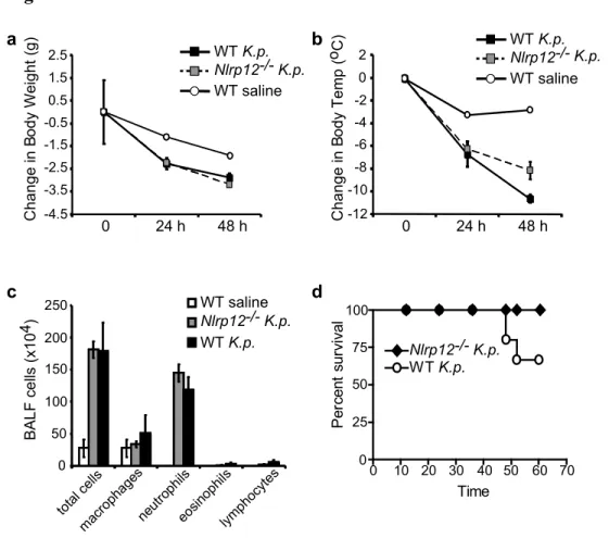
Figure 3.6. In vivo challenge of Nlrp12-/- mice with Klebsiella pneumoniae………...74
Figure 3.7. Analysis of BMDC cell surface markers on WT and Nlrp12-/- BMDC……...75
Figure 3.8. Antigen presentation assays comparing WT and Nlrp12-/- BMDC………76
Figure 3.9. Nlrp12-/- mice exhibit attenuated migration of peripheral dendritic cells to draining lymph nodes………...77
Figure 3.10. Cell surface expression of CCR7 & CXCR4 on WT and Nlrp12-/- BMDC…...78
Figure 3.11 Nlrp12-/- BMDCs exhibit attenuated migration toward lymph node homing chemokines………..79
Figure 3.12. Nlrp12-/- BMDCs display altered activation of ERK and noncanonical NF-κB following CCR7 activation………..80
Figure A1.1. NLRP12 suppresses noncanonical NF-κB activation………...102
Figure A1.3. The NBD and LRR domains of NLRP12 mediate NIK binding………..104
LIST OF ABBREVIATIONS
ASC: apoptosis-associated speck-like protein containing a CARD domain BAFF: B cell activating factor
BALF: Bronchoalveolar lavage fluid
BMDC: bone marrow derived dendritic cells C-terminal: carboxy-terminal
CAPS: Cryopyrin-Associated Periodic Syndromes CARD: activation and recruitment domain
CATERPILLER: caspase activation and recruitment domains [CARD], transcription enhancer, R [purine]-binding, lots of leucine repeats
CCL19: chemokine (C-C motif) ligand 19 CCL21: chemokine (C-C motif) ligand 21 CCL5: chemokine (C-C motif) ligand 5 CCR7: chemokine (C-C motif) receptor 7 CD40L: CD40 ligand
CFA: Complete Freund’s Adjuvant
CFSE: Carboxyfluorescein succinimidyl ester CHS: contact hypersensitivity
CIITA: MHC class II transcriptional activator
CINCA: Chronic infantile neurologic cutaneous articular syndrome COX-2: cyclooxygenase-2
CXCR4: chemokine (C-X-C motif) receptor 4 DC: Dendritic cell
DSS: dextran sodium sulfate
EAE: experimental autoimmune encephalitis ERK: extracellular signal-regulated kinase
FCAS: Familial cold auto-inflammatory syndrome FITC: fluorescein isothiocyanate
GA: Geldanamycin
H&E: hematoxylin and eosin Ha: hemagglutinin
Hsc70: heat shock cognate 70 Hsp70: heat shock protein 70 Hsp90: heat shock protein 90 i.p.: intraperitoneal
IFN-β: interferon-beta IKK: IκB kinase
IL-1β: interleukin 1 beta IκB: inhibitor of NF-κB
IKK: inhibitor of NF-κB kinase
IRAK1: interleukin-1 receptor-associated kinase IRF: interferon regulatory factor
LRR: leucine-rich repeats
LTβR: lymphotoxin beta receptor
MAPK: mitogen-activated protein kinase
MAVS: mitochondrial anti-viral signaling protein MDP: muramyl dipeptide
MOG: myelin oligodendrocyte glycoprotein MPO: myeloperoxidase
MSU: monosodium urate
MWS: Muckle-Wells Syndrome N-terminal: amino-terminal
NALP: NACHT domain-, leucine-rich repeat-, and pyrin- containing protein NBD: nucleotide binding domain
NF-κB: nuclear factor kappa B
NF-κB2: nuclear factor kappa B2, noncanonical NF-κB NIK: NF-κB inducing kinase
NLR: nucleotide binding domain leucine-rich repeat
NLRP12: nucleotide binding domain leucine-rich repeat containing a pyrin 12 NLS: nuclear localization signal
NOD: nucleotide oligomerization domain
NOMID: Neonatal-onset multisystem inflammatory disease Ova: Ovalbumin
PGD2: prostaglandin D2
PRR: pattern recognition receptor R protein: disease resistance protein RIG-I: retinoic acid inducible gene 1 RIP2: receptor-interacting protein kinase 2 RLH: RIG-like helicases
s.c.: subcutaneous
SDS-PAGE: sodium dodecyl sulfate polyacrylamide gel electrophoresis TAK1: transforming growth factor beta-activated kinase 1
TH1: T helper type 1
TH2: T helper type 2
TIR: Toll/IL-1β receptor TLR: Toll-like receptor
TNFα: tumor necrosis factor alpha WT: wildtype
CHAPTER 1: INTRODUCTION
Portions of section B, part 2 have been adapted from,
Lich JD, Arthur JC, and Ting JP. Cryopyrin: in from the cold. Immunity 24, 241-3 (2006). © 2006 Elsevier Inc.
A. INTRODUCTION
Vertebrates utilize the immune system to protect themselves against pathogens. Over the course of evolution, they have developed two systems of immune detection – the innate and adaptive immune systems. All vertebrates employ the innate immune system, however, only jawed vertebrates employ both innate and adaptive immune systems. Adaptive immune cells, such as T and B lymphocytes, can express nearly an unlimited number of antigen-specific receptors that are generated through somatic rearrangement. After activation
through these receptors, lymphocytes undergo clonal expansion to exponentially increase the number of antigen-specific lymphocytes. This generates effector cells capable of clearing the infection, as well as memory cells that are poised to react quickly upon secondary exposure to the same antigen. Activation and expansion of an adaptive immune response occurs over several days. In contrast, an innate immune response can be initiated within minutes to hours and shapes the ensuing adaptive immune response. Accordingly, the innate immune system is considered the first line of defense against pathogens.
Nearly two decades ago it was predicted that the immune system utilizes germline encoded receptors to rapidly detect and alert the host to invading pathogens. Janeway hypothesized that the immune system employs pattern-recognition receptors (PRR) to detect conserved microbial products, termed pathogen associated molecular patterns (PAMPs), and distinguish “self” from “non-self” 1. Matzinger hypothesized that invading pathogens cause the host to produce danger signals that alert the adaptive immune system to infection 2,3. Both theories have proven to be correct.
the first member identified in a family of eleven PRR called Toll-like receptors (TLR). TLRs are type I transmembrane glycoproteins that recognize pathogen products through their extracellular leucine-rich repeat (LRR) domain. Their cytosolic TIR (Toll/IL-1β Receptor) domain recruits cytosolic adaptor proteins including MyD88, TIRAP/Mal, TRAM, and TRIF to transduce downstream signals leading to activation of the NF-κB and mitogen-activated protein kinase (MAPK) pathways 5-7. This signaling induces the secretion of
pro-inflammatory cytokines and upregulation of costimulatory molecules important for shaping the ensuing adaptive immune response. All members of the TLR family recognize conserved microbial structures such as viral double stranded RNA 8, the Gram negative bacterial cell wall component lipopolysaccharide (LPS) 9-11, flagellin – a component of the bacterial motility apparatus flagellum 12, and unmethylated CpG motifs in bacterial DNA 13. TLRs localize to the plasma membrane or within endosomes, however, many bacteria and viruses invade the host cell’s cytosol. Thus it was believed that there similar molecules might reside in the intracellular and cytosolic compartments.
ancient family of immune defense genes. In addition, several members of this newly discovered family had previously been linked to human immune and autoinflammatory diseases 17 (see section A.3). In response to pathogen products and endogenous danger signals, NLR proteins trigger signaling pathways that can enhance or suppress immune responses, such as those mediated by the innate immune Toll-like receptors (TLRs)18. However, many questions remain regarding how NLR proteins function at the biochemical level and what physiologic response is evoked by their activation.
In this dissertation I discuss two major findings regarding the NLR protein NLRP12: an evolutionarily conserved mechanism that controls the stability and function of NLRP12, as well as an in vivo role for NLRP12 in bridging innate and adaptive immunity by
controlling dendritic cell migration.
1. Discovery of the NLR family
Within the past decade, we and others have discovered the NLR family as a large evolutionarily conserved gene family that serves an important role in innate and adaptive immunology 14. Many NLR family members with known function participate in the innate immune response, such as sensing pathogenic insult and regulating inflammatory signaling and cell death. In addition, several NLR gene products have recently been shown to affect adaptive immune responses. At least half of the NLR genes encode proteins with functions that remain elusive or not yet studied. For this reason it is exciting to consider what will be discovered in the near future within the field of NLR biology.
CIITA, the MHC class II transcriptional activator and founding member of the NLR gene family (see section B.1). Approximately 20 genes are present in humans, and all encode a putative central nucleotide binding domain and C-terminal leucine rich repeats (LRR). We named this family CATERPILLER, an acronym for caspase activation and recruitment domains [CARD], transcription enhancer, R [purine]-binding, lots of leucine repeats 15. Others have named this group or subgroups of these genes NOD (nucleotide oligomerization domain) 19, NOD-LRR 20, PYPAF 21, PAN 22 and NALP (NACHT domain-, leucine-rich repeat-, and pyrin- containing protein) 23. Recently a unifying nomenclature has been
adopted for the NLR family, designated “NLR”, as well as individual NLR family members. The gene symbol for each family member begins with “NLR” plus an additional letter
signifying the subfamily to which the individual NLR member belongs. The subfamily is based upon the N-terminal effector domain: NLRA, NLR family, acidic domain containing; NLRB, BIR domain containing; NLRC, CARD domain containing; NLRP, pyrin domain containing; NLRX, N terminal domain with no known homology. Within the subfamily, the individual NLRs are numbered sequentially, i.e. NLRP1, NLRP2, etc. CIITA, NAIP, NOD1 and NOD2 have retained their original names in addition to their new NLR designation 14. Please refer to Table 1.1.
2. NLR domain organization
interactions are utilized by NOD1/NLRC1 and NOD2/NLRC2 to bind receptor-interacting protein kinase 2 (RIP2) and elicit downstream NF-κB activation 24-26. Several NLRs utilize the N terminal pyrin domain in homotypic interactions with the adaptor ASC (apoptosis-associated speck-like protein containing a CARD domain) to activate caspase-1 27. The N terminal domain of several NLRs acts as a dominant negative when overexpressed (Lich, J.D. unpublished data), raising the possibility that unidentified splice variants may serve to negatively regulate the activity of these NLRs.
The C-terminus of NLR molecules is comprised of a varying number of leucine rich repeats (LRR), which are involved in autoregulation, ligand recognition, and protein-protein interactions. LRRs are defined by repeating units of LxxRxxL (‘x’ being any amino acid) and each unit is a structural motif of 20-30 amino acids forming a beta strand-turn-alpha helix 41,42. In both plant R proteins and mammalian NLRs, truncation of the LRRs can yield a constitutively active molecule, suggesting the LRRs maintain the NLR protein in an auto-inhibited state until an activating stimulus is received 43-46. Evidence of a direct interaction between the LRRs of NLR proteins and pathogens or pathogen products is sparse 32. In plants, however, yeast two-hybrid experiments have detected an interaction between the LRR-like region of Pi-ta, a rice R gene, and its cognate avirulence effector from the rice blast fungus Magnaporthe grisea 47. However, such interactions have only been demonstrated in artificial systems and not verified with endogenous protein. Thus further studies are required to determine whether or not NLRs are activated through direct ligand binding.
3. Associations with human disease
Of the CARD-containing NLRs, mutations in NOD1 and NOD2 are the best studied. Mutations in NOD2 have been linked to Crohn’s disease, an inflammatory disease of the intestines, and Blau’s syndrome, a familial granulomatous disease characterized by inflammation of the eyes, joints and skin 26,49,50. Mutations in NOD1 are associated with numerous inflammatory disorders such as inflammatory bowel disease (IBD), asthma, and sarcoidosis 51,52.
Several pyrin-containing NLRs have been linked to human inflammatory disorders. Mutations in NLRP3/Cryopyrin are associated with a group of dominantly inherited
autoinflammatory disorders that are referred to as cryopyrinopathies or Cryopyrin-Associated Periodic Syndromes (CAPS). CAPS is comprised of three syndromes, listed as least to most severe: Familial cold auto-inflammatory syndrome (FCAS)53, Muckle-Wells syndrome (MWS)53,54 and Neonatal-onset multisystem inflammatory disease (NOMID) / Chronic infantile neurologic cutaneous articular syndrome (CINCA)55,56. Symptoms of these syndromes include recurrant rash, fever/chills, joint pain, deafness, systemic amyloidosis, central nervous system inflammation, mental retardation, and bone deformities. Recently, mutations in the most closely related NLR to NLRP3, NLRP12, have been linked to
B. MECHANISM OF ACTION
NLR proteins organize and assemble into multi-protein complexes to assert their function(s). Based upon these functions, NLRs can be categorized into three groups. It should be noted, however, that many NLRs likely have overlapping functions and should only be loosely categorized into these three groups. CIITA is the sole member of the first group and serves as a transcriptional coactivator at the promoter of MHC class II genes (reviewed in 62). The second group contains NLRP1, NLRP3, NLRC4, and NAIP/NLRB1. These NLRs assemble into multi-protein complexes with ASC and caspase-1 to promote IL-1β processing and secretion (reviewed in 63). They also perform an important but less studied role in promoting distinct forms of cell death (reviewed in 64). The third and final group, the signaling NLRs, includes NOD1, NOD2, NLRX1 and NLRP12. These NLR proteins fine-tune inflammation by enhancing or suppressing distinct arms of inflammatory signaling pathways, such as those leading to interferon regulatory factor (IRF) and NF-κB 65-67.
1. CIITA, the MHC class II transactivator
The founding member of the NLR family, CIITA, controls constitutive and cytokine-induced activation of MHC class II genes. However, while CIITA drives expression of MHC class II genes, it does not directly bind DNA 48. Instead, it acts as a transcriptional
coactivator by organizing other proteins in the appropriate spatial orientation to interact with and induce transcription from MHC class II promoters 68. MHC class II promoter
multi-protein complex and recruit CIITA to MHC class II promoters, forming the MHC class II enhanceosome and promoting MHC class II transcription 76. While these transcription factors are constitutively expressed, transcription of MHC class II genes is not induced in the absence of CIITA 48. CIITA organizes and stabilizes this complex of transcription factors, chromatin modifiers (including Brahma-related gene 1 (BRG-1)) and the requisite
transcriptional machinery (histone acetyltransferases, CREB binding protein (CBP)/p300, CBP/p300 associated factor (pCAF), steroid receptor coactivator 1 (SRC-1), the TATA-binding protein (TBP), TATA associated factors), and transcriptional elongation factors necessary for MHC class II gene expression 62,77,78.
CIITA is encoded by MHC2TA, which was identified by complementation cloning of an MHC class II negative RJ 2.2.5 cell line 48. MHC2TA is expressed in a cell type and differentiation state specific manner that mirrors MHC class II expression. MHC2TA is epigenetically activated in response to IFNy through histone acetylation and chromatin remodeling 79,80 and epigenetically silenced through DNA hypermethylation 81. MHC2TA transcription is controlled by at least three different promoters based upon cell type 82.
Furthermore, deletion of 140 amino acids containing the GTP binding domain increases CIITA nuclear export and association with the nuclear export protein CRM1 89. GTP binding is also required for CIITA self-association 36,37 and activation of the MHC class II promoter
38-40
.
Post-translational modifications are important in modulating CIITA activity, as unmodified CIITA is not recruited to the MHC class II enhanceosome 90. CIITA activity is inhibited through phosphorylation by ERK1/2 91 and protein kinase A (PKA), such as upon prostaglandin E (PGE) treatment 92. Phosphorylation of CIITA may signal its nuclear export, as mutating these phosphorylation sites 93 or inhibiting ERK1/2 causes retention of CIITA in the nucleus and prevents association with and nuclear export via CRM1 91. Ubiquitination of CIITA, however, enhances the ability of CIITA to activate MHC class II gene expression 94.
2. Inflammasome-forming NLRs
Similar to the requirement for CIITA to form higher multiprotein structures to coordinate MHC class II expression , several other NLR proteins have also been shown to form protein complexes termed the inflammasome, which functions in inflammatory cytokine activation 95. Assembly of this molecular platform leads to processing and release of the potent pro-inflammatory cytokine, IL-1β, and related cytokines, IL-18 and IL-33. The inflammasome includes the core components ASC and caspase-1. In the absence of either of these core components, pro-IL-1β is not processed into its active form 95.
Most inflammasomes studied to date contain the core components ASC and caspase-1. However, the particular NLR involved in inflammasome formation appears to be
promotes IL-1β processing and secretion in response to muramyl dipeptide and anthrax lethal toxin of Bacillus anthracis 96. The NLRC4 inflammasome is formed in response to multiple Gram-negative bacteria expressing flagellin 97 while NLRP3 inflammasomes have been shown to respond to a variety of pathogen and host derived molecules. Recently an additional layer of complexity has been revealed by the discovery of an inflammasome complex consisting of both NOD2 and NLRP1. This heterogenous inflammasome was formed in response to B. anthracis and the bacterial cell wall component muramyl dipeptide (MDP) 98. Given the well defined role of NOD2 in NF-κB activation, it is tempting to speculate that this heterogeous inflammasome couples transcriptional activation of
inflammatory genes with IL-1β production. Further research will likely reveal the existence of other heterogenous inflammasomes and may explain the apparent overlap between certain elicitors and NLR proteins.
The NLRP3 inflammasome
The NLRP3 inflammasome is arguably the best studied among the NLR family. Therefore it provides a model for understanding the molecular mechanisms underlying inflammasome formation and activation leading to IL-1β and IL-18 production. A collection of dominantly inherited human autoinflammatory disorders are associated with mutations in NLRP3. Interestingly, symptoms are relieved by IL-1β neutralization, suggesting that excessive and improperly regulated NLRP3 inflammasome activation and downstream IL-1β production underly these disorders.
mediated predominantly by the IL-1β converting enzyme caspase-1. Treatment of
macrophages with LPS results in the production of high levels of pro-IL-1β that accumulate in secretory lysosomal structures 99. Yet, the release of mature IL-1β is very inefficient in the absence of a second signal. This second signal can be provided by ATP, which activates the ion-gated channel P2X7. This triggers the rapid activation of caspase-1 and subsequent processing and release of bioactive IL-1β 100. The role of P2X7 in IL-1β release has been well documented over the years. However, it was not until the discovery of NLRP3 that the molecular mechanisms began to be revealed.
NLRP3, formerly known as Cryopyrin, PYPAF1 or NALP3, is encoded by the NLRP3 gene, formerly known as CIAS1, and is a pyrin-containing member of the NLR family of genes 101. Hoffman et al. first identified point mutations within exon 3 of NLRP3 that segregate with Muckle-Wells Syndrome (MWS) and Familial Cold Autoinflammatory Syndrome (FCAS) 53, two inflammatory diseases characterized by fever, rash, and excessive IL-1β production. However, until recently, the role of NLRP3 in these disorders has
remained elusive. In 2006, four groups described an important role for NLRP3 in promoting IL-1β maturation, leading to the release of this potent pro-inflammatory cytokine 102-105.
Because R848 and R837 are purine analogs, it is possible that these compounds stimulate members of the P2X or P2Y family of purine receptors, similar to ATP stimulation. Alternatively, NLRP3 may “sense” these agonists within the cytoplasm in a manner
analogous to the recognition of the bacterial cell wall component muramyl dipeptide (MDP) by NOD2.
Further support for NLRP3 in bacterial recognition lies in the finding that NLRP3 is also required for IL-1β secretion in response to the Gram-positive bacteria Staphylococcus aureus and Listeria monocytogenes (Lm). NLRP3 displays a level of specificity, as it is not required for caspase-1 activation and IL-1β release in response to Salmonella typhimurium or Francisella tularensis 103. A previous report demonstrated that NLRC4, another member of the NLR family, is required for IL-1β release in response to Salmonella typhimurium 106. These findings support the general assumptions that distinct NLR family members respond to different stimuli.
The reports described above indicate a role for NLRP3 in caspase-1 activation and IL-1β maturation in response to a variety of stimuli. A question is undoubtedly raised regarding how NLRP3 can respond to such diverse stimuli with no shared molecular
permits vesicular import of the NLRP3 inflammasome, thus allowing it to colocalize with caspase-1 and pro-IL-1β. This supports the observation that inflammasome components are released from stimulated macrophages along with IL-1β. Interestingly, iPLA2 activity also leads to the production of cytoplasmic lipids such as lysophospholipids and arachidonic acid. This has prompted the suggestion that NLRP3 may respond to lipid second messengers generated by phospholipase activity 99.
In addition to the stimuli described above, NLRP3 mediates IL-1β maturation and secretion induced by endogenous danger signals and crystalline particles including asbestos
108
, silica 108-110, aluminum hydroxide 111-114, and fibrilar amyloid-β 115. The first of these found to activate the NLRP3 inflammasome are monosodium urate (MSU) and calcium pyrophosphate dehydrate (CPPD) crystals 104, the deposition of which lead to gout. In the case of MSU and CPPD, IL-1β maturation occurred in the presence of a P2X7 inhibitor, suggesting NLRP3 responds to signals other than those initiated from this ATP receptor. IL-1β processing and secretion was blocked by the microtubule inhibitor colchicine, suggesting that cytoskeletal events, such as endocytosis or vesicle trafficking, are required for NLRP3-mediated IL-1β release. This observation was further supported by the finding that
destabilization of the lysosomal membrane in response to phagocytosis of crystalline
3. Signaling NLRs
In addition to NLR proteins that comprise the inflammasome, a smaller but growing number of NLRs function in controlling pro- and anti-inflammatory signal transduction. Signaling NLRs include NOD1, NOD2, NLRX1, and NLRP12 (see section C for greater detail on NLRP12). These NLRs enhance or suppress inflammation by fine-tuning
inflammatory signaling pathways such as NF-κB, MAPK, and IRF3/7 65-67. By controlling these inflammatory pathways, signaling NLRs not only control the innate immune response, but also influence the ensuing adaptive immune response 117-120.
NOD1 and NOD2
Both NOD1 and NOD2 were identified before the discovery of the NLR gene family. NOD1 was cloned in 1999 by two groups interested in identifying CARD-containing proteins resembling the pro-apoptotic protein APAF-1 24,25. NOD2 was identified based upon its homology to NOD1 26. Their importance in human immunity was evident as mutations in these genes could be linked to Crohn’s disease, Blau syndrome, and inflammatory bowel disease 26,49,50. While NOD1 and NOD2 were identified based upon their homology to the pro-apoptotic protein APAF-1, it was soon found that NOD1 and NOD2 act as cytosolic molecular sensors that promote host resistance to a variety of bacteria by activating inflammatory signaling pathways.
moiety found in both Gram positive and negative bacteria, NOD1 responds to a moiety found predominantly in Gram negative bacteria. The minimal structure required to activate NOD1 is GlcNAc-MurACc-LAla-c-D-Glu-meso-diaminopimelic acid (GM-triDAP)121, whereas NOD2 responds to MDP and the minimal component GlcNAc-MurNAc-LAla-D-isoGlen (GM-Di)122,123. NOD1 and NOD2 clearly display a high level of specificity, however, neither have been shown to directly bind these bacterial products.
NOD1 and NOD2 activate multiple pro-inflammatory signaling pathways that result in the secretion of inflammatory cytokines and chemokines like IL-6, IL-8 and TNFα and antimicrobial peptides including defensins. Upon activation, NOD1 and NOD2 are recruited to the plasma membrane 124,125, self-oligomerize and recruit the CARD-containing kinase RIP2 through homotypic CARD interactions 126,127. RIP2 is activated by K63-ubiquitination
which recruits transforming growth factor-β-activated kinase 1 (TAK1) 128-130. Signaling can proceed through the mitogen activated protein kinases (MAPK) p38, JNK and ERK, as well as to NF-κB via ubiquitination of IKKγ/NEMO, the regulatory subunit of the IKK (inhibitor of κB (IκB) kinase) complex, phosphorylation of IκB, and nuclear translocation of NF-κB 26,131,132. NOD2-mediated NF-κB activation can be suppressed by A20 130, an enzyme with both ubiquitinating and de-ubiquitinating machinery that suppresses NF-κB downstream of pro-inflammatory receptors 133.
The importance of NOD1 in protecting the host from bacterial infection is supported by numerous in vitro and in vivo studies. NOD1 induces an inflammatory response by activating NF-κB upon stimulation with Bacillus species133, Shigella flexneri134,
enteroinvasive Escherichia coli 135, Listeria monocytogenes 127, and Campylobacter jejuni
136
Helicobacter pylori, but not to infection with nonpathogenic cagPAI-negative Helicobacter
137
. This finding suggests that NOD1 may participate in distinguishing between commensal and noncommensal flora in the gut, where NOD1 is highly expressed in intestinal epithelial cells. NOD1 can also drive antigen-specific T cell responses and antibody responses, but the mechanism involved remains unknown 119.
NOD2 typically activates NF-κB and promotes an inflammatory response upon recognition of pathogens, some of which include Streptococcus pneumoniae138,
Mycobacterium tuberculosis139, Staphylococcus aureus140, and Listeria monocytogenes (Lm)117. Interestingly, NOD2 may also suppress inflammatory pathways under certain conditions. NOD2 suppresses NF-κB activation when stimulated in concert with TLR2, an extracellular TLR that like NOD2, recognizes peptidoglycan. Accordingly, IL-12 secretion is reduced, as is the ensuing TH1 response118. Perhaps NOD2 cooperates with TLR2 to
promote a TH2 response and enhanced clearance of extracellular bacteria. Other groups,
however, have found a synergism upon co-stimulation of NOD2 and TLRs 141-143. Thus in vivo studies utilizing whole bacteria may provide a clearer view in characterizing the role of NOD2 in host defense.
intestinal defensin expression 145. It is not fully understood whether or not decreased defensin expression contributes to the pathology of Crohn’s disease, or how mutations in NOD2 contribute to the pathology of Crohn’s disease.
NLRX1
NLRX1 is a newly characterized NLR that suppresses anti-viral signaling. Intracellular viral RNA is sensed by RIG-like helicases (RLH) that associate with
mitochondrial anti-viral signaling protein (MAVS) to activate IRF3 and NF-κB. This results in the production of type 1 interferon and inflammatory cytokines that are essential for anti-viral defenses, but when dysregulated can cause excess inflammation and tissue damage in the host 146.
NLRX1 was identified as a member of the NLR family in 2003 15, however, its physiologic role remained uncharacterized until 2008 147. NLRX1 is ubiquitously expressed in human cells and cell lines and encodes an unclassified N-terminal domain, central
nucleotide binding domain, and C-terminal leucine-rich repeats. It contains a mitochondrial targeting sequence in the N-terminus that targets it to the mitochondrial outer membrane147. NLRX1 is the first NLR, with the exception of CIITA, that requires localization to a
particular organelle.
response is independent of the extracellular viral RNA sensor TLR3 and requires
C. NLRP12 / MONARCH-1
1. NLRP12 identification and expression
NLRP12, formerly named RNO, PYPAF7, and Monarch-1, is a pyrin-containing NLR protein expressed in cells of myeloid lineage. A partial 3’ portion of the gene encoding NLRP12 was first identified in the HL60 human leukemic cell line. This gene was
upregulated when these cells were stimulated with nitric oxide, thus it was first named rno – Regulated by Nitric Oxide 149. The full-length gene product was subsequently cloned two groups: Our group named this gene Monarch-1 150 and the other group named it PYPAF7 151. In 2008, the HUGO Gene Nomenclature Committee approved the designation NLRP12 for this gene14.
NLRP12 encodes an intracellular protein with an N-terminal pyrin domain, a central nucleotide binding domain, and C-terminal leucine-rich repeats. The full-length human cDNA has a 3189-bp open reading frame (accession no. AY116204) encoded by 10 exons. There are also four known splice forms of NLRP12 150, however it remains unknown if these splice forms are differentially expressed and/or serve different functions from the full-length product. In humans, NLRP12 is expressed exclusively in cells of myeloid lineage –
granulocytes including neutrophils and eosinophils, monocytes/macrophages, and immature dendritic cells 149-151. NLRP12 expression is upregulated by nitric oxide, yet it is
downregulated in response to pathogens, pathogen products, and inflammatory cytokines
149,150,152
. Downregulation of NLRP12 after TLR stimulation is achieved, at least in part, by binding of B lymphocyte-induced maturationprotein-1 (Blimp-1) to the NLRP12 promoter
153
2. NLRP12 as a negative regulator of inflammation
The expression of NLRP12 is restricted to immune cells and its expression is downregulated in response to pathogens, pathogen products, and inflammatory cytokines, thus we and others predicted that NLRP12 functions in regulating inflammation and immunity. Early studies, however, describe conflicting roles for NLRP12. One study
describes that NLRP12 co-localizes with ASC and activates NF-κB and caspase-1, leading to
IL-1β secretion 151. This is reminiscent of other pyrin-containing NLRs that regulate IL-1β processing by forming an inflammasome with ASC. Another report describes that NLRP12 can control expression of classical and non-classical MHC I genes 150. Again, this is
reminiscent of CIITA, an NLR that is essential for the expression of MHC II genes. However both studies relied upon overexpression in non-immune cells. Thus they provide little information about the function of NLRP12 in cells that naturally express the gene product. They do, however, imply a complex role for NLRP12.
3. Molecular mechanisms of NLRP12-mediated NF-κB suppression
Biochemical studies in our lab have revealed that NLRP12 suppresses
pro-inflammatory cytokine and chemokine production downstream of TLRs by targeting multiple points in the NF-κB pathway 152,154. Stimulation through TLRs leads to the recruitment of cytoplasmic adaptor proteins, such as MyD88, that then recruit the kinase IRAK1. IRAK1 becomes activated through autophosphorylation and accumulates in a hyperphosphorylated form, leading to downstream NF-κB activation 155,156. Exogenous expression of NLRP12 reduces IRAK1-induced activation of an NF-κB luciferase reporter plasmid, suggesting that NLRP12 may intersect the NF-κB pathway by affecting IRAK1 signaling. Indeed, minutes after TLR stimulation, NLRP12 7associates with IRAK1 and prevents the accumulation of hyperphosphorylated IRAK1 152. Current literature support that the loss of
hyperphosphorylated IRAK1 would hinder downstream signaling leading to NF-κB 157. As NLRP12 expression declines upon TLR stimulation but returns within 24 hours in THP-1 monocytes (Arthur, J.C. unpublished data), this suggests a possible role for NLRP12 in the resolution of inflammation and may even play a role in the transition from innate to adaptive responses.
4. Canonical and noncanonical pathways of NF-κB activation
NF-κB represents a family of dimeric transcription factors that mediates cellular responses during inflammatory conditions. NF-κB subunits include RelA (also known as p65), RelB, c-Rel, NF-κB1 (p105/p50) and NF-κB2 (p100/p52) 160. The latter two are expressed as large precursors that must be proteolytically cleaved to their corresponding smaller and active forms 161,162.
NF-κB activation occurs through two distinct pathways, referred to as canonical and noncanonical (Figure 1.1). The canonical pathway proceeds rapidly upon activation of pro-inflammatory receptors, such as TLRs. In this pathway, RelA/p50 heterodimers are
sequestered in the cytoplasm in an inactive state by a family of inhibitors of NF-κB (IκB). Activation is mediated through a large IκB kinase (IKK) complex comprised of the
regulatory subunit IKKγ/NEMO, and two catalytic subunits, IKKα and IKKβ. This complex can be activated by a wide range of upstream kinases and serves to phosphorylate IκB, leading to its degradation. Newly liberated RelA/p50 heterodimers then rapidly translocate to the nucleus to regulate the activation of early inflammatory genes. One of these genes is NF-κB2/p100, whose gene product must be processed to its active form p52 through the noncanonical pathway 163-165.
In contrast to the canonical pathway, the noncanonical NF-κB pathway displays slower kinetics and tighter regulation. It is commonly activated downstream of TNF receptor superfamily members such as BAFF, LTβR and CD40 158,159. While initial canonical NF-κB activation NF-κB1/p105 processing to p50 is constitutive, processing of NF-κB2/p100 to p52 is inducible and relies upon the activity of NF-κB inducing kinase, NIK 166,167. In this
subsequent processing of p100 to its active form p52. Nuclear translocation of RelB/p52 dimers results in the activation of a different set of inflammatory genes that support the ongoing immune response 168-170.
5. NLRP12 targets NIK to control the noncanonical NF-κB pathway
Detailed analysis of the canonical and noncanonical NF-κB pathways in THP-1 cells has revealed that while elevated expression of NLRP12 moderately suppresses the canonical NF-κB pathway, NLRP12 nearly abolishes activation of the noncanonical NF-κB pathway. In THP-1 cells expressing elevated levels of NLRP12 and stimulated with TLR agonist followed by CD40L, nuclear translocation of the canonical NF-κB subunits RelA and p50 proceeds normally. However, processing of NF-kB2/p100 to p52 and nuclear translocation of p52 is nearly absent in these cells. To exert this effect, NLRP12 targets NIK, the sole kinase responsible for activation of the noncanonical pathway. NLRP12 associates with NIK and induces its degradation via the proteasome, a major protein degradation pathway 154. Accordingly, in NLRP12-silenced cells, NIK and p52 levels are elevated (Lich, J.D., unpublished data). This results in increased expression p52-regulated cytokines and chemokines including CXCR4, CXCL12 and CXCL13 154 (see Appendix 1).
6. Conclusions
(Figure 1.1). Consequently, NF-kB2/p100 to p52 processing is abolished, and p52-regulated genes are suppressed until NLRP12 expression fades. The noncanonical NF-κB pathway is triggered later than the canonical NF-κB pathway, and often in response to a second signal through TNF receptor superfamily members. In this manner, the noncanonical pathway drives later events in innate immunity. In addition, in antigen presenting cells like
monocytes/macrophages and dendritic cells, activation of noncanonical NF-κB may come from the interaction with cell-surface molecules on T cells, indicating that this alternative pathway is also intimately involved in the transition from innate to adaptive immunity.
Based on these statements, two key hypotheses are raised.
1). In order for the noncanonical pathway to operate, NLRP12 must be tightly regulated in a manner that allows NIK to function. We hypothesize that NLRP12 activity is controlled at the level of protein stability.
2). Because NLRP12 is expressed in antigen presenting cells, we hypothesize that NLRP12 affects adaptive immune responses in vivo.
In this dissertation we provide support for these two hypotheses. First we provide a mechanism by which the stability of the NLRP12 protein is regulated in human monocytes. This is through an evolutionarily conserved mechanism involving the chaperone Hsp90. Second, we reveal that NLRP12 affects immunity in vivo by controlling dendritic cell
Table 1.1. Human NLR family members
NLR name Alternative names Protein accession
CIITA NLRA, MHC2TA, C2TA NP_000237
NAIP NLRB1, BIRC1, CLR5.1 NP_004527
NOD1 NLRC1, CARD4, CLR7.1 NP_006083
NOD2 NLRC2, CARD15, CD, BLAU, IBD1, PSORAS1, CLR16.3 NP_071445
NLRC3 NOD3, CLR16.2 NP_849172
NLRC4 IPAF, CARD12, CLAN, CLR2.1 NP_067032
NLRC5 NOD27, CLR16.1 NP_115582
NLRP1 NALP1, DEFCAP, CARD7, CLR17.1 NP_127497
NLRP2 NALP2, PYPAF2, NBS1, PAN1, CLR19.9 NP_060322
NLRP3 CIAS1, PYPAF1, NALP3, CLR1.1, Cryopyrin NP_004886
NLRP4 NALP4, PYPAF4, PAN2, RNH2, CLR19.5 NP_604393
NLRP5 NALP5, PYPAF8, MATER, PAN11, CLR19.8 NP_703148
NLRP6 NALP6, PYPAF5, PAN3, CLR11.4 NP_612202
NLRP7 NALP7, PYPAF3, NOD12, PAN7, CLR19.4 NP_996611
NLRP8 NALP8, PAN4, NOD16, CLR19.2 NP_789781
NLRP9 NALP9, NOD6, PAN12, CLR19.1 NP_789790
NLRP10 NALP10, PAN5, NOD8, PYNOD, CLR11.1 NP_789791
NLRP11 NALP11, PYPAF6, NOD17, PAN10, CLR19.6 NP_659444
NLRP12 NALP12, PYPAF7, RNO2, PAN6, CLR19.3, Monarch1 NP_653288
NLRP13 NALP13, NOD14, PAN13, CLR19.7 NP_789780
NLRP14 NALP14, NOD5, PAN8, CLR11.2 NP_789792
Figure 1.1. NLRP12 suppresses noncanonical NF-κB activation
Proteasome degradation
RelB RelB TRAFs
TRAFs
MAP3K
MAP3K NIKNIK
p50 p50 p65
p65 p100p100
p50 p50 p65 p65
p50 p50 p65 p65
"Canonical pathway” "Noncanonical pathway”
RelB RelB p52 p52 P
P
NLRP12
p100 p100 IkBa
IkBa P P
CHAPTER 2: HSP90 ASSOCIATES WITH MONARCH-1/NLRP12 AND REGULATES ITS ABILITY TO PROMOTE DEGRADATION OF NF-κκκκB
INDUCING KINASE
This research was originally published in the Journal of Immunology.
Arthur JC, Lich JD, Aziz RK, Kotb M, and Ting JP. Hsp90 associates with Monarch-1 and regulates its ability to promote degradation of NF-κB inducing kinase.
J Immunol 179, 6291-6 (2007).
A. ABSTRACT
Monarch-1/NLRP12, is expressed in myeloid cells and functions as a negative
regulator of inflammation by inducing proteasome mediated degradation of NF-κB inducing
B. INTRODUCTION
Inflammation is a dynamic protective response that must be controlled at both the initiation and resolution phase as improper regulation underlies many human diseases. Nucleotide Binding Domain- Leucine Rich Repeat (NLR) proteins (previously known as CATERPILLER, NOD-LRR or NACHT-LRR) play a critical role in this regulation
19,23,67,101
. The importance of NLR proteins in controlling inflammation is highlighted by strong linkage of the NLR proteins CIITA, NOD2, and NALP3 to human immunodeficiency and autoinflammatory diseases 171. Yet despite the critical role of NLR proteins in human health, relatively little is known regarding their molecular regulation.
NLR proteins share a strong structural and functional homology to a class of disease resistance (R) proteins that comprise a major immune response system in the plant kingdom. These plant proteins function as molecular sensors that mediate the containment of a broad range of pathogens including bacteria, viruses, fungi, parasites, nematodes, and insects 16,172. Recent evidence suggests that a critical component in R protein mediated defense responses is Hsp90. This evolutionarily conserved molecular chaperone associates with a subset of proteins, deemed “client proteins,” to promote their maturation and stability
The Hsp90 chaperone cycle is a multi-step process where client proteins first form an early complex with Hsp70. An intermediate complex then forms with the incorporation of Hsp90. Within this intermediate complex, the client protein is transferred from Hsp70 to Hsp90. Finally, Hsp70 dissociates from the complex and the client protein remains bound to Hsp90 in an activation-competent state (reviewed in 173). Pharmacologic inhibition of Hsp90 prevents the transfer of client proteins to Hsp90 and stalls this chaperone cycle at the
Hsp70 and is degraded by the proteasome 174-176. Notably, the majority of known Hsp90 client proteins are signaling molecules such as kinases and transcription factors (reviewed in
177
).
NLRP12 is an understudied NLR protein with a unique function. Previously, we demonstrated that NLRP12 serves as a novel attenuating factor of inflammation by
destabilizing NIK, which results in suppression of noncanonical NF-κB activation 178. In the
C. MATERIALS AND METHODS
Cell lines: HEK293T cells (GenHunter) were maintained in DMEM (Gibco) supplemented with 10% fetal calf serum (FCS) and 100mg/ml penicillin and 100mg/ml streptomycin. Undifferentiated THP-1 cells were maintained in RPMI (Gibco) supplemented with 10% FCS, 1mM sodium pyruvate, 0.1mM nonessential amino acids, 100mg/ml
penicillin and 100mg/ml streptomycin. THP-Ha-NLRP12 and THP-EV cells have been previously described 178.
Primary cell isolation: Peripheral blood mononuclear cells (PBMC) were isolated from whole blood (American Red Cross) using a ficol hypaque gradient (ICN-Cappel). To enrich human primary adherent monocytes, PBMC were plated in serum-free RPMI (Gibco) and allowed to adhere for 2 hours at 37°C; at this time non-adherent cells were removed and adherent cells were washed with sterile PBS in triplicate. Cells were cultured in RPMI supplemented as described above.
Two-dimensional gel electrophoresis and mass spectrometry: HEK293T cells seeded in 10 cm plates were transfected with 5 µg Flag-tagged NLRP12 or pcDNA control vector. Twenty-four hours later the cells were lysed in 0.5% CHAPS and protein complexes
immunoprecipitated for 18 hours with M2-agarose. Protein complexes were dissociated in an 8M urea solution and individual proteins separated by two-dimensional gel
electrophoresis. Briefly, protein eluate was loaded on18-cm immobilized pH gradient strips (pH 4–7) and separated by pI for a total of 58,000 V-h. The strips were transferred to SDS-PAGE gels (10%; 19 x 18 cm) and the proteins were then separated by molecular weight. Silver stained gels were analyzed and spots unique to precipitates derived from NLRP12 transfected cells were excised from the gel and analyzed by MALDI-MS as described
previously 180. Protein identities were established using the MASOCT search engine (Matrix Sciences) with the following settings: peptide mass tolerance of 0.1 Da, zero missed cleavage sites, one fixed modification of carboxymethyl cysteine, one variable modification of
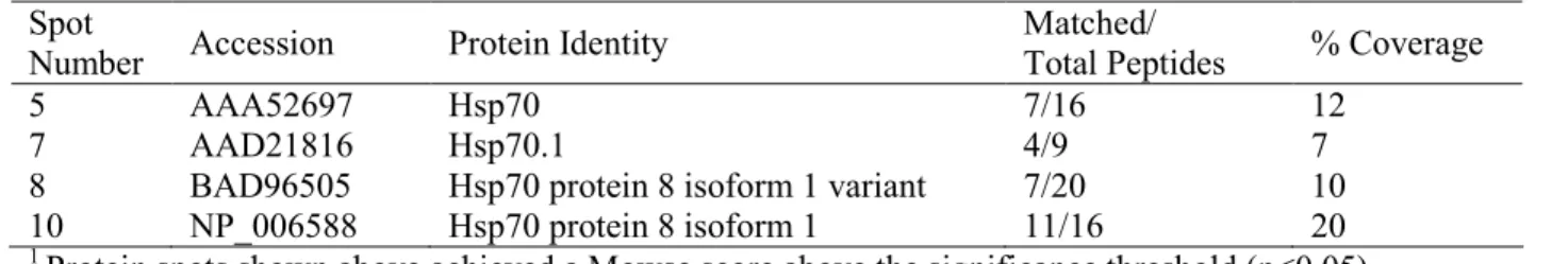
methionine oxidation, and no restrictions on protein molecular weight or pI. The protein identities reported received a Mowse score greater than the significance threshold (p<0.05).
Immunoprecipitations and Western Blot analysis: HEK293T cells seeded in 6-well plates were transfected with 1µg Ha-tagged NLRP12 plasmid described previously using Fugene 6 (Roche). Twenty hours later the cells were lysed in 1% TX-100, 150mM NaCl, 50mM Tris pH 8 supplemented with protease inhibitor cocktail (Roche). Samples were immunoprecipitated with 2µg of the indicated antibody and rotated end-over-end for 18 h.
nitrocellulose (BioRad), probed with the indicated primary antibody and visualized by chemiluminescence (Pierce).
To examine stably expressed proteins in THP-1 cells, THP-Ha-NLRP12 and THP-EV cells were seeded at a density of 8x10⁶ per 25cm2 tissue culture flask. To examine
endogenous NLRP12 protein, THP-1 cells were seeded at a density of 2x10⁷ in 75cm2 tissue culture flasks; primary adherent monocytes were seeded at 1x10⁷ in 25cm2 flasks. Samples
D. RESULTS
NLRP12 interacting proteins include Hsp70.
NLR proteins assemble into large, multi-protein complexes that serve as functional platforms to promote downstream activities such as transcription regulation and IL-1β
processing 101. To begin to identify the protein complexes formed by NLRP12, HEK293T cells were transfected with Flag-NLRP12 or a pcDNA control vector and cellular lysates were immunoprecipitated with anti-Flag antibodies. Captured protein complexes were resolved by two-dimensional gel electrophoresis. The gels were stained and protein spots unique to NLRP12 transfected cells were identified (Figure 2.1A). These spots were excised and processed for MALDI-MS (Table 2.1). Protein identities were determined by comparing the resulting peptide mass fingerprints to the MASCOT search engine. Among the proteins that achieved high confidence scores were members of the Hsp70 family.
Mammalian Hsc/Hsp70 binds to a wide range of newly synthesized proteins in unstressed cells 181. To confirm the association of NLRP12 with Hsp70, we performed co-immunoprecipitation experiments. HEK293T cells were transfected with Ha-NLRP12 or pcDNA control vector, and endogenous Hsp70 complexes were immunoprecipitated from cellular lysates. Western blots were then probed with anti-Ha to detect NLRP12 (Figure 2.1B, lanes 1-2). In agreement with the results obtained from two-dimensional gels (Figure 2.1A), NLRP12 co-precipitated with endogenous Hsp70. Identical results were obtained from THP-1 monocytes stably transfected with Ha-tagged NLRP12 (THP-Ha-NLRP12) (Figure 2.1B, lanes 3-4). As NLRP12 is expressed exclusively in cells of myeloid lineage
150,151
2.1B, lane 5).
NLRP12 associates with endogenous Hsp90.
The molecular chaperone Hsp70 is an essential component of the Hsp90 multi-chaperone complex. This complex aids in the maturation and stabilization of a select set of client proteins, predominantely signaling molecules 177. We identified Hsp70 as a NLRP12-interacting protein, and combined with the important role of Hsp90 in plant R protein
function, we hypothesized that NLRP12 also associates with Hsp90. To test this hypothesis, we transfected HEK293T cells with Ha-NLRP12 or pcDNA control vector and endogenous Hsp90 containing complexes were immunoprecipitated. Western blots were then probed with anti-Ha to detect NLRP12. NLRP12 co-precipitated with endogenous Hsp90 but not in control samples employing an isotype matched antibody (Figure 2.2). Importantly, this association was also found in THP-Ha-NLRP12 monocytic cells, demonstrating that NLRP12 forms molecular complexes with Hsp90 in a more relevant model system (Figure 2.2, lanes 3-4).
Hsp90 inhibition alters the association of NLRP12 with Hsp70 and Hsp90.
Pharmacologic inhibition of Hsp90 prevents the transfer of client proteins to Hsp90. This then leads to the accumulative association of client proteins within Hsp70 complexes
173
. The observation that NLRP12 associated with both Hsp70 and Hsp90 led us to
To test this, we treated THP-Ha-NLRP12 cells with GA and then immunoprecipitated endogenous Hsp70 or Hsp90 complexes at multiple time points over a 6 h period (Figure 2.3A). Western blots were probed with anti-Ha to detect co-precipitated NLRP12. After 2 h of GA treatment, NLRP12 was barely detectable in Hsp90-containing complexes. Notably, NLRP12 levels decreased with GA treatment, suggesting that the stability of NLRP12 is dependent upon Hsp90 activity. However, since NLRP12 levels were comparable at 1 h and 6 h post-GA treatment (lanes 2 and 5) the loss of NLRP12/Hsp90 complex formation was due to Hsp90 inhibition and not due to decreased levels of NLRP12 protein.
In sharp contrast to its association with Hsp90, the association of NLRP12 with Hsp70 increased after 2 h of GA treatment and strengthened throughout the 6 h time course (Figure 2.3B). This agrees with other studies where GA treatment causes an increase in association of the Hsp90 client protein with Hsp70 182-184. Also in line with previous reports
185-188
, a minimal increase in Hsp70 levels was detected upon inhibition of Hsp90. These results indicate that Hsp90 inhibition leads to the accumulation of NLRP12 within Hsp70 containing molecular complexes and supports our hypothesis that NLRP12 is processed through the Hsp90 chaperone cycle.
In addition to Hsp70, we also analyzed the association between NLRP12 and Hsc70. In contrast to Hsp70, which is induced upon cell stress, Hsc70 is constitutively expressed and performs multiple functions distinct from Hsp70 189. Also in contrast to Hsp70, the
association between NLRP12 and Hsc70 is dependent upon Hsp90 activity. Taken together, these results demonstrate that NLRP12 is a substrate of the Hsp90 multi-chaperone complex.
Endogenous NLRP12 stability in THP-1 cells and primary monocytes is dependent upon Hsp90 activity.
In addition to altering the dynamic association of client proteins with Hsp70 and Hsp90, inhibition of Hsp90 also results in degradation of the client protein 173. Consistently, Western blot analysis of cellular lysates indicated that NLRP12 levels decreased upon Hsp90 inhibition. In fact, in THP-Ha-NLRP12 cells, NLRP12 protein levels were dramatically reduced after only 1 h of treatment with GA (Figure 2.3). This was also observed upon treatment of THP-Ha-NLRP12 cells with Radicicol, an Hsp90 inhibitor that is structurally dissimilar and chemically unrelated to GA (Figure 2.4A) 190.
To ensure that the effect of Hsp90 inhibition on NLRP12 levels was not due to overexpression of a tagged protein, we next analyzed endogenous NLRP12 levels in wild type THP-1 monocytes. In agreement with the results obtained with tagged NLRP12, endogenous NLRP12 protein levels were significantly reduced after 1 h of treatment with GA (Figure 2.4B), demonstrating that NLRP12 stability is regulated by Hsp90.
h of treatment. Control samples using normal rabbit serum in the immunoprecipitation confirmed the specificity of the NLRP12 band. Together these data demonstrate that in human monocytes, NLRP12 stability is dependent upon Hsp90 activity.
Hsp90 inhibition results in proteasome-mediated degradation of NLRP12. It is generally believed that Hsp90 inhibition leads to proteasome mediated degradation of Hsp90 client proteins 174-176. To determine if the reduction of NLRP12 protein levels upon GA treatment was due to proteasome mediated degradation, we treated THP-Ha-NLRP12 cells with GA in the presence or absence of the proteasome inhibitor, MG132. In agreement with the results presented above, NLRP12 protein levels declined after 1 h of treatment with GA. In contrast, however, NLRP12 protein levels remained stable in cells that were pre-treated with proteasome inhibitor (Figure 2.5). No change was
observed in the cellular levels of TAK1, demonstrating that these treatments were not globally affecting signaling molecules. In addition, no change was observed in cellular levels of Hsp90 or actin under these treatment conditions. Together, these results
demonstrate that Hsp90 controls NLRP12 stability and upon Hsp90 inhibition, NLRP12 is degraded via the proteasome.
Hsp90 regulates NLRP12-induced NIK degradation.
Recently, we demonstrated that NLRP12 suppresses the production of
role of Hsp90 in NLRP12-induced NIK degradation, NIK and NLRP12 were co-expressed in HEK293T cells in the presence or absence of GA. NIK levels were then monitored by Western blot analysis. In cells expressing NIK alone, inhibition of Hsp90 caused a slight reduction in NIK levels (Figure 2.6A, lane 2). This agrees with an earlier report indicating that NIK is an Hsp90 client protein and NIK levels are reduced upon Hsp90 inhibition 191. In agreement with our previous report, co-expression of NLRP12 and NIK resulted in the near ablation of NIK protein (Figure 2.6A, lane 3). However, GA treatment restored NIK levels to those observed in cells treated with GA in the absence of NLRP12 (Figure 2.6A, compare lanes 2 and 4), thus demonstrating that Hsp90 activity is required for NLRP12-induced NIK degradation.
E. DISCUSSION
NLR proteins are rapidly emerging as important mediators of innate and adaptive immune signaling. Yet, despite recent reports describing the physiologic role of NOD2, NALP3, and IPAF in the response to numerous ligands, relatively little is known concerning the molecular events that regulate these proteins. Recently, we demonstrated that the NLR protein, NLRP12, functions as a negative regulator of NF-κB activation through its
association with NIK 178. In this report we show NLRP12 requires Hsp90 for both its stabilization as well as its negative regulatory activity.
Hsp90 is a highly conserved chaperone molecule that plays a critical role in the stability and function of many signaling proteins. These client proteins generally follow a pathway where upon translation they associate with Hsp70 to achieve proper folding conformation. The client protein is then transferred to Hsp90 where it is held in a
functionally active state. Upon the addition of Hsp90 inhibitors, the client protein no longer associates with Hsp90, but instead remains in a complex with Hsp70 and undergoes
proteasome-mediated degradation 177. In this study, we show that NLRP12 follows the same mechanism as reported Hsp90 client proteins. We found that upon Hsp90 inhibition with GA, NLRP12 proteins dissociated from Hsp90 and, simultaneously, accumulated within Hsp70 complexes. As a result, NLRP12 protein levels rapidly decreased due to degradation via the proteasome.
Nicotiana benthamiana, virus-induced gene silencing of Hsp90 results in the loss of resistance mediated by R proteins PRF (against P. syringae), RX (against potato virus X), and N (against tobacco mosaic virus) 195,196. Thus, Hsp90 is critical for disease resistance in plants as this chaperone regulates both the stability and function of multiple R proteins.
Similar to these R proteins, in this report we demonstrate that, in addition to
regulating NLRP12 stability, Hsp90 also controls NLRP12 function. Although NLRP12 still bound NIK, it no longer induced NIK degradation in the presence of an Hsp90 inhibitor. Thus, while Hsp90 is required for the negative regulatory function of NLRP12, it is not required for NLRP12 to form molecular complexes with NIK. A similar observation has been made for the Hsp90 client protein, Raf. In this report, inhibition of Hsp90 reduced cytoplasmic Raf levels but did not prevent Raf from binding downstream signaling proteins
197
. These results suggest that Hsp90 activity is required for the processing steps that function downstream of NLRP12-NIK complex formation to promote degradation of the kinase. Future studies will elucidate if Hsp90 regulates the association if NLRP12-NIK complexes with ubiquitin conjugating enzymes and/or the proteasome degradation complex.
In summary, in this report we utilized an unbiased proteomic approach to show that NLRP12 associates with Hsp70, and a further investigation shows that it also associates with Hsp90. These two heat shock proteins regulate protein stability, and Hsp90 in particular is required for the stability of numerous inflammatory signaling molecules 177,191. The effect of Hsp90 on NLRP12 was studied using specific Hsp90 inhibitors, and was observed not only in experiments investigating NLRP12 protein derived from transfected expression plasmids, but also with endogenous NLRP12 protein in both a monocytic cell line and primary human monocytes. Our results indicate that Hsp90 protects NLRP12 from proteasome-mediated degradation, and further regulates the function of NLRP12. These findings suggest an evolutionary conserved regulation of mammalian NLR proteins by Hsp90 that is strikingly similar to the regulation of plant R proteins.
Table 2.1 Summary of Hsp70 proteins identified by MALDI-TOF MS
Spot
Number Accession Protein Identity
Matched/
Total Peptides % Coverage
5 AAA52697 Hsp70 7/16 12
7 AAD21816 Hsp70.1 4/9 7
8 BAD96505 Hsp70 protein 8 isoform 1 variant 7/20 10
10 NP_006588 Hsp70 protein 8 isoform 1 11/16 20
1
Protein spots shown above achieved a Mowse score above the significance threshold (p<0.05)
Figure 2.1
Figure 2.2
Figure 2.3
Figure 2.4
Figure 2.5
Figure 2.6
CHAPTER 3: NLRP12 CONTROLS ADAPTIVE IMMUNE RESPONSES BY REGULATING DENDRITIC CELL MIGRATION
This research was originally submitted to Nature in November of 2008.
It was submitted to Science in a revised form in April of 2008 and is currently under review. Arthur JC, Lich JD, Ye Z, Allen IC, Gris D, Schneider M, O’Connor BP, Moore CB, Morrison A, Sutterwala FS, Bertin J, Liu Z, and Ting JP. NLRP12 controls adaptive immune
A. ABSTRACT
Long-term immuno-reactivity is a salient feature of protective immunity as well as destructive autoimmune and hyperinflammatory responses. The establishment of this reactivity relies largely on the ability of dendritic cells (DCs) to orchestrate innate and adaptive immune pathways 200,201. Under homeostatic conditions, these cells survey peripheral tissues, collect local antigens, and then migrate to secondary lymphoid organs. Under inflammatory conditions the migration to draining lymph nodes intensifies and these cells become highly potent activators of antigen specific T cells 200, 202. In this study, we described an unexpected role for a nucleotide-binding domain leucine rich repeat (NLR) protein, NLRP12 (formerly Monarch-1154), an NLR linked to atopic dermatitis58 and
hereditary periodic fever57. We show that Nlrp12 is expressed in DCs and is required for the adaptive immune response to hapten-induced contact hypersensitivity, a model of atopic dermatitis, and experimental autoimmune encephalitis (EAE), a model of multiple sclerosis. The inability of Nlrp12-/- cells to induce adaptive immunity is not due to failure in antigen
B. INTRODUCTION
NLRs constitute a large gene family of 20-30 members that are preserved from plants to humans. NLR proteins have rapidly emerged as important components of inflammation and immunity. To date, at least five NLR proteins are predicted to form a large protein complex termed the inflammasome 95. This complex functions in caspase-1 activation and
subsequent release of bioactive IL-1β and related cytokines. Other NLR proteins have been
shown to function as regulators of inflammatory signals such as NF-κB 118,147,203. We
C. MATERIALS AND METHODS
Mice: An Nlrp12 targeting vector was electroporated into 129SvEvBrd Lex-1 embryonic stem (ES) cells and homologous recombinant ES cells were microinjected into C57BL/6 blastocysts. Chimeras were backcrossed to C57BL/6 mice (Jackson Laboratories) for 9 generations and PCR of genomic tail DNA confirmed germline transmission. Nlrp3 -/-mice204 were also backcrossed onto C57BL/6 for 9 generations. OT-II mice, which express the OVA323–339-specific TCR transgene on the C57BL/6 background,were obtained from M.
Croft (La Jolla Institute of Allergyand Immunology). All mice were maintained in specific pathogen free housing at the University of North Carolina (UNC) at Chapel Hill. All
experiments were performed with age- and sex- matched mice between 6-12 weeks old. All studies were conductedin accordance with the National Institutes of Health Guide forthe Care and Use of Laboratory Animals as well as the InstitutionalAnimal Care and Use Committee guidelines of UNC Chapel Hill.
Genotyping: Genomic tail DNA was isolated from Nlrp12+/- crosses and amplified by PCR with the following primers: F1 CCCACAAAGTGATGTTGGACTG-3’, F2 5’-GCAGCGCATCGCCTTCTATC-3’, R1 5’-GAAGCAACCTCCGAATCAGAC-3’.
Generation of bone marrow derived dendritic cells (BMDC): Dendritic cells were generated from bone marrow precursors as previously described205. Surface marker
phenotype was determined by single-color flowcytometry as described205,206. Staining was quantified with a CyAn ADP flow cytometer (Beckman Coulter), and analyzed using FlowJo software (Tree Star Inc.).