Regulation of Epithelial Morphogenesis by the
Drosophila
Folded gastrulation Signaling Pathway
Alyssa Julia Manning
A dissertation submitted to the faculty of the University of North Carolina at Chapel Hill in partial fulfillment of the requirements for the degree of Doctor of Philosophy in the
Department of Biology.
Chapel Hill 2013
Approved by:
Victoria Bautch
Bob Goldstein
Mark Peifer
Stephen Rogers
ii
Abstract
ALYSSA JULIA MANNING: Regulation of Epithelial Morphogenesis by the Drosophila
Folded gastrulation Signaling Pathway (Under the direction of Stephen Rogers)
Understanding morphogenesis, the set of processes by which cells are rearranged
and change shape to form organs and other higher-order structures, is crucial to our
knowledge of biology. I have used the Folded gastrulation (Fog) signaling pathway necessary
for Drosophila epithelial folding to study the principles of morphogenesis. During
gastrulation, a signal from the secreted protein Fog is received by cells of the presumptive
mesoderm. Then the G protein, Concertina (Cta), signals through the canonical Rho axis to
induce actin-based apical constriction and invagination of these cells. This pathway is also
active during several other epithelial folding events throughout development. We have
developed a cell culture model to study Fog signaling and used it to discover a GPCR, Mist,
which is a Fog receptor.mist RNA is specifically expressed in cells known to undergo Fog
signaling. We show that the transcription factor Snail is necessary for mist expression in the
mesoderm. We have also made a deletion allele which disrupts mist expression. This allele
causes ventral midline defects and improper invagination of mesodermal cells, which shows
that mist is also required for proper gastrulation movements. We also investigated GPRK2
and Kurtz which act as negative regulators of Mist signaling in cell culture. Alterations of
Kurtz levels in wing discs disrupt their folding, similarly to core Fog pathway components.
These data reveal that the GPCR Mist controls the location and timing of epithelial
iii
Table
of Contents
List of Tables ... v
List of Figures ... vi
List of Abbreviations ... vii
Chapter 1 ... 1
Abstract ... 1
Introduction ... 1
Interactions between Fog pathway members ... 5
Induction of the Fog pathway ... 11
Other regulatory mechanisms ... 17
Conclusions ... 21
Chapter 2 ... 23
Abstract ... 23
Introduction ... 23
Results ...25
Discussion ... 37
Materials and methods ... 38
Supplemental text ... 44
iv
Chapter 3 ... 53
Abstract ... 53
Introduction ... 53
Results ...56
Discussion ... 62
Methods ... 64
Chapter 4 ... 67
A model for Fog signaling in cell culture ... 67
New insights into Fog pathway actions in vivo ... 68
Future studies ... 69
The importance of studying Fog signaling ... 70
v
List of Tables
vi
List of Figures
Figure 1-1. The Fog Signaling Pathway ... 3
Figure 1-2. Known Inputs into the Fog Signaling Pathway ... 9
Figure 2-1. Mist acts as a Fog receptor in cell culture. ... 26
Figure 2-2. mist RNA is expressed specifically in the ventral furrow downstream of Snail .. 28
Figure 2-3. Mist is zygotically required for gastrulation. ... 31
Figure 2-4. mistYO17 gastrulation phenotypes are specific to Mist activity. ... 32
Figure 2-5. Mist and Fog regulate wing and leg morphogenesis. ... 35
Supplemental Figure 2-1. mist RNAi reduces Mist protein levels in S2R+ cells and S2 cells do not express endogenous Mist ... 45
Supplemental Figure 2-2. mist RNA remains in mesoderm after invagination and is only modestly affected by loss of twist ... 46
Supplemental Figure 2-3. Mist antibody specifically recognizes Mist in embryos ... 47
Supplemental Figure 2-4. mistYO17 is a deletion disrupting both mist and rok ... 48
Supplemental Figure 2-5. The mistYO17 allele expresses mist RNA under UAS control ... 49
Supplemental Figure 2-6. mist RNAi disrupts mesoderm invagination ... 50
Supplemental Figure 2-7. rok disruption does not cause mistYO17 gastrulation phenotypes. . 51
Supplemental Figure 2-8. A9-GAL4 drives exogenous construct expression in a subset of wing disc cells. ... 51
Figure 3-1. S2R+ cells respond quickly to Fog treatment... 57
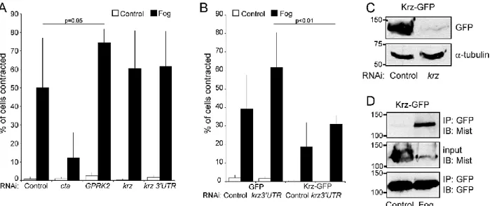
Figure 3-2. Krz and GPRK2 have a modest attenuating effect on Fog-induce S2R+ cell contraction and Krz interacts with Mist in a Fog-dependent manner ... 59
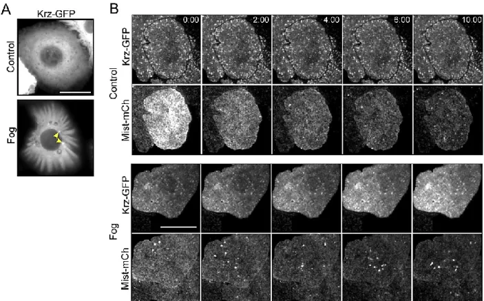
Figure 3-3. Krz and Mist may be internalized following Fog treatment ... 60
vii
List of Abbreviations
Abl
Abelson kinase
Cta
Concertina
Fog
Folded gastrulation
GAP
GTPase activating protein
GBE
Germ band extension
GDP
Guanine diphosphate
GEF
Guanine nucleotide exchange factor
GPCR
G-protein coupled receptor
GRK
G-protein coupled receptor kinase
GTP
Guanine triphosphate
Krz
Kurtz
Mist
Mesoderm invaginating signal transducer
MT
Microtubule
PMG
Posterior midgut
Rok
Rho kinase
Sqh
Spaghetti squash
TF
Transcription factor
VF
Ventral furrow
VFF
Ventral furrow formation
Chapter 1
Introduction
The Fog signaling pathway: Insights into signaling in morphogenesis
This chapter is in preparation as a review article.
Abstract
A complex interplay between many inter- and intracellular signaling molecules, along
with extrinsic cues such as temperature and cellular tension, controls morphogenesis during
animal development. The Drosophila Folded gastrulation pathway is one of the most
extensively studied examples of signal transduction pathways controlling morphogenesis. It is
used reiteratively during epithelial folding events and all of its core components are known. In
this review, I discuss principles of morphogenesis and signaling gleaned through in-depth
examination of this pathway. I also consider various regulatory mechanisms and the system’s
relevance to mammalian development. I propose future directions which will continue to
broaden our knowledge of morphogenesis across taxa.
Introduction
Epithelial morphogenesis, or the process through which simple sheets of cells are
rearranged and change shape to form mature structures and organs, has recently become an
area of intense focus in the field of developmental biology, e.g. (Spear & Erickson, 2012; Nelson
& Gleghorn, 2012; Suzuki, et al., 2012). A key morphogenetic movement which occurs in almost
all multicellular organisms is the folding or bending of flat epithelial sheets to form more
complex structures. These changes are often driven at least in part by actin- and myosin-based
2
pathways regulating this process is the Drosophila Folded gastrulation (Fog) pathway in which
all the crucial steps are known, from initiation by transcription factors (TFs) to the mechanics of
cell shape changes. This pathway, which drives apical constriction, therefore allows
examination of some of the intricacies of protein signaling during development in vivo.
Many stereotypical signaling mechanisms exist in the Fog pathway, including patterned
induction of gene expression by TFs, G-protein coupled receptor (GPCR) to G-protein signaling,
and actin rearrangement induced by the Rho GTPase axis. The Fog pathway also reveals some
novel insights, such as how multiple signaling pathways can be integrated into a single outcome
and that GPCRs, among their many other functions, have morphogenetic roles. While certain
aspects of the Fog pathway have been worked out in great detail, many questions still remain.
What mechanisms recruit signaling components apically? How are Fog pathway components
spatially and temporally patterned and what role does this patterning play in development?
Which mechanisms regulate the attenuation of Fog signaling? We will explore these questions
in this review.
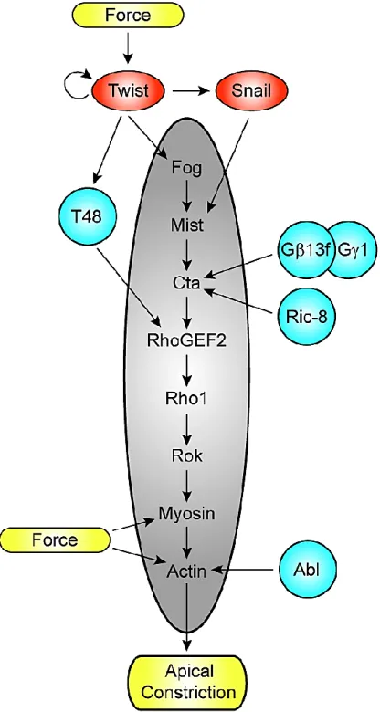
The Fog pathway, diagrammed in Figure 1-1, begins with the specific expression of Fog in
subsets of cells fated for actomyosin-based shape changes. Fog is a large secreted protein that is
thought to signal primarily as an autocrine factor (Costa, et al., 1994). The Fog signal is
transmitted across the plasma membrane by the GPCR Mesoderm invaginating signal
transducer (Mist), a member of the secretin family of GPCRs, to a G-protein of the G12/13
family, Concertina (Cta) (Parks & Wieschaus, 1991)(Chapter 2). In turn, RhoGEF2, a Dbl family
RhoGEF; the small GTPase Rho1; and the Rho effector, Rho Kinase (Rok) are all activated
(Barrett, et al., 1997; Dawes-Hoang, et al., 2007). Rok phosphorylates the regulatory light chain
of non-muscle myosin II to induce contraction of the apical actomyosin network in the cells that
receive the Fog signal. While the ligand, Fog, is not conserved outside of Drosophila and the
receptor, Mist, is not conserved outside of insects, the axis of signaling from G proteins
3
development and disease (Figure 1-1) (Waterhouse, et al., 2011). For example, lysophosphatidic
acid and sphingosine 1-phosphate are membrane lipid derivatives known to signal through
GPCRs, the G12/13 family, RhoGEFs, RhoA, and various effectors in mammals (Xiang, et al.,
2013; Suzuki, et al., 2009). These pathways are able to modulate various cytoskeletal and cell
Figure 1-1. The Fog Signaling Pathway. Fog is a large secreted protein which acts as a ligand for Mist, a seven pass transmembrane GPCR. In its ligand-free state Mist is predicted to interact with inactive, GDP-bound Cta. Once Fog binds Mist, it likely stimulates Cta’s exchange of GTP for GDP, which allows Cta to dissociate from its trimer partners, G and G. Cta-GTP binds to RhoGEF2 which can then act as a GEF for Rho1. In its GTP-bound form Rho1 then activates Rok. Finally, the regulatory light chain of non-muscle myosin II, Spaghetti squash, is phosphorylated by active Rok to induce apical actomyosin network contraction in the cells which receive the Fog signal. Boxed are vertebrate components of Rho axis signaling which act in a similar manner to induce actomyosin cytoskeleton rearrangements. In
4
shape changes, including neurite outgrowth and retraction, tumor cell invasion, as well as
angiogenesis.
The Fog pathway is active in many morphogenetic events in Drosophila development,
with known roles in ventral mesoderm and posterior midgut (PMG) invagination during
gastrulation, imaginal disc folding during larval development, salivary gland internalization in
mid-embryogenesis, and morphogenesis of the central nervous system during late
embryogenesis (Costa, et al., 1994; Nikolaidou & Barrett, 2004; Ratnaparkhi & Zinn, 2007). In
all of these cases Fog induces apical constriction, except in the CNS where the cellular results of
Fog’s action are not known. Apical constriction, along with other concomitant shape changes, in
cells of the ventral mesoderm, PMG, and salivary gland eventually results in complete
internalization of these cell groups. The folds of imaginal discs only invaginate as far as to form
U-shaped folds within the plane of the tissue.
During VFF there are two phases of apical constriction: a stochastic, nonproductive
phase, when individual cells contract and relax without any overall reduction in apical area, and
a concerted, coordinated phase, when individual cells undergo ratchet-like reductions in apical
area which are much more stable (Sweeton, et al., 1991; Martin, et al., 2009). Actin and myosin
periodically coalesce and these concentrations tend to move toward the center of a cell (Martin,
et al., 2009). Via these actomyosin contractions, the plasma membrane is pulled inward.
During random constriction the membrane relaxes to its original position when actomyosin
coalescences are disassembled. Once the concerted phase of constriction begins, membrane
deformations are stabilized to reduce apical cell area. This pulsatile mode of cellular
constriction has also been observed in other contracting groups of cells in the Drosophila
embryo (Solon, et al., 2009).
In addition to the conserved nature of the signaling components, these cell shape
changes are similar to morphogenetic processes in mammals (Sweeton, et al., 1991; Schoenwolf
5
resembles neural tube formation in vertebrates. In both cases, a subset of epithelial cells within
a flat sheet undergoes apical constriction to invaginate and form a tube sealed off from the
surrounding epithelium (Copp & Greene, 2010). When these processes are disrupted
Drosophila eggs don’t hatch; in humans debilitating congenital defects such as spina bifida or
anencephaly can occur, sometimes leading to death. Working out the intricacies of the Fog
signaling pathway and its resulting cell and tissue movements will ultimately lead to a more
profound understanding of our own development and greater potential for medical
interventions in disease states.
Interactions between Fog pathway members
Ligand and receptor
A discussion of the core components of the Fog signaling pathway must begin with Fog
itself. Embryos lacking Fog, the secreted ligand initiating the pathway, display disorganized VF
cell apical constriction, though most mesodermal cells do eventually internalize (Costa, et al.,
1994). Major problems arise in the next steps of development since PMG cells do not invaginate
and improper germ band extension (GBE) leads to a twisted body axis. All embryos mutant for
fog die before emerging from the egg. Embryos lacking fog in subsets of cells that cross the VF
have a distinct divide between apically constricting cells (wild-type) and non-constricting cells
(fog mutant) (Costa, et al., 1994). This experiment suggests that the Fog signal does not diffuse
farther than a couple of cell widths, consistent with Fog being a fairly large protein predicted to
be glycosylated.
The most recent addition to our knowledge of Fog signaling is the discovery of a
receptor, Mist, which can function downstream of Fog (Chapter 2). Mist is a GPCR with a large
extracellular domain, appropriate for interacting with a large ligand such as Fog. This discovery
was made possible by the development of a cell culture model for studying Fog signaling.
Drosophila S2R+ cells plated on a substrate of Concanavalin A respond to exogenously added
6
Many avenues of study not possible using whole animals have been opened by the development
of this model system. The discovery of Mist both answers old questions and raises new ones.
fog and mist transcription are both precisely regulated in space and seem to be under
independent control, with overlapping but not completely coincident expression patterns
(Dawes-Hoang, et al., 2007)(Chapter 2). This redundancy helps explain how the formation of
Fog-induced epithelial invaginations is so regular within the complex developmental dynamics
of wild-type animals.
Ubiquitous overexpression of Fog in the early embryo results in a normal VF and no
precocious apical constriction (Dawes-Hoang, et al., 2007). This can now be explained by mist’s
restriction to ventral and posterior cells and its upregulation at the end of cellularization when
VF invagination normally begins (Chapter 2). The opposite is also true, with ubiquitous Mist
expression not significantly disrupting gastrulation presumably due to Fog’s spatial restriction.
Adding complexity to the situation, however, is that ubiquitous Fog overexpression results in
apical flattening in cells outside the VF (Morize, et al., 1998; Dawes-Hoang, et al., 2007).
Perhaps there is a low level of Mist in dorso-lateral cells which allows flattening but does not
reach the threshold for full apical constriction. There is also the possibility of multiple Fog
receptors working either redundantly with, in concert with, or differently from Mist in the same
or different tissues. There may be a second receptor in cells outside the VF and PMG
invaginations in the early embryo which responds to Fog by inducing apical flattening
specifically. Another possibility is a redundant receptor in other tissues, though it is not likely in
the VF and PMG given the similarities of mist and fog zygotic phenotypes (Chapter 2). Mist may
have an obligate coreceptor, in which case missing either one of the pair would phenocopy a
complete lack of receptor. One possibility for a receptor working with or in parallel to Mist is
the GPCR CG31660, which was found by genetic screening to play a role during the
morphogenetic movements of gastrulation (Mathew, et al., 2009). The precise actions of this
7
The recent discovery of a receptor connecting Fog and Cta across the plasma membrane
in the well-studied Fog signaling pathway creates an easily manipulated system for examining
GPCR activity in vivo. This pathway can be further studied in Drosophila cell lines to add to our
picture of GPCR signaling. Combining the genetic malleability and drug susceptibility of the fly
embryo with live imaging and other microscopic techniques will allow fine detail of the
interactions between components of G-protein signaling pathways, and the developmental
results thereof, to be examined. Mist being the primary example of G-protein signaling in
morphogenesis, it will be extremely important to learn all that we can from this system.
Heterotrimeric G-protein signaling
Among all of the known Fog pathway components, Cta was discovered first and yet
comparatively little is known about it (Parks & Wieschaus, 1991). Embryos lacking maternal Cta
have very similar gastrulation phenotypes to fog or mist zygotic mutants. Cta is required to
organize myosin apically in the contractile VF cells, though it is not necessary for apical actin
(Fox & Peifer, 2007). However, Cta is expressed much more broadly throughout embryogenesis
than are Fog and Mist, and likely has roles outside the VF and PMG. One possible
Fog-independent role of Cta is in maintenance of cortical cytoskeletal stability throughout the
blastoderm (Kanesaki, et al., 2013).
In the early embryo, ubiquitous expression of constitutively active Cta or injection of
cholera toxin, which activates Cta, phenocopies ubiquitous expression of Fog, including apical
flattening of all cells (Morize, et al., 1998). This result suggests that Fog-dependent apical
flattening works through Cta, though as mentioned above it may not work through Mist.
Receptor-specific Cta activation and subcellular localization in certain cells may help restrict
which downstream effectors are activated and therefore which cellular pathways are tiggered.
Unfortunately, no method for visualizing endogenous Cta has been developed, making it
difficult to learn about this protein in more detail. A reliable antibody to Cta or replacement of
8
open up a wealth of new information about how G-proteins function during development in
vivo.
G proteins function with Gs and Gs in obligate heterotrimers. G13f and G1 have
been suggested as partners for Cta during gastrulation, as embryos lacking either have the same
gastrulation and cuticle phenotypes as those lacking Cta (Figure 1-2) (Schaefer, et al., 2001;
Wang, et al., 2005; Izumi, et al., 2004). They are also the most widely expressed and
subunits during embryogenesis. G proteins are generally thought to be the primary signal
transducing members of heterotrimeric G-proteins, but it is now well established that and
subunits can signal independently of Gs (reviewed in (Clapham & Neer, 1997). Additionally,
Gs have been reported to require chaperone-like cofactors, such as Ric-8, for proper
localization (Figure 1-2) (Wang, et al., 2005). Embryos lacking Ric-8 have disrupted VF apical
constriction which results in similar cuticle phenotypes to embryos from cta mutant mothers
(Wang, et al., 2005; Kanesaki, et al., 2013). Ric-8 is also necessary for apical myosin
accumulation and cortical tension during VFF (Kanesaki, et al., 2013). It will be interesting to
further investigate the roles of these three essential co-factors in epithelial morphogenesis.
The Rho signaling axis
The intracellular signaling components of the Fog pathway fit into the well-established
Rho signaling axis which leads from activation of a G12/13 family member to actin cytoskeletal
rearrangement, e.g. (Somlyo & Somlyo, 2000). Some vertebrate members of this pathway are
listed in boxes in Figure 1-1. Cta, RhoGEF2, Rho1, Rok, myosin, and actin are present in all cells
in Drosophila early embryos and imaginal discs (Parks & Wieschaus, 1991; Barrett, et al., 1997;
Hacker & Perrimon, 1998; Mizuno, et al., 1999; Kiehart, et al., 1990; Warn & Magrath, 1983).
They are all supplied maternally to embryos, as well, which speaks to their importance during
the early stages of development. However, these proteins are apically localized specifically in
9
10
their limited subcellular localization help give developmental control to their downstream
effects. This section aims to highlight some of the important points we have learned about how
this pathway enacts cell shape changes from studying Fog signaling and what we can potentially
learn from further examining Rho axis signaling in Drosophila.
RhoGEFs, and RhoGEF2 in our case, act as signal concentrators within the cell,
specifying and amplifying the outcome of Rho activation. Maternal RhoGEF2 mutant
gastrulation phenotypes are much stronger than those of either zygotic fog or maternal cta
mutants, with no mesoderm or posterior endoderm internalization at all (Hacker & Perrimon,
1998; Barrett, et al., 1997). Also unlike fog and cta mutants, RhoGEF2 mutants have defects in
both actin and myosin accumulation at the apical sides of VF cells (Fox & Peifer, 2007). There
must be another pathway feeding into the activation of RhoGEF2 in the VF which is somewhat
additive with the input from Fog-Mist-Cta. (Some possibilities will be discussed in the “Other
inputs into Fog-induced cell shape change” section below.)
Rho1 acts in early embryos and cell culture to organize both the actin and myosin
networks, with Cta upstream of its action on myosin (Fox & Peifer, 2007). Disruption of Rho1
function in early embryos by exogenous expression of an inactive version mimics the loss of
RhoGEF2 (Barrett, et al., 1997; Hacker & Perrimon, 1998). Embryos with disruptions in
RhoGEF2 or Rho1 do exhibit apical constriction but not in a coordinated or concerted fashion.
However, Rho1 and RhoGEF2 maternal mutants have noticeably different phenotypes, with
Rho1 mutants having more and varied cell shape defects throughout embryogenesis (Barrett, et
al., 1997; Magie, et al., 1999). These results are complicated by the requirement for Rho1 during
egg formation, but do suggest that Rho1 can be activated by other RhoGEFs in addition to
RhoGEF2 or by other mechanisms (Magie, et al., 1999). Overall, RhoGEF2 and Rho1 do not
seem to be absolutely necessary for actin and myosin rearrangement but act to organize and
11
Rho1, RhoGEF2, and zipper (encoding the heavy chain of myosin II) all interact
genetically during leg and wing morphogenesis, during imaginal disc folding and/or limb
eversion (Halsell, et al., 2000; Nikolaidou & Barrett, 2004). Fog, Mist, and Cta have all been
implicated in these processes as well (Nikolaidou & Barrett, 2004)(Chapter 2). Improper
expression levels or patterns of Fog pathway components in wing imaginal discs leads to
stochastic folding of the epithelium. Proliferation, specification, and polarity of discs do not
seem to be altered when the Fog pathway is disrupted, but the tissue’s normal growth forces
once flat epithelial sheets to fold within the confines of the disc without proper patterning
information (Nikolaidou & Barrett, 2004). These data confirm again that patterning and
specificity of Rho activation is crucial during morphogenesis. Techniques for imaging imaginal
disc development live have been developed (Aldaz, et al., 2010). Additionally, Förster
Resonance Energy Transfer (FRET) probes for the activity of the GTPase Cdc42 have been used
in live Drosophila embryos (Kamiyama & Chiba, 2009). A combination approach could be
taken using a Rho1 biosensor to further investigate the protein’s activity downstream of Fog
activation in wing discs. The prospect of using an initially flat tissue with increasing complexity
and dynamics such as the imaginal disc for studying Rho activation at high resolution in vivo is
exciting.
Induction of the Fog pathway
Transcription factors
There are several factors that contribute to the expression pattern of Fog pathway
components, as well as initiation and organization of the pathway itself. First, transcriptional
control of certain Fog pathway members can influence pathway activation within developmental
space and time. We know the most detail about this topic relative to ventral furrow formation
(VFF). During egg production, a nuclear gradient of the Dorsal TF is maternally set, with
highest levels on the ventral side of the egg (Roth, et al., 1989). The cells that receive the highest
helix-12
loop-helix family, and Snail, a zinc finger TF (Leptin & Grunewald, 1990). Twist in the ventral
mesoderm reinforces both its own expression and Snail expression (Ip, et al., 1992). Twist and
Snail are each independently required for both mesoderm specification and the morphogenetic
movements of gastrulation, though they have slightly different phenotypes (Figure 1-2)(Leptin,
1991). twist single mutants retain some ability to accumulate myosin and constrict VF cells,
though they are never able to transition to the coordinated, productive phase of apical
constriction (Martin, et al., 2009). Twist is required to stabilize actomyosin-based constrictions,
perhaps due in part to an ability to respond to force (see “Mechanical inputs” below). snail
mutants do not undergo visible myosin coalescence, though some mesodermal cells are
eventually internalized, suggesting that Snail is required for the initial stages and coordination
of apical constriction (Martin, et al., 2009). In snail twist double mutants VF cells don’t
accumulate myosin apically, contract, or form an invagination suggesting that these two TFs
together are necessary to transcribe key molecules involved in all steps of VF cell shape change
(Leptin, 1991; Martin, et al., 2009).
Some of these transcriptional targets are known. Twist activates the transcription of fog
and T48, a single pass transmembrane protein that acts to apically localize RhoGEF2 during
VFF (see “Other inputs into Fog-induced cell shape change” below; Figure 1-2) (Morize, et al.,
1998; Kolsch, et al., 2007). Snail’s only known target necessary for gastrulation is mist (Figure
1-2; Chapter 2). fog mRNA and mist mRNA have similar localizations in wild type embryos,
with enrichments along the ventral side and the posterior end of the embryo. One marked
difference between them is that mist RNA is present in a continuous stripe while fog RNA
exhibits a gap between its mesodermal and endodermal patches. fog RNA in twist mutant
embryos and mist RNA in snail mutant embryos both lose expression in the ventral mesoderm
while retaining it in the PMG (Seher, et al., 2006)(Chapter 2). An independent set of TFs is
probably required in the PMG. These somewhat independent and overlapping patterns suggest
13
Mist being a transcriptional target of Snail clarifies several previously unexplained
results. First, ectopic Fog expression in wild-type or fog mutant embryos induces a VF to form
in its normal location (Morize, et al., 1998). Twist is not required for this to occur. In snail
mutants, though, ectopic Fog expression fails to induce flattening of VF cell apices (Morize, et
al., 1998; Dawes-Hoang, et al., 2007). Mist may be the Snail target required for apical
flattening, at least in VF cells. Second, the stochastic phase of VF apical constriction occurs in
twist but not snail mutants (Martin, et al., 2009). Twist, T48, and, importantly, Fog are not
required for random cellular constrictions, but a Snail target is. This could be explained by
spontaneous agonist-free excitation of Mist, which is a property of many GPCRs (reviewed in
(Smit, et al., 2007). Overlapping expression of Snail and Twist patterning expression of Mist
and Fog is a novel mechanism for robustly controlling the location and timing of a
developmentally important signaling pathway.
Outside of the VF we don’t know the transcriptional regulators controlling Fog pathway
members. The Fork head TF is necessary for salivary gland primordium apical constriction and
invagination (Myat & Andrew, 2000). As Fork head is also expressed at the extreme ends of the
early embryo, it may also be involved in PMG invagination, though it has not been specifically
implicated in controlling Fog signaling in either of these processes (Weigel, et al., 1989). Fog
and Mist expression patterns in the wing imaginal disc are complex and don’t follow any known
TF patterns (Chapter 2). They are likely under combinatorial control of many TFs in this tissue.
Downstream players in the Fog pathway are maternally deposited in embryos and are widely
expressed in other tissues. Their localized activity rather than expression is likely the
determining factor in localized signal transduction.
Mechanical inputs
Another mode of control feeding into Fog signaling is mechanical force (Figure 1-2). As a
flat sheet of cells folds the apically constricting cells produce force which pulls on neighboring
14
can experience mechanical strain. We don’t know all of the implications of these forces yet, but
there are some ideas in the literature. For instance, stress across the apical surfaces of cells
undergoing Fog signaling could increase the membrane tension enough to reduce endocytosis,
leaving more competent or active Mist on the membrane for signaling (Driquez, et al., 2011).
Conversely, apical-basal shortening toward the end of furrow invagination could result in a
reduction in total cell volume, cell surface area, and membrane tension leading to an increase in
endocytosis and termination of signaling.
As mentioned previously, VF cell contraction occurs in two phases: a random
unproductive period of contraction and then a coordinated period that forms an epithelial fold
(Sweeton, et al., 1991; Martin, et al., 2009). The trigger that allows for the change from the
stochastic phase to the collective phase is not yet known, but it has been suggested that this
transition occurs when a threshold of strain builds up across the tissue (Martin, et al., 2010).
This mechanical strain may feed directly into the actomyosin network. It will be interesting to
further study the interactions between signaling and mechanics during these contractions and to
investigate their roles in other organisms.
Force could also feed less directly into Twist, Fog, and T48 expression, as Twist protein
expression seems to be positively correlated with the mechanical deformation of cells during
GBE (Farge, 2003). Just after gastrulation, large scale tissue rearrangements comprising GBE
produce compressive forces on the dorsal side of the embryo and stretching forces on the ventral
side. Physically disrupting GBE movements reduces Twist expression, but artificial force on
these disturbed embryos can rescue Twist levels (Desprat, et al., 2008). (While Twist is no
longer required for Fog signaling at GBE, it is still necessary for proper mesoderm
differentiation (Leptin, 1991).) Similarly, Snail is required for apical myosin localization in the
VF, but artificial indentation of snail mutant embryos can rescue myosin localization and
15
There is evidence for mechanical strain influencing RNA transcription, cytoskeletal
dynamics, and tissue movements in many systems. For instance, formation of the head fold in
the chick embryo, an epithelial folding event, exerts significant forces on the surrounding tissues
(Varner, et al., 2010). Application of ectopic forces to embryo explants undergoing this process
alters their morphogenetic movements. We don’t yet know how forces are involved in most
tissues where Fog signaling is active, but we can use this pathway and its resulting epithelial
invaginations to investigate the problem in a very detailed manner. The early Drosophila
embryo and imaginal discs can be mechanically manipulated and methods have already been
developed to do so, e.g. (Farge, 2003). The embryo is a relatively simple, yet 3-dimentional in
vivo system in which we can modulate gene activity and mechanical stress in combination.
Insights about the interaction between these two inputs into the Fog signaling pathway will
likely be broadly applicable to many developmental processes.
Subcellular localization
We know that much of the signal transduction within the Fog pathway must occur at or
near the apical surface of contractile cells in order to restrict actomyosin contraction to cell
apices, but we know very little about how this is achieved. fog mRNA is focused apically in the
PMG and imaginal discs, and mist mRNA is apical in imaginal discs (Dawes-Hoang, et al.,
2007)(Chapter 2). Fog protein localizes to puncta, presumably vesicles, in the apical portion of
PMG cells during invagination, suggesting that it may be apically secreted (Dawes-Hoang, et al.,
2007). Mist protein is present in discrete puncta on the apical surface of VF cells during
invagination (Chapter 2). Localized translation and directional trafficking likely contribute to
the apical localization of these proteins. Specific association of Cta with apically concentrated
Mist in cells undergoing Fog signaling may help to restrict Cta to the apical domain, but this is
not yet known.
Before gastrulation, RhoGEF2 localizes to the basal ends of cellularization furrows but is
16
VF cells, but not in lateral and dorsal cells, RhoGEF2 then becomes apically concentrated before
constriction occurs. This striking relocalization may, at least in part, be caused by directional
transport of RhoGEF2 on dynamic plus-ends of microtubules (MTs) (Rogers, et al., 2004).
Activation of Cta causes RhoGEF2 to dissociate from MTs, possibly allowing for RhoGEF2 to
associate with Cta itself, interact with lipids in the plasma membrane, and activate Rho1. MTs
in the blastoderm epithelium are generally thought to be oriented with their plus-ends basally,
the reverse orientation to that which would bring RhoGEF2 to the apical surface (Harris &
Peifer, 2005). The MT arrays in many interphase Drosophila cells are acentrosomal, however,
so there may be mixed polarity MT arrays or short MTs along apical cell surfaces which may
contribute to localization of Fog signaling components (Rogers, et al., 2008). Alternatively,
RhoGEF2’s association with MT plus-ends could be a mechanism for keeping it basally localized
before Cta activation. The orientation and dynamics of MTs in contractile cells in vivo should be
examined in greater detail in order to determine whether and how they play a role in localizing
these signaling components.
Myosin localizes apically during VFF, PMG invagination, salivary gland invagination,
and imaginal disc folding (Nikolaidou & Barrett, 2004; Zhang & Ward, 2011). Myosin is
concentrated basally during early cellularization, but, unlike RhoGEF2, it is both lost from the
basal surface and enriched apically only in VF cells. This accumulation during VFF does not
occur in embryos lacking Fog, Mist, Cta, RhoGEF2, or Rok, suggesting that a complete Fog
pathway is required for establishment or maintenance of the apical myosin network (Nikolaidou
& Barrett, 2004; Dawes-Hoang, et al., 2007)(Chapter 2).
The major determinant of epithelial apical behavior in most organisms is the apical PAR
complex, made up of Par-6, Par-3/Bazooka, and aPKC, which must be in place for apically
restricted events to occur properly (reviewed in (Goldstein & Macara, 2007). These apical
proteins likely have direct as well as indirect roles in organizing Fog. In the early Drosophila
17
(Müller & Wieschaus, 1996). Bazooka, through recruitment of several partner proteins, localizes
G proteins apically in Drosophila neuroblast cells (Siegrist & Doe, 2005). A similar
mechanism may help localize Cta. The PAR complex also interacts with the proteins that set up
subapical adherens junctions in the early embryo. These cell-cell contacts are necessary for
tissue cohesion during gastrulation (Müller & Wieschaus, 1996; Dawes-Hoang, et al., 2007).
Adherens junction proteins move from their normal subapical localization to a more extreme
apical localization in the VF cells just before apical constriction (Dawes-Hoang, et al., 2007).
We do not know how much influence their location along the apical-basal axis has on the ability
of cells to invaginate in the VF, though adherens junction migration is known to be a driving
force in Drosophila dorsal epithelial folding (Wang, et al., 2012).
The transmembrane protein Crumbs is also a major player in apical membrane identity
and recruitment of proteins to the apical region of cells (Assémat, et al., 2008). During salivary
gland invagination, Rho1 activity in the invaginating cells is required for crumbs transcription
and for crumbs mRNA and protein apical localization (Xu, et al., 2008). Crumbs, in turn, helps
to organize the apical domain of these cells, leading to proper actomyosin constriction
downstream of Rho1. How Crumbs- and PAR complex-induced polarity interacts with other
signaling complexes is a convoluted matter and will likely take years more work to figure out.
The strict localization and restricted timing of Fog signaling offer a good system with which to
study these interactions.
Other regulatory mechanisms
Negative regulation of Fog signaling
Several other signaling pathways or proteins have been shown or hypothesized to feed in
to the Fog pathway at various points. One thoroughly unknown aspect of the Fog pathway is
how the contractile signal is terminated. The mRNAs or proteins of pathway members may be
turned over to terminate signaling. mist RNA is present in the presumptive mesodermal cells
18
mesodermal cells shortly after the VF has invaginated (Costa, et al., 1994). If there is no
activating ligand there should be no pathway activation, whether other pathway components are
competent for signaling or not. Translational or transcriptional regulation may not be rapid
enough for termination of the signal in VFF, as mesoderm internalization only lasts about ten
minutes. Other Fog pathway-dependent morphogenetic processes probably occur on a longer
time scale, though.
GPCR signaling is canonically terminated by phosphorylation of the C-terminal tail of
ligand-bound GPCRs by G-protein Coupled Receptor Kinases (GRKs). Once phosphorylated,
GPCRs are bound by -Arrestins which can induce receptor internalization, cause receptor
degradation, compete for GPCR binding with Gs, and potentially activate independent
signaling cascades. Vertebrate genomes encode many GRKs and -Arrestins, some of which are
visual system specific and some of which are utilized more generally across tissues. Drosophila
only has one non-visual GRK and one -Arrestin, GPRK2 and Kurtz (Krz) respectively (Cassill,
et al., 1991; Roman, et al., 2000). GPRK2 is required maternally for egg production (Schneider
& Spradling, 1997). However, some of the few eggs laid by GPRK2 mutant mothers display
disrupted gastrulation phenotypes suggesting a possible role in regulating VFF and PMG
invagination. Eggs lacking Krz also display cuticle phenotypes suggestive of gastrulation defects
(Tipping, et al., 2010). Alteration of levels of either protein in wings also causes morphological
defects (Molnar, et al., 2011). These data raise the possibility that GPRK2 and/or Krz could play
a role in termination of Fog signaling. Investigation of the roles of GPRK2 and Krz in this
pathway could allow us to more precisely determine how and when signal termination is
achieved during other morphogenetic signaling events.
There are a few canonical molecules which terminate Rho axis signaling in many
contexts: Rho GTPase activating proteins (GAPs) and myosin phosphatase. RhoGAPs accelerate
the inherent GTPase activity of Rho proteins, increasing the ratio of inactive to active Rho.
19
Fog signaling, Rho1 activity is restricted to the apical sides of cells (Simões, et al., 2006). In
these cells RhoGEFs remain apical while RhoGAPs are baso-lateral. The complementary
localization of these regulatory proteins organizes Rho1 activation and also allows for its
deactivation promptly after termination of an activating signal. However, we don’t know
whether or which GAPs are acting in Fog signaling or how they may contribute to signaling
dynamics.
Myosin phosphatase removes the activating phosphates from regulatory myosin
subunits. Rok can phosphorylate both myosin light chain to activate it and phosphorylate
myosin phosphatase to inactivate it, a twofold way of maintaining myosin activity (Amano, et al.,
2010). When negative regulation is not exerted on myosin phosphatase, it can act to
downregulate myosin activity. The role of this deactivation mechanism in Fog signaling is not
yet known.
There may be other contributing factors to the termination of Fog signaling. For
instance, Par-6 has been found to negatively regulate Rho in several contexts, and therefore Rho
activation within an apical PAR domain must overcome this local downregulation (Goldstein &
Macara, 2007). Also, changes in membrane trafficking could influence aspects of signaling such
as Mist presentation on the apical plasma membrane and secretion of Fog. Alteration of
membrane tension during cell shape change may also influence the ability of the actomyosin
cytoskeleton to pull against the plasma membrane. These questions may be difficult to
approach in vivo, but are ideal problems to solve using a cell culture model of apical
constriction.
Other inputs into Fog-induced cell shape change
There are several other accessory proteins that have been shown genetically or
mechanistically to influence Fog signaling but don’t fit into a well-defined category. First, the
single pass transmembrane protein T48, a Twist transcriptional target, is expressed along the
20
Leptin, 1991). Interestingly, it is required for organized VF invagination but is not even
expressed in the PMG (Kolsch, et al., 2007). T48 is necessary for proper apical localization of
RhoGEF2 in the VF. It also helps to organize the transition of adherens junctions from
subapical to apical localization in VF cells as constriction begins. Just as Fog and Cta aren’t
absolutely required for mesoderm internalization, neither is T48, but embryos lacking both Cta
and T48 do not form a VF at all. T48 may act as an accessory protein in Fog signaling or in a
parallel pathway, though the mechanism of its influence is not yet known.
MTs have been implicated in working with the actin cytoskeleton in order to enact cell
shape changes during morphogenesis, potentially in nuclear positioning or membrane
trafficking, (e.g. (Suzuki, et al., 2012). Within the cytoplasm actin regulatory proteins could also
influence the organization or formation of the apical contractile array during Fog-induced cell
shape changes. For instance, the formin Diaphanous (Dia) is an actin filament elongation factor
which is also a Rho effector in several systems (reviewed in (Young & Copeland, 2010).
Embryos lacking maternal dia have defects in coordinating apical constriction in the VF so that
only a subset of cells constrict (Homem & Peifer, 2008).
One actin regulator with a more defined role in VFF is Ableson kinase (Abl), a
non-receptor tyrosine kinase that interacts directly with the actin cytoskeleton (Figure 1-2) (Van
Etten, et al., 1994). Abl is present apically in all cells during early embryogenesis and is
enriched and activated in VF and PMG invaginations (Fox & Peifer, 2007). Embryos lacking Abl
zygotically have similar gastrulation defects to those lacking Cta maternally. They have
uncoordinated VF cell contraction with disorganized apical networks of actin, but do internalize
most, if not all, mesodermal cells. The double mutant phenotype of abl and cta is much
stronger than either alone, and resembles RhoGEF2 mutants. Abl likely acts parallel to Cta to
coordinate the actin and myosin networks in apically constricting cells. Loss of Abl and
Abl-related gene in mice leads to strong neural tube closure defects, implicating a similar molecular
21
interaction between G-protein signaling and actin regulatory proteins in Rho activation and cell
shape change should be more deeply studied, with VFF being a great model.
Conclusions
In this review I have summarized our current understanding of the Fog signaling
pathway and discussed known and potential inputs into the ultimate cell shape changes which
occur in cells undergoing Fog signaling. Drosophila morphogenesis, specifically VFF, has long
been used as a simplified model for vertebrate morphogenesis and signaling for several decades.
Many wide-reaching paradigms have been discovered and investigated in depth using this
model, not the least of which is the complement of physical cell shape changes which occur
during apical constriction. Additionally, quantification of different aspects of VF cellular
contraction in wild-type and perturbed embryos has allowed us to analyze how physical forces
are coupled to cellular contractions and ultimately to tissue-scale movements (Martin, et al.,
2010; Driquez, et al., 2011). The intimate integration of multiple signaling pathways to trigger a
single outcome has become clearer in recent years as well, with the study of how cell polarity
affects cell shape and Rho signaling (Xu, et al., 2008). Fog signaling is also a pioneer model for
GPCR-G-protein signaling in morphogenesis (Chapter 2).
The mechanistic interactions between known players in Fog-activated morphogenetic
events do need more attention in the coming years. We still have a lot to learn from this system
in terms of spatial and temporal regulation, for example. The complementary patterns of Fog
and Mist expression throughout Drosophila development in combination with all of the
accessory proteins required for normal tissue invagination give us a hint as to the level of robust
control required by evolution for development. I predict that one of the main questions moving
forward will be how the timing of Fog signaling is regulated, which will likely lead to the
discovery of more auxiliary players. We still know nothing of Fog signal termination. Our
22
to the understanding of signaling and morphogenesis in our own development and will continue
Chapter 2
Regulation of epithelial morphogenesis by a G-protein coupled receptor, Mist,
and its ligand, Fog
This chapter represents a manuscript in revision. The experiments were designed by me, Mark Peifer, and my advisor, Stephen Rogers, and were carried out by me, Kimberly Marston, and Mark Peifer. The manuscript was written by me, Kimberly Marston, Mark Peifer, and my advisor, Stephen Rogers.
Abstract
Epithelial morphogenesis is essential for shaping organs and tissues and for establishment
of the three embryonic germ layers during gastrulation. Much of our understanding of how
epithelial morphogenesis is governed by developmental patterning mechanisms has come
from studies of gastrulation in Drosophila. We developed a novel assay to recapitulate
morphogenetic shape changes in individual cultured cells, and used RNAi-based screening
to identify Mist, a Drosophila G-protein coupled receptor, which acts as a receptor for the
secreted ligand Folded gastrulation in cultured cells. Mist plays a role in Fog-dependent
embryonic morphogenesis, and its zygotic expression is regulated by the transcription factor
Snail. Our data show how a cell fate transcriptional program can act through a ligand-GPCR
pair to provide spatial regulation of epithelial morphogenesis.
Introduction
During embryogenesis, the developmental program sculpts sheets of epithelial cells
to build organs, define tissue compartments, and establish the embryonic body plan. Forces
driving these tissue-level rearrangements are produced by the actin and myosin cytoskeleton
24
adherens junctions (Pilot & Lecuit, 2005; Kasza & Zallen, 2011). Cell and tissue shape
changes are regulated by a complex interplay between maternally supplied proteins and
patterned zygotic gene expression. Understanding how developmental patterning organizes
cytoskeletal processes with spatial precision is a key question in the field of developmental
biology (Leptin, 1995).
G-protein coupled receptors (GPCRs) are one of the largest groups of proteins found in
the human genome, yet there are few examples of GPCRs regulating morphogenesis. Genetic
analyses in Drosophila have revealed a possible example involving a pathway that triggers
epithelial folding via apical constriction during gastrulation and salivary gland invagination
downstream of the secreted protein Folded gastrulation (Fog) (Costa, et al., 1994; Sawyer, et
al., 2010). This pathway is thought to involve a GPCR, as the Gα12/13 homologue Concertina
(Cta) is an integral component of the pathway. GPCR-independent activities of G-proteins
also can regulate the cytoskeleton (Parks & Wieschaus, 1991; Izumi, et al., 2004; Wilkie &
Kinch, 2005); thus it is unclear whether GPCRs are involved in initiating apical constriction.
Downstream of Fog, Cta is thought to activate RhoGEF2, which is recruited to the
apical membrane by the transmembrane protein T48(Rogers, et al., 2004; Kolsch, et al.,
2007). RhoGEF2 then activates the small GTPase Rho1 to recruit and stimulate cytoskeletal
contractile machinery, including Rho kinase (Rok), non-muscle myosin II, and actin, thereby
inducing apical constriction (Barrett, et al., 1997; Hacker & Perrimon, 1998; Dawes-Hoang,
et al., 2007; Kolsch, et al., 2007). This pathway is best characterized during gastrulation
where it initiates formation of both the ventral furrow (VF), to internalize mesoderm, and
the posterior midgut (PMG), to internalize endoderm (Sweeton, et al., 1991). It has served as
a powerful paradigm for morphogenesis from the level of gene expression to cytoskeletal
regulation. Fog is thought to act as a ligand to initiate this signaling pathway, but a receptor
for Fog has remained elusive despite 20 years of genetic and cell biological analysis (Costa,
25
Results
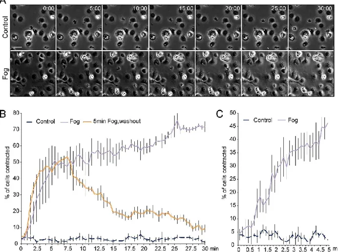
Mist acts as a Fog receptor in cell culture
We developed a novel functional genomic approach to identify Fog receptors by
reconstituting the pathway in a cell-based assay. We previously found that activating the
downstream effector Rho1 in cultured Drosophila S2 cells induces a characteristic contracted
morphology (Rogers, et al., 2004). We engineered S2 cells to express Fog, and used
conditioned medium from these cells to screen several immortalized Drosophila cell lines for
a contractile response. S2R+ cells exhibited robust contraction in response to Fog, including
actin rearrangement and increased levels of phosphorylated myosin regulatory light chain
(Spaghetti squash; Sqh), while S2 cells and several other epithelial-derived cell lines failed to
respond (Figure 2-1A). RNAi-mediated depletion of proteins known to act in the epithelial
folding pathway, including Cta, RhoGEF2, or Rho1, prevented Fog-induced S2R+ cell
contraction, indicating that we had recapitulated this morphogenetic cascade in cultured
cells (Figure 2-1B).
To identify a receptor that acts downstream of Fog, we performed a targeted RNAi
screen, individually depleting the 138 known and predicted GPCRs in the Drosophila
genome (Table 2-S1) (Brody, 2000; Broeck, 2001) and looking for cell contraction in
response to Fog. Among the candidates, only two independent dsRNAs corresponding to the
uncharacterized gene CG4521 (methuselah-like 1) consistently blocked Fog-induced
contraction (Figure 2-1C). This gene, designated here as mesoderm-invagination signal
transducer (mist), encodes a predicted GPCR of the secretin family. Mist is predicted to have
a large N-terminal extracellular domain characteristic of this family, seven
membrane-spanning helices, and a cytoplasmic C-terminal domain (Figure 2-1D). We generated
antibodies to Mist that recognized a single protein band on immunoblots of S2R+ cells that
was depleted by treatment with mist dsRNA (Figure 2-S1A). Consistent with the hypothesis
26
levels of Fog on the plasma membrane when treated at 4°C to block endocytosis when
compared to the Fog trapped by S2R+ cells expressing GFP alone (Figure 2-1E, F).
We next addressed whether Mist is sufficient to confer Fog responsiveness to
otherwise nonresponsive cells. S2 cells have undetectable levels of Mist and do not respond
to Fog (Figure 2-1C, 2-S1B). However, ectopic expression of full-length Mist endowed these
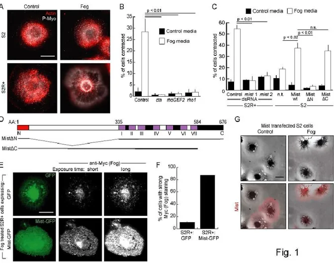
Figure 2-1. Mist acts as a Fog receptor in cell culture. A. S2R+ and S2 cells treated with control- or Fog-conditioned media and stained for actin (red) and phosphorylated myosin (Sqh) (P-Myo; white).
B. Percentage of S2R+ cells contracted in response to treatment with control- or Fog-conditioned media after RNAi knockdown of known Fog pathway components. C. Percentage of cells contracted in response to control or Fog treatment after mist knockdown (in S2R+ cells) or overexpression of Mist constructs (in S2 cells). n.t.: not transfected. n.s.: not significant. D. Mist predicted structure. Top: 37aa signal sequence (red), 298aa extracellular domain (white), 7 predicted transmembrane domains (purple, numbered with Roman numerals), and a 93aa intracellular domain (black). Extracellular loops are white and intracellular loops black. Bottom: Mist truncations used in C. E. S2R+ cells expressing GFP or Mist-GFP were treated with Fog at 4°C and stained for Myc (Fog). Short and long exposures of Myc staining are shown. F. Percentage of GFP or Mist-GFP transfected S2R+ cells with strong Myc (Fog) staining after treatment with Fog at 4°C. G. S2 cells transfected with untagged Mist,
27
cells with the ability to contract upon treatment with Fog (Figure 2-1C, G). To define the
domains required for Fog responsiveness we created Mist deletion constructs that retain the
signal sequence but lack the predicted N-terminal extracellular domain (MistN), or lack the
cytoplasmic domain (MistC) (Figure 2-1D). MistN failed to confer Fog responsiveness
upon S2 cells, indicating that the extracellular domain of Mist is required for Fog signaling
(Figure 2-1C). In contrast, MistC did confer Fog responsiveness on S2 cells, indicating the
C-terminus is not essential for activating downstream effectors (Figure 2-1C, D). This result
is not surprising, as it has been shown that some G subunits are primarily activated by
intracellular loops of GPCRs (Cronshaw, et al., 2010). Together these data demonstrate that
Mist is required for Fog signaling in cultured Drosophila cells and that the large extracellular
domain of Mist is necessary, perhaps acting as a ligand-binding surface.
Mist is essential for Drosophila gastrulation
While these data indicate that Mist can act as a Fog receptor, they do not reveal
whether Mist mediates the effects of Fog in vivo. Fog was originally identified as a secreted
protein that triggers the early embryonic movements of gastrulation (Costa, et al., 1994).
Thus, we tested the hypothesis that Mist acts as a Fog receptor to induce mesoderm
invagination. We first examined whether mist is expressed at the right time and place to act
in the Fog pathway. mist mRNA is present in the blastoderm, suggesting a maternal
contribution (Figure 2-2A). Just prior to mesoderm invagination mist mRNA is strongly
elevated specifically along the ventral side and posterior end of the embryo, corresponding
to the VF and PMG primordia (Figure 2-2B-C’). It is also expressed in the cells between
these two regions but remains at much lower levels in all other cells. fog RNA differs slightly
in its expression pattern, notably lacking expression in the region between the VF and PMG
(Figure 2-4F) (Costa, et al., 1994). mist mRNA expression remains strong in the mesoderm
28
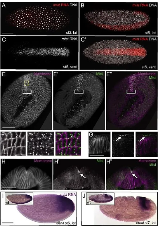
Figure 2-2.mist RNA is expressed specifically in the ventral furrow downstream of Snail. A-C’.
Fluorescent in situ hybridization for mist RNA (red) in wild-type embryos counterstained for DNA (white). A. Pre-blastoderm stage embryo. B-C. Blastoderm stage embryos before VF apical
constriction. C. mist channel alone from C’. Anterior is to the left in this and all other embryo figures.
E-H’’. Mist protein (green) in cross sectioned embryos undergoing VF invagination--ventral is to the top and membranes are marked in magenta. E-E’’. Grazing apical cross section.F. Enlarged images of boxed area from E-E’’. G. Onset of apical constriction. H-H’’. Continuation of apical constriction. Arrows and brackets: Mist is enriched apically in cells of the ventral furrow. I-J.in situ hybridization to mist RNA in snail mutant embryos. I. Blastoderm stage embryo. J. Gastrulating embryo.
29
localized to the apical contractile surfaces of VF cells (Figure 2-2E-H’’). Our antibody
controls suggest that weak punctate cytoplasmic staining seen in all cells is likely
background (Figure 2-S3). The elevation of mist mRNA in contractile cells of the VF and
PMG primordia just prior to gastrulation is consistent with a role for Mist in the regulation
of morphogenesis.
To investigate how the mist expression pattern is formed we looked to the embryonic
dorso-ventral axis specification pathway, which is initiated by the maternally supplied
Dorsal transcription factor. Dorsal acts through the zygotic transcription factors Twist and
Snail, both of which are independently required for VF invagination (Zusman & Wieschaus,
1985; Leptin & Grunewald, 1990). fog is a known transcriptional target of Twist in early
embryos, but Snail targets involved in VF invagination remain unclear (Simpson, 1983;
Boulay, et al., 1987; Costa, et al., 1994; Seher, et al., 2006). We thus tested the hypothesis
that mist might be a Twist or Snail target gene. Wild-type embryos exhibited robust
expression of mist mRNA in the VF and PMG from cellularization through germ band
extension (Figure 2-2A and 2-2F insets). When we crossed snail heterozygous parents, 25%
of embryos, likely snail homozygous mutants, lacked mist expression in the VF but retained
expression in the PMG (Figure 2-2F, 2-S2B). In contrast, most embryos from twist
heterozygous parents exhibited wild-type mist expression with only a few lacking VF
expression, presumably because Twist enhances Snail expression in the mesoderm (Figure
2-S2B, C) (Leptin, 1991). These data are consistent with mist being an embryonic target of
Snail.
To test whether Mist functions in vivo during gastrulation, we created a mutant
affecting mist expression by imprecise excision of a P-element inserted in the mist 5’UTR
(Figure 2-S4A). This generated a small deletion, which we call mistYO17 (Figure 2-S4B).
mistYO17 lacks the promoter, upstream regulatory region, and part of the 5’UTR of mist. It
30
protein SmG, the ribosomal subunit RpS19a, the unannotated gene CG9777, and the
maternally supplied Fog pathway component rok (see Supplemental Text for further
description of the mutant). The mistYO17 mutation is zygotically embryonic lethal and
embryos hemizygous for this mutation exhibit a significant reduction of mist mRNA
throughout gastrulation (Figure 2-3A and 2-S4C). Further, like other Fog pathway mutants
(Nikolaidou & Barrett, 2004; Dawes-Hoang, et al., 2007), mistYO17 mutant embryos have
reduced apical recruitment of non-muscle myosin heavy chain (Zipper; Zip) within VF cells
and uncoordinated VF apical constriction (Figure 2-S4D, E).
fog hemizygous mutants exhibit defects in internalization of mesodermal and PMG
cells which result in changes to the morphology of the ventral midline (Figure 2-3C, E)
(Costa, et al., 1994). These defects are not seen in wild-type embryos (Figure 2-3B, E). In
crosses yielding 25% mistYO17 mutant embryos, we saw clear defects in the internalization of
Twist-expressing mesoderm cells or morphology of the ventral midline in slightly more than
a quarter of the embryos, suggesting that mist or one of the other genes deleted in mistYO17 is
critical for this process (Figure 2-3D, E). We then used in situ hybridization for mist to
genotype individual embryos. As expected, embryonic progeny of mistYO17 heterozygous
females and wild-type males either had wild-type patterned mist expression or very little
mist expression (presumptive mistYO17/Y; Figure 2-4A, B and 2-S5A). More than 80% of
embryos with wild-type mist expression showed no gastrulation defects, while 95% of
embryos with weak mist RNA staining exhibited either a failure to fully invaginate
mesoderm cells or a defect in the ventral midline, correlating mist expression with
embryonic phenotype (Figure 2-4A, B, and D).
To test the hypothesis that mistYO17 gastrulation phenotypes are solely due to the lack
of mist, we examined whether restoration of mist expression could rescue the observed
defects. To do so, we took advantage of the fact that although the mistYO17 allele deleted the
31
Activating Sequence (UAS) and minimal promoter directed toward the mist coding region
(Figure 2-S4B). This allowed us to express mist under control of specific GAL4 drivers from
the endogenous locus. We confirmed that this allele precisely expressed mist mRNA and
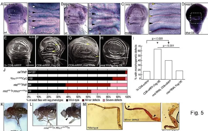
Figure 2-3. Mist is zygotically required for gastrulation. A. in situ hybridization for mist RNA in
mistYO17/Y embryos. Corresponding stages of wild-type embryos are shown in insets. B.-D’’. Actin
(white) and Twist (red) stained embryos showing the range of gastrulation defects seen. B-B’’. Wild-type embryos. C-C’’. fogS4/Y embryos. D-D’’. mistYO17/Y embryos. C. and D. Ventral midline defects.
C’. and D’. Single Twist positive cells not internalized (arrowheads). C’’. and D’’. Sheets of Twist positive cells not internalized (brackets). E. Quantification of gastrulation phenotypes for stage 6-8 embryos as pictured in B-D’’. rok2 gastrulation phenotype distribution is not significantly different
from wild-type, while mistYO17, fogS4, and mistYO17;tub-rok distributions vary from wild type
32
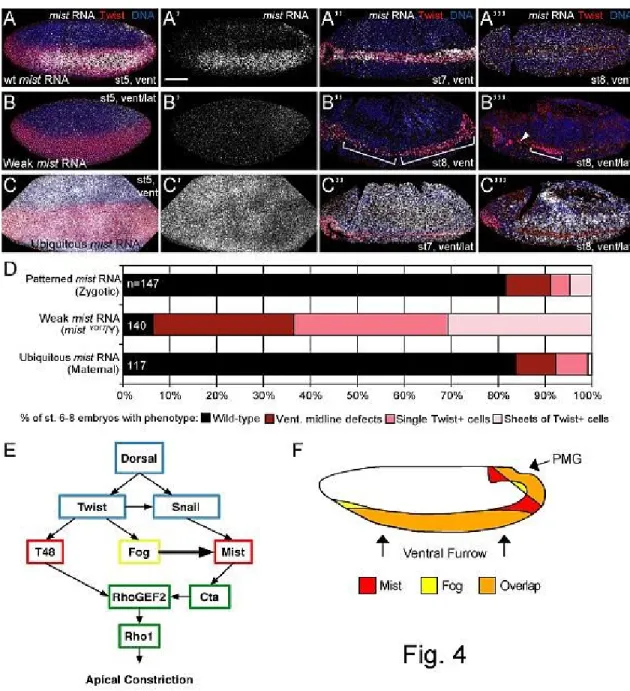
Figure 2-4. mistYO17 gastrulation phenotypes are specific to Mist activity. A-C’’’. Fluorescent in situ
hybridization for mist RNA (white) seen in embryos from crosses shown in fig. S7. Twist antibody (red) reveals presumptive mesoderm and DNA is shown in blue. A.-A’’’. Wild-type embryos showing patterned mist mRNA expression. B.-B’’’. mistYO17/Y embryos showing loss of patterned mist
expression. C.-C’’’. Embryos expressing ectopic mist mRNA uniformly (driven maternally). A.-C. Cellularization stages. A’.-C’. mist RNA alone from embryos in A.-C. A’’.-C’’. Early germ band extension stages. A’’’.-C’’’. Late germ band extension stages. D. Quantification of gastrulation phenotypes as pictured in Fig. 3, B-D’’. Embryos with weak mist RNA expression have a higher frequency of gastrulation defects compared to wild-type embryos and embryos expressing ubiquitous
33
protein by using a driver which is activated in the posterior compartment of each segment in
the later embryo (engrailed-GAL4; Figure 2-S3 and 2-S5B). We then crossed mistYO17
heterozygous females containing a maternally expressed GAL4 driver to wild type males
(Figure 2-S5A). This cross results in GAL4 being loaded into eggs during their formation in
the ovary and remaining into embryogenesis. The progeny of this cross had high level
ubiquitous expression of mist RNA throughout most of embryogenesis (Figure 2-4C).
Strikingly, these embryos showed normal gastrulation in proportions similar to those with
wild-type mist RNA expression (Figure 2-4D). While these embryos had ubiquitous mist
expression, they still have properly patterned Fog which presumably allows for the normal
organization of their morphogenesis. Interestingly, embryos ubiquitously expressing fog but
with presumed localized mist expression also form a fairly normal VF (Morize, et al., 1998).
To further confirm that loss of mist alone can cause gastrulation defects, we injected
mist dsRNA into preblastoderm embryos and compared them to fog dsRNA- and control
dsRNA-injected embryos. Control injected embryos rarely exhibited morphogenetic defects,
while more than 50% of mist dsRNA injected embryos displayed disorganization of the
ventral midline and/or failure of mesoderm invagination (Figure 2-S6A, D-F). These defects
resemble those of fog dsRNA injected embryos, fog mutants, as well as mistYO17 mutants
(Figure 2-S6B, C, and G, and 2-3C, D). Together, these data suggest that mist is the gene
responsible for the mistYO17 gastrulation defects and that Mist is necessary for Drosophila
gastrulation.
Of the other genes disrupted in mistYO17, only rok has been shown to have a role in
morphogenesis. Therefore, it was imperative to test whether gastrulation defects in mistYO17
embryos are due to rok loss of function. Previous analysis revealed that rok is not zygotically
embryonic lethal, which suggests that mistYO17 defects are not solely caused by loss of Rok
(Winter, et al., 2001). We found that flies hemizygous mutant for rok do not exhibit