5-HT
2C
Receptor
Structures
Reveal
the
Structural
Basis
of
GPCR
Polypharmacology
YaoPeng,1,2,3,13JohnD.McCorvy,4,13KasperHarpsøe,5,13KatherineLansu,4ShuguangYuan,6PetrPopov,7,8LuQu,1,3 MengchenPu,1 TaoChe,4 LouiseF.Nikolajsen,1,5 Xi-PingHuang,4,9 YiranWu,1 LingShen,1,10
WaldenE.Bjørn-Yoshimoto,5 KangDing,1,10 DanielWacker,4 GyeWonHan,7 JianjunCheng,1 VsevolodKatritch,7,8 AndersA.Jensen,5 MichaelA.Hanson,11 SuwenZhao,1,10 DavidE.Gloriam,5 BryanL.Roth,4,9,12,*
RaymondC.Stevens,1,7,10,*andZhi-JieLiu1,2,3,10,14,*
1iHumanInstitute,ShanghaiTechUniversity,Shanghai201210,China
2Yunnan Key Laboratory of Stem Cell and Regenerative Medicine, Institute of Molecular and Clinical Medicine, Kunming Medical University,
Kunming650500,China
3NationalLaboratoryofBiomacromolecules,InstituteofBiophysics,ChineseAcademyofSciences,Beijing100101,China 4DepartmentofPharmacology,UniversityofNorthCarolinaatChapelHill,ChapelHill,NC27599,USA
5DepartmentofDrugDesignandPharmacology,UniversityofCopenhagen,Universitetsparken2,2100Copenhagen,Denmark
6LaboratoryofPhysicalChemistryofPolymersandMembranes,EcolePolytechniqueFe´ de´ raledeLausanne(EPFL),CHB3495(Baˆ timentCH)
Station 6, Lausanne 1015, Switzerland
7DepartmentsofBiologicalSciencesandChemistry,BridgeInstitute,MichelsonCenter,UniversityofSouthernCalifornia,LosAngeles,
CA 90089, USA
8Moscow Institute of Physics and Technology, Dolgoprudny 141700, Russia
9NationalInstituteofMentalHealthPsychoactiveDrugScreeningProgram(NIMHPDSP),UniversityofNorthCarolinaatChapelHill,
ChapelHill,NC27599,USA
10SchoolofLifeScienceandTechnology,ShanghaiTechUniversity,Shanghai201210,China 11GPCR Consortium, San Marcos, CA 92078, USA
12DivisionofChemicalBiologyandMedicinalChemistry,EshelmanSchoolofPharmacy,UniversityofNorthCarolinaatChapelHill,
ChapelHill,NC27599,USA
13Theseauthorscontributedequally 14Lead Contact
*Correspondence:[email protected](B.L.R.),[email protected](R.C.S.),[email protected](Z.-J.L.) https://doi.org/10.1016/j.cell.2018.01.001
SUMMARY
Drugs frequently require interactions with multiple
targets—via a process known as
polypharmacol-ogy—to achieve their therapeutic actions. Currently,
drugs targeting several serotonin receptors, including
the 5-HT
2Creceptor, are useful for treating obesity,
drug abuse, and schizophrenia. The competing
challenges of developing selective 5-HT
2Creceptor
ligands or creating drugs with a defined
polypharma-cological profile, especially aimed at G
protein-coupled receptors (GPCRs), remain extremely
diffi-cult. Here, we solved two structures of the 5-HT
2Creceptor in complex with the highly promiscuous
agonist ergotamine and the 5-HT
2A-Creceptor-selec-tive inverse agonist ritanserin at resolutions of 3.0 A˚
and 2.7 A˚, respectively. We analyzed their respective
binding poses to provide mechanistic insights into
their receptor recognition and opposing
pharmaco-logical actions. This study investigates the structural
basis of polypharmacology at canonical GPCRs
and illustrates how understanding characteristic
patterns of ligand-receptor interaction and
acti-vation may ultimately facilitate drug design at multiple
GPCRs.
INTRODUCTION
For effective G protein-coupled receptor (GPCR) drug discov-ery, some degree of receptor selectivity is essential to avoid deleterious ‘‘off-target’’ interactions with related GPCRs and other druggable targets (e.g., ion channels, kinases, enzymes, and so on) (Elkins et al., 2016). As GPCRs frequently have conserved orthosteric binding pockets, drugs targeting these sites often interact with multiple molecular targets; the pro-cess whereby drugs bind to many targets is known as polypharmacology.
Indeed, creating effective medications which are selective (e.g., ‘‘magic bullets’’) is not only difficult but also frequently unsuccessful, particularly for complex CNS disorders where the etiologies may be multifactorial and polygenic (Boyle
et al., 2017). In fact, drugs with a polypharmacological basis
(e.g., ‘‘magic shotguns’’) are frequently more effective thera-peutics (Roth et al., 2004), as exemplified by the atypical anti-psychotics clozapine (Clozaril) and aripiprazole (Abilify), which interact with numerous GPCRs (Jacobson et al., 2014; McCorvy
and Roth, 2015; Roth et al., 2004; Shapiro et al., 2003).
Addi-tionally, carazolol and tiotropium exert their therapeutic effects by interacting with multiple adrenergic (Cherezov et al., 2007;
Moukhametzianov et al., 2011) and muscarinic acetylcholine
RESULTS
Overall Structure of Agonist- and
Inverse-Agonist-Bound 5-HT2C
The human 5-HT2C was crystallized with a thermostabilized apocytochrome b562RIL (BRIL) fused to the third intracellular loop (IL3) and a single C360N7.45 thermostabilizing mutation (superscripts denote amino acid position as described by
Balles-teros and Weinstein, 1995) (Figure S1). In both structures, ERG
and RIT are bound in the presumed orthosteric site and also engage a potential extended binding site encompassing the extracellular portions of transmembrane (TM) helices III, V, VI, and VII as well as extracellular loop 2 (EL2) (Figure 1A). Although disordered in other solved serotonin receptor crystal structures
(Liu et al., 2013; Wacker et al., 2013, 2017b; Wang et al.,
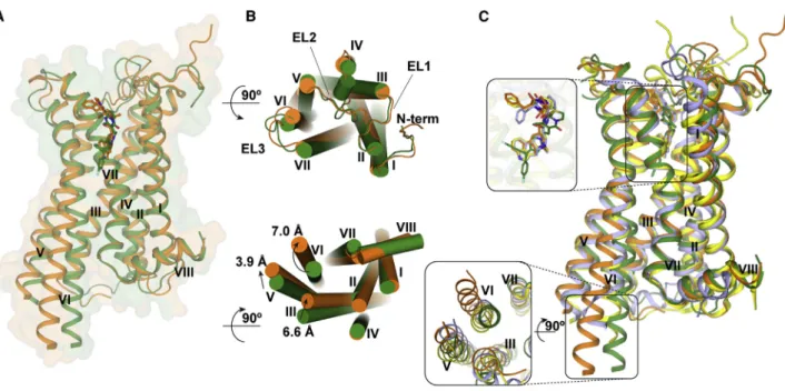
2013), all extracellular loops are well resolved in both 5-HT2C crystal structures. Superposition of the ERG and RIT complexes shows shifts of 7.0 A˚, 3.9 A˚, and 6.6 A˚ at the intracellular ends of helices VI, V, and III, respectively, indicating that they represent different conformational states of the receptor (Figure 1B).
Compared to the active and inactive state structures of b2-adrenergic receptor (b2AR) (Rasmussen et al., 2011; Wacker
et al., 2010), 5-HT2C-RIT resembles the inactive state
conforma-tion of b2AR, whereas 5-HT2C-ERG shows all ‘‘active-like features’’ exemplified by the active state of b2AR (Figure S2).
Superposition of the 5-HT1B, 2B, 2C-ERG and 5-HT2C-RIT
(Wacker et al., 2013; Wang et al., 2013) structures shows an
increased opening on the intracellular ends of helices V and VI in the order 5-HT2B-ERG < 5-HT1B-ERG < 5-HT2C-ERG compared to 5-HT2C-RIT (Figures 1C andS2).
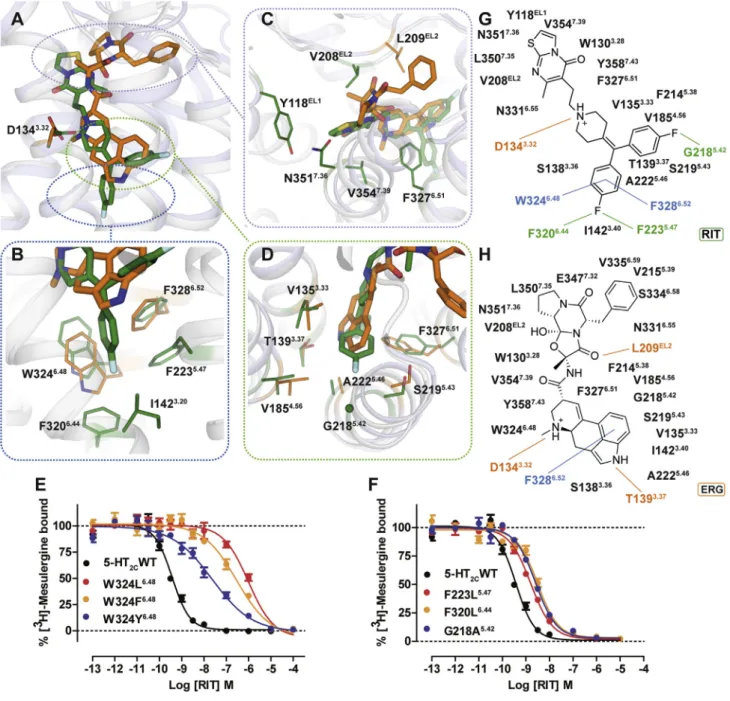
Different Binding Modes of ERG and RIT
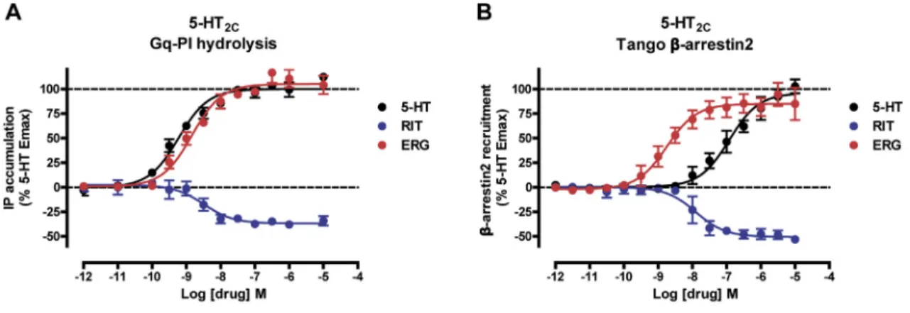
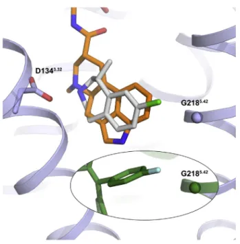
ERG and RIT have different chemical scaffolds with distinct 5-HT2Cactivity: ERG acts as an agonist, whereas RIT is an in-verse agonist for both Gaq-inositol phosphate accumulation and b-arrestin2 recruitment (Figure S3). These differential effi-cacies are mirrored by their distinctive binding modes as observed in the 5-HT2Ccrystal structures, where the only com-mon interaction is the salt bridge between the protonated nitro-gen of the ligand and the conserved aspartate, D1343.32—a canonical interaction for aminergic and many other GPCRs (
Fig-ure 2A). Compared to the ERG-bound structure, RIT binds
approximately one helical turn deeper into the TM bundle (
Fig-ure 2B), which is outside of the recognized orthosteric site of
other solved aminergic GPCR structures (Venkatakrishnan
et al., 2013). By contrast, ERG’s shallower binding pose allows
an aromatic interaction only with F3286.52and van der Waals (vdW) contact with W3246.48(Figures 2B and 2H).
The deep binding pose of RIT in the 5-HT2Cis characterized by one of the 4-fluorophenyl groups encased in a hydrophobic pocket between helices V and VI, where it interacts via halogen-aromatic interactions with F2235.47 and F3206.44 and aromatic edge-to-facep-pstacking interactions with residues F3286.52and W3246.48, the purported ‘‘toggle switch’’ important for GPCR activation (Figures 2B and 2G) (Preininger et al., 2013). We validated RIT’s binding pose by mutating W3246.48, F2235.47 and F3206.44, all of which decreased RIT’s affinity (Figures 2E and 2F;Table S2). The W324L6.48mutation especially reduced
with improved efficacy for complex disorders (Besnard
etal.,2012).
Structure-baseddrugdesignapproacheshavefacilitatedthe creation of selective GPCR drugs (Wang et al., 2017) with improved therapeutic profiles, as exemplified by the recent discoveryofm-opioidselectiveGprotein-biasedagonists( Man-gliketal.,2016).Structure-guidedapproaches,perforce,require high-resolutioncrystalstructuresinordertoexploit ligand-bind-ingpocket interactions atthetargeted GPCR(Wacker etal.,
2017a; Wang et al., 2017). However, structure-guided drug
designwillnottypicallypredictacompound’soff-targetactions. Ultimately, structure-based polypharmacological drug design willrequiremanyhigh-resolutionGPCRstructureswithvarious
chemotypes to illuminate how polypharmacology might be
achievedatmultipledefineddrugtargets.
The5-HT2Cserotoninreceptor(5-HT2C)isavalidatedtarget for anti-obesity medications as illustrated by the selective 5-HT2Cagonistlorcaserin(Belviq).The5-HT2Cisalsoapotential therapeutictargetfordepression,schizophrenia,drugaddiction, andotherdisorders(McCorvyandRoth,2015;Palaciosetal.,
2017;Pogorelovetal.,2017).Creatingselective5-HT2Cagonists
isextremelychallenging,however,asoff-targetagonistactivity at theclosely related 5-HT2A and 5-HT2B receptors leads to lysergic acid diethylamide (LSD)-like hallucinations (Nichols, 2016)andcardiacvalvulopathy(Roth,2007),respectively. More-over,the5-HT2CexhibitsseveralRNA-editedisoforms, where thenon-editedisoform (INI) displayshigh constitutiveactivity thatcanbeexploited toquantify aninverse agonist’sactivity
(Barkeretal.,1994).
To date,only two serotoninreceptor structureshave been solved:the5-HT1Bandthe5-HT2B,bothincomplexwith ergot-amine(ERG)(Wackeretal.,2013,2017b;Wangetal.,2013)and, most recently, 5-HT2B in complex with LSD (Wacker et al.,
2017b).ERG isa naturallyoccurringergotanti-migrainedrug
thatcontains an ergoline nucleus, a common chemotypefor manydrugs includingLSD,bromocriptine,methysergide,and lisuride. ERG, however, has a complex polypharmacological profilewithserioussideeffects,includingcardiacvalvulopathy via5-HT2Bserotoninreceptor agonism,whichlimits its wide-spreaduseasananti-migrainemedication.Incontrast,ritanserin (RIT)isaselective5-HT2receptorinverseagonistthathasbeen previouslyinvestigatedasanadjunctforantipsychotic medica-tions(DenBoeretal.,2000).RITcontainsa 4-benzylidenepiper-idine core scaffold, which is also found in the promiscuous antipsychoticclozapine.The4-benzylidenepiperidineisaknown privileged scaffold with applications for exploiting multiple GPCRstoyieldadesiredpolypharmacologicalprofile(Garland
andGloriam,2011).
Here,wepresentthestructuresofthe5-HT2CINIisoformin
complexwithboththepolypharmacologicalagonistERG and
theselectiveinverseagonist RITinorderto clarifythe struc-turalfeaturesresponsibleforGPCRpolypharmacology.Tothis end,werevealtheactive-likestate5-HT2Creceptorstructure
with ERG and, for comparison, an inactive state with RIT.
Knowledgeofthesecrystalstructureswillfacilitatethebasisof
chemotype-specific recognition, inverse agonism, and
RIT affinity by >1,000-fold, whereas W324F6.48and W324Y6.48 mutations, which preserve the aromatic character of this residue, had substantially less effects on RIT binding affinity (Figure 2E;
Table S2). These mutations support the hypothesis that the
4-flu-orophenyl moiety is dependent on p-p stacking interactions below the commonly recognized aminergic orthosteric site and that these interactions with W3246.48 are apparently driving RIT’s deep binding pose.
The second 4-fluorophenyl group of RIT occupies essentially the same site as the indole of ERG and is lined by V1353.33, T1393.37, V1854.56, S2195.43, A2225.46, and F3276.51(Figure 2D), with the aromatic ring systems positioned orthogonally to one another in the binding pocket (Figures 2A and 2D). The second 4-fluorophenyl group also apparently ‘‘pushes’’ against the back-bone of helix V at residue G2185.42(Figure 2D). Mutation of this residue to G218A5.42attenuates RITs binding affinity more than 10-fold (Figure 2F) but only has a modest effect on ERG affinity, further supporting the differential binding poses (Table S2).
The thiazolopyrimidine of RIT, which stems from the charged nitrogen of the 4-benzylidenepiperidine core scaffold, is located orthogonally to the ergoline ring in the 5-HT2C-ERG structure and is positioned toward helices II and VII interacting with Y118EL1, V208EL2, F3276.51, N3517.36 and V3547.39 (Figure 2C). The ligand contacts at EL2 also differ between the 5-HT2C-RIT and 5-HT2C-ERG structures as RIT has only hydrophobic vdW con-tacts, but ERG engages in a hydrogen bond with the backbone of L209EL2(Figure 2C). Finally, ERG’s terminal benzyl moiety
ex-tends much further toward the extracellular loops of the receptor than RIT making vdW contacts with residues L209 in EL2 and V2155.39, S3346.58, and V3356.59at the topmost turns of helices V and VI (Figure 2H).
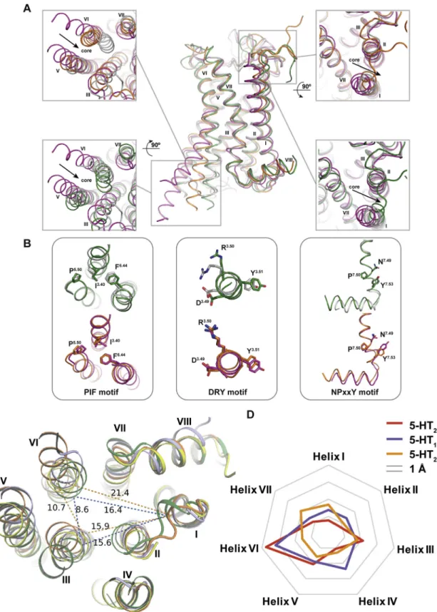
Conformational Changes between Agonist- and Inverse
Agonist-Bound 5-HT2C
Comparison of the 5-HT2C-ERG and 5-HT2C-RIT crystal struc-tures also provides key insights into activation-related confor-mational changes in 5-HT2C. Importantly, the intracellular end of the helix VI in 5-HT2C-ERG is tilted outward by 7.0 A˚ and helix III is shifted inward by 6.6 A˚ compared to the 5-HT2C-RIT struc-ture (Figure 1B, bottom panel). Hydrogen bonds between the highly conserved D1343.32 and Y3587.43 are observed in both structures (Figure 3A). A comparison of the structures reveals an overall 1–2 A˚ binding pocket compaction with inward shift of helices V, VI, and VII around the ergoline moiety of ERG (
Fig-ure 1B, top panel). Such compaction of the binding pocket is
expected for ergolines and the endogenous agonist 5-hydroxy-tryptamine (5-HT) (Wang et al., 2013), both of which are less bulky than the 4-(diphenylmethylene)-piperidine core of the inverse agonist RIT. These helix movements are accompanied by rotamer switches in the conserved P5.50-I3.40-F6.44 (P-I-F) motif and a shift of the W3246.48‘‘toggle switch’’ in helix VI (
Fig-ure 3B), representative of active-state-like structures at biogenic
amine and other GPCRs (Venkatakrishnan et al., 2013; Wacker
et al., 2013). As mentioned above, the inverse agonist RIT binds
Figure 1. Overall Architecture of 5-HT2C-ERG and -RIT and Their Comparison with 5-HT1B,2B-ERG
(A) ERG- (agonist) and RIT- (inverse agonist) bound 5-HT2Care shown as orange and green cartoons. ERG and RIT are shown as orange and green balls and sticks, respectively. The helices are indicted with I through VIII.
(B) The top panel is the extracellular view. The bottom panel is the intracellular view (loops are omitted for clarity). The arrows show helical shifts as indicated (distance measured by the Caatoms of I3036.27, A2455.69, and R1573.55) between the 5-HT2C-ERG and 5-HT2C-RIT structures.
(C) Superposition of the following structures: 5-HT2C-ERG (orange), 5-HT2C-RIT (green), 5-HT1B-ERG (light blue; PDB: 4IAR), and 5-HT2B-ERG (yellow; PDB: 4IB4).
Figure 2. Different Binding Modes of ERG and RIT in 5-HT2C
(A–D) Superposition of the 5-HT2C-ERG (orange) and RIT (green) ligand binding pockets with an overview and close-up views of the orthosteric and extended binding sites. (A) Overall distinctive binding modes of ERG and RIT observed in 5-HT2C. (B) The deeper binding pose of RIT and shallower binding pose of ERG. (C) Key interactions around RIT’s thiazolopyrimidine as well as ERG’s ergoline ring and terminal benzyl moiety. (D) The second 4-fluorophenyl group of RIT occupies the same site as the indole of ERG and positions orthogonally to each another in the binding pocket.
(E) W3246.48
appears to be a major determinant of RIT’s binding mode as measured by binding affinity loss at mutations. Binding affinity is partially recovered by the conservative mutation W324F6.48
. (F) Mutations of residues F2235.47
, F3206.44
, and G2185.42
also show RIT affinity loss, but less compared to W3246.48 .
(G and H) Schematic representation of RIT (G) and ERG (H) contacts with the 5-HT2C, respectively. Lines indicate interactions types: orange, polar, salt bridges, and hydrogen bonds; blue, aromatic contacts; and green, dipolar interactions.
deeper than most class A GPCR ligands in the TM helical bundle where one of its 4-fluorophenyl groups forms tight interaction with W3246.48, I1423.40, and F3206.44side chains thus apparently preventing the conformational changes in these key activation microswitches. Furthermore, the 4-fluorophenyl group of RIT is in close proximity to I1423.40apparently facilitating an outward shift of the intracellular end of helix III (Figure 1B, bottom panel), which potentially explains the inverse agonist activity of RIT at this receptor isoform.
To test this hypothesis, functional studies were performed with mutants of these key microswitch residues in 5-HT2Cmeasuring Gaq activation and b-arrestin2 recruitment. Mutations of the ‘‘toggle switch’’ W3246.48 and F3206.44, which are part of the P-I-F trigger motif, selectively abolish RIT’s Gaqinverse agonism without affecting RIT’sb-arrestin2 inverse agonist activity (
Fig-ure 3C) indicating that these microswitch residues are mainly
involved in the Gaq activation process. Furthermore, the I142A3.40mutation of the P-I-F motif also selectively abolishes
RIT’s Gaqinverse agonism and the I142F3.40mutant transforms RIT into a Gaqpartial agonist with little agonist effect on arrestin recruitment (Figure 3C). This mutant I142F3.40 likely imparts additional aromatic stacking properties to RIT, indicating that the P-I-F motif is important for RIT’s inverse agonist profile (
Fig-ure 3C). These results support a model whereby the
4-fluoro-phenyl moiety of RIT stabilizes an inactive state of the receptor via interference with the ‘‘toggle switch’’ W6.48 and ‘‘trigger motif’’ P5.50-I3.40-F6.44and that these microswitch residues are critical for inverse agonist activity and the Gaq activation process, in general, at the 5-HT2C.
Multiple ERG-Serotonin Receptor Structure Complexes Reveal Determinants of Polypharmacology
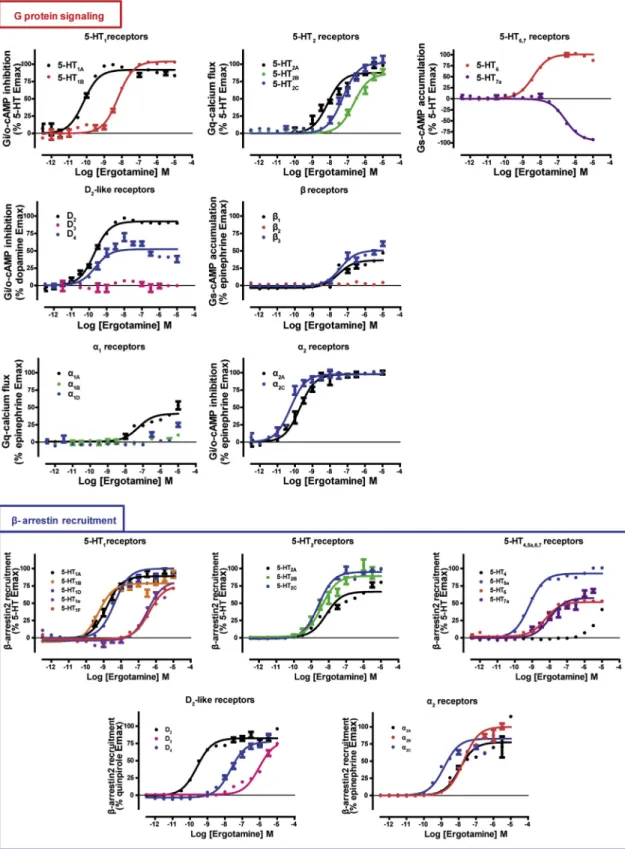
To illuminate ERG’s polypharmacology, we first assessed its binding affinity across the aminergic receptor family. ERG shows appreciable affinity (Ki< 10mM) for nearly 70% of all human ami-nergic GPCRs with low or sub-nanomolar affinity for fifteen of
Figure 3. Conformational Changes between 5-HT2C-ERG and -RIT Structures and Mutagenesis Validation
(A) ERG (orange sticks) and RIT (green sticks) in the binding pocket of 5-HT2C. Key residues in 5-HT2C-ERG and 5-HT2C-RIT are shown in orange and green sticks, respectively. Hydrogen bonds between D1343.32
and Y3587.43
are shown by the dashed line. (B) Conformational changes of I1423.40
and F3206.44
in the P-I-F motif and the W3246.48
‘‘toggle switch’’ in helix VI. (C) Mutations of W3246.48
and F3206.44
completely abolish RIT’s Gaqinverse agonism, yet retainb-arrestin2 inverse agonism. Mutations of I1423.40selectively abolish RIT’s Gaqinverse agonism and instead show weak agonism.
is ideal for smaller aliphatic residues such as leucine, valine, or isoleucine (Figures 4A and 4B), likely contributing to ERG’s shared binding pose among GPCRs.
RIT Structure Reveals Determinants of 5-HT2Subtype
Selectivity
Although RIT is selective for 5-HT2-family receptors, it contains a 4-benzylidenepiperidine moiety, which is a variant of a known 4-arylpiperidine GPCR privileged structure (Garland and
Glo-riam, 2011) (Figure 5A). The 5-HT2C-RIT crystal structure reveals
that D3.32, W6.48, and at least one aromatic residue at positions 5.47 and/or 6.52 are important for binding of this privileged struc-ture. Within class A GPCRs, this sub-site is conserved in most aminergic receptors, all four opioid receptors and the melanin-concentrating hormone receptor 1 (MCR1) (Figure 5B). Like the ergoline scaffold of ERG, the 4-benzylidenepiperidine privileged structure found in RIT ensures binding to a wide range of recep-tors thereby providing a molecular basis for use of this privileged structure as a starting point for polypharmacological design. Indeed, this is nicely illustrated by the polypharmacology of clozapine binding to nearly thirty aminergic receptors (Roth
et al., 2004; Yadav et al., 2011a) and by cinnarizine binding to
fifteen different aminergic receptors and them-opioid receptor
(Figure 5C). By contrast, RIT binds almost exclusively to 5-HT2
receptors with >100-fold preference over any other tested
GPCR with the exception of the H2 histamine receptor
(19-fold) (Figure 6A;Table S3).
To uncover the molecular basis for RIT’s 5-HT2receptor selec-tivity, despite containing the embedded promiscuous 4-benzyli-denepiperidine privileged structure, we identified residues responsible for RIT’s selectivity and designed a series of muta-tions. We exchanged the differing binding site residues in 5-HT2Cwith those of 5-HT1Athat demonstrated low affinity for RIT (Figure 6A). The most pronounced effects occurred for the two mutants G218S5.42(60-fold) and V354N7.39(425-fold), both of which are unique to 5-HT2A-C subtypes (Figure 6B; Table S5). In accordance with the structure showing only non-specific interactions, the other mutations, e.g., S138C3.36, showed small or negligible effect on RIT affinity. As previously mentioned, G2185.42 and V3547.39 engage the second 4-fluorophenyl of RIT and the thiazolopyrimidine, both of which are structural mod-ifications stemming from the 4-benzylidenepiperidine core scaffold that are unique to RIT (Figure 6C). Furthermore, the importance for residues 5.42 and 7.39 conferring 5-HT2 selec-tivity is supported by the fact that 5-HT1A, which contains S5.42 and N7.39 at these positions, has a1,300-fold lower af-finity for RIT than does 5-HT2Cbut sub-nanomolar affinity for ERG (Table S3). In fact, 5-HT4, which does not demonstrate any appreciable RIT affinity at all, contains larger residues at both 5.42 and 7.39 positions (Cys and Leu, respectively). To confirm the notion that residues 5.42 and 7.39 are more impor-tant for driving RIT subtype selectivity rather than ligand promis-cuity, we also sought to test clozapine at these mutations, which also contains an embedded 4-benzylidenepiperidine privileged structure. Interestingly, clozapine is only modestly affected (4-fold or below) by any of the tested binding site mutants
(Table S5). These results clarify that RIT’s 4-fluorophenyl
and thiazolopyrimidine interactions with respective residues those receptors (Table S3). Further characterization of ERG
activityrevealedadiversefunctionalprofileofeitherGprotein agonist activityor b-arrestin2 recruitmentactivity, or insome casesboth(FigureS4),indicatingthatERGpossessesfunctional
selectivity across the aminergic GPCRome. However, ERG
possessed no apparent Gprotein-dependent agonist activity attheD3dopamine,a1B,a1Dorb2adrenergicreceptorsdespite appreciableaffinity indicatingantagonism at thesereceptors. Interestingly,ERGwasaninverseagonistat5-HT7whereinverse agonismhasbeenpreviouslyreportedforthestructurallyrelated ergolineanalog LSD (Wacker et al.,2013). Tofully illuminate ERG’spolypharmacologicalprofile,ERGwasthenscreenedat 320non-olfactoryhumanGPCRsviaab-arrestin2recruitment assay (Kroeze et al., 2015), which unexpectedly revealed apparentopioidreceptoragonistactivity(FigureS5; TableS4).
Touncoverthe molecularbasis forERG’s highaffinity and polypharmacologicalprofileataminergicGPCRs,weanalyzed thebinding modesof ERG atthe 5-HT1B, 2B, 2C-ERG crystal structurecomplexes(Wacker etal.,2013;Wangetal.,2013). ERGsharesacommonbindingmodeatallthreeofthese recep-tors,wheretheonlydifference istheorientationofthebenzyl substituentin5-HT1B-ERGcomplex(Figure 1C).Inregions in
which ERG packs most tightly and has polar interactions—
aroundtheergolinescaffold—allreceptorsdemonstratinghigh ERGaffinityexhibithighlyconservedresidueproperties(Figures 4Aand4B,leftpanel).Theergolinecoreissurroundedbyfour heliceswherehelicesIIIand VIsandwich theplanar sidesof theergolinecore,whereastheothertwo edgesareenclosed byhelicesVand VII.InadditiontoD3.32,which formsasalt bridge to most aminergic ligands, positions 3.33, 3.36, 3.37, 5.42,and5.46areinveryclosecontactwithERGinallthree structuresand arenot ableto accommodatesignificant ERG affinitywhentherearesignificantlylargeraminoacidspresent atthese residue positions. Additionally, the conserved T3.37 residue and semi-conserved A/S/T5.46 residues form either
vdW interactions and/or a hydrogen bond to ERG’s indole
N-H.Threeadditional helixVIpositions (6.48,6.51, and6.52)
contribute favorable vdW and aromatic interactions where
alanine or leucine mutations of these residues only lead to reducedbutnotabrogatedERGbindingat5-HT2C(TableS2) and5-HT1B,2B(Wangetal.,2013).Therefore,theergolinecore isrecognizedbyninekeyresidues,eightofwhichhavespecific conservedaminoacidpropertiestoenablebinding.Allofthese propertiesare presentin all receptors that demonstratehigh ERGaffinity(Figures4Aand4B,leftpanel).
ForERG’scyclictripeptideandbenzylsubstituents,the struc-turesdisplayonlynon-specificside-chainvdWcontacts,which tolerateahighdegreeofflexibilityanddiversityofaminoacids. OneexceptiontothisistheconservationofapositioninEL2 (L209in5-HT2C)ofallreceptorsdemonstratinghighERGaffinity, wherea small aliphatic residue such as a leucine, valine, or isoleucineisalmostalwayspresent.Interestingly,the homolo-gous EL2 residue found in 5-HT2B (L209EL2) and in 5-HT2A (L229EL2)hasbeenrecentlystudiedasadeterminantforligand residence time, which contributes to b-arrestin recruitment
(Wackeretal.,2017b).Similarlyobservedinthe5-HT2C
G2185.42and V3547.39, which are residues exclusive to 5-HT2 re-ceptors, are primary determinants of RIT’s 5-HT2selectivity.
DISCUSSION
Here, we determined agonist- and antagonist-stabilized struc-tures of the 5-HT2C receptor, a molecular target important for
drugs that can treat diseases as diverse as obesity, schizophrenia, and drug abuse. Importantly, the 5-HT2C-ERG and -RIT structures not only reveal the molecular determinants for selective ligand binding across GPCRs but also reveal a structural basis for promiscuous ligand ergotamine binding across several receptor subtypes (e.g., serotonin, dopamine, adrenergic, histamine, muscarinic, and opioid). We anticipate that our findings will
Figure 4. Polypharmacological Profile of ERG
(A) The chemical structure of ERG highlighting the ergoline core (brown), tripeptide (blue), and benzyl portions (purple) in the structures of 5-HT2C(orange), 5-HT1B (light blue; PDB: 4IAR), and 5-HT2B(yellow; PDB: 4IB4). ERG is shown as thick sticks, and the protein backbone is represented as a cartoon and the side chains of relevant amino acids as thin sticks. For clarity, the Ca-carbon of G5.42 is depicted as a small sphere.
(B) Mean values of ERG affinities (Ki) in nanomolars from radioligand competition binding assays represent the mean from experiments performed in duplicate (see alsoTable S3). Sequence alignments of the three subsites: gray background, conservation of positions and residue types critical for ERG binding; SN, short and negative; IH, intermediate length and hydrophobic; SU, small and unbranched; VSP, very small or small and polar; HF, hydrophobic with a maximum size of Phe; HA, hydrophobic and aliphatic. Receptors are listed in order of decreasing ERG affinity (Kicolor scale green over white to orange) but separated into aminergic and non-aminergic receptors.
S6), but also will provide a primer for understanding how drugs like clozapine interact with multiple targets, ultimately facilitating a structure-guided polypharmacological approach to drug design.
Figure 5. Polypharmacology of the 4-Benzylidenepiperidine Privileged Structure
(A) The interactions between the 4-benzylidenepiperidine of RIT and its surrounding residues in the 5-HT2C-RIT structure.
(B) Sequence alignments of the privileged structure subsites with the number of ligands identified in a search for GPCR ligands in ChEMBL containing the privileged structure and Ki< 1mM. Conservation of positions critical for binding is shown in a gray background, and conservation is assessed by the residue properties crucial for interaction; 1AR, aromatic residue in at least one of the two positions.
(C) Examples of ligands identified in the search with confirmed GPCR targets (Ki< 1mM) are listed, and the privileged structure is highlighted in red in the chemical structures. For cinnarizine and clozapine, the hits from ChEMBL (Bento et al., 2014) have been supplemented with hits from the PDSP Kidatabase (Yadav et al., 2011a).
ultimately prove fundamental for not only the design of
selective 5-HT2C ligands, such as lorcaserin (Belviq), which, similarto RIT,has G2185.42 asamajorselectivitydeterminant
To unveil the structural basis of GPCRs’ polypharmacology, we identified ten key conserved amino-acid residues critical for ERG promiscuity by using sequence alignments of residues that interact with the ergoline core and benzyl sub-sites at eigh-teen aminergic receptors that show high ERG affinity (<30 nM). In fact, deviation from these conserved amino acid types reliably predicted decreased ERG affinity, dependent on position and type of amino acid present. To estimate the full target profile of ERG, we performed a sequence alignment focused on the ten ERG-interacting residues at all class A GPCRs and found that ERG’s polypharmacology is not confined to aminergic GPCRs but also extends to the delta-opioid receptor (d), which has a semi-conserved binding site and, thus, lower affinity. These ten conserved amino-acid residues can now be used to guide struc-ture-based design of polypharmacological ligands at these receptor types, especially for computational-based methods that can predict known and unexpected drug targets (Besnard
et al., 2012; Chaudhari et al., 2017). Additionally, we identify
two positions for determining RIT 5-HT2 selectivity, with V3547.39having the largest effect on selectivity (425-fold;Table
S5), which should be avoided in design of compounds with poly-pharmacology profiles. This is nicely illustrated by examining cyproheptadine (Periactin), whose structure is based on the same 4-(diphenylmethylene)-piperidine core as RIT but lacks the thiazolopyrimidine (Figure 5C) interacting with V3547.39. Cyproheptadine displays reduced selectivity between aminergic receptors (https://pdsp.unc.edu/databases/kidb.php) and within the 5-HT receptors, e.g., the 5-HT2Cversus 5-HT1A selec-tivity is reduced from 1,288-fold for RIT (Table S3) to 4- to 26-fold for cyproheptadine (Toll et al., 1998). Taken together, our anal-ysis of the 5-HT2C-ERG and -RIT structures in comparison with the previously published 5-HT1B/2B-ERG structures reveal highly conserved receptor sub-pockets and ligand sub-structures responsible for GPCR polypharmacology in contrast to the inter-actions that confer ligand selectivity.
The successful polypharmacological drugs, however, depend on a diverse range of incorporated pharmacological activity (e.g., agonism, biased agonism, inverse agonism) at each respective target. As mentioned, clozapine binds to nearly 30 aminergic receptors at which clozapine mainly demonstrates
Figure 6. Ritanserin’s Selectivity on 5-HT2Receptors
(A) Mean values of RIT affinities (Ki) in nanomolars for radioligand competition binding assays from experiments performed in duplicate. Sequence alignment of the RIT binding site residues in the eleven serotonin receptors for which RIT’s binding affinity was determined. Conservation of residue type by 5-HT2Cis indicated by a gray background, and mutated positions are indicated with black boxes.
(B) The unique residue types in positions 5.42 and 7.39 of the 5-HT2A-Creceptors (see A) appear as major determinants of the RIT selectivity for these receptor subtypes as binding affinity decreased when mutated in 5-HT2Cto the corresponding residues of 5-HT1A, i.e., G218S
5.42
and V354N7.39
. Data represent mean±
SEM of three independent experiments performed in duplicate. (C) The interactions between RIT and G2185.42
and V3547.39
antagonist activity (Roth et al., 2004). However, clozapine has also been shown to demonstrate functional selectivity at 5-HT2A receptor (Yadav et al., 2011b) as well as inverse agonism at 5-HT7 receptor (Thomas et al., 1998). Therefore, incorporating functional selectivity or biased agonism into a drug’s polypharmacological profile can often lead to novel ave-nues of therapeutic potential, as observed with aripiprazole, a polypharmacological drug that demonstrates functional selec-tivity (Shapiro et al., 2003; Tuplin and Holahan, 2017). The 5-HT2C-RIT and the ERG crystal structures shed light on such a strategy with RIT’s and ERG’s opposing pharmacological action leading to different activation states of the 5-HT2C. We identified key areas in both the P-I-F and the W6.48 ‘‘toggle’’ microswitch trigger motifs that appear to be mainly involved in the 5-HT2C-Gaq activation process and not necessarily involved inb-arrestin recruitment, which may serve as a starting point for pathway-selective drugs. In addition, we also show that RIT interferes with these microswitch motifs to produce
Gaq-dependent inverse agonism, which appears to be an
important mechanism for stabilizing the inactive state of the receptor. Targeting these motifs may represent a potential mechanism for the design of novel inverse agonists, especially aimed at 5-HT2receptors where current inverse agonists aimed at this receptor subtype (e.g., pimavanserin) are indicated for schizophrenia and psychosis-associated with Parkinson’s dis-ease (Meltzer and Roth, 2013).
It is worth noting that the non-edited INI isoform of the 5-HT2C was selected to study the ligand-receptor interactions and its indications to GPCR polypharmacology. There are signaling dif-ferences among isoforms, and studying other 5-HT2Cisoforms could result in different pharmacological profiles for the specific receptor subtype. However, because the binding pocket resi-dues are identical in the edited isoforms, our inferences are not necessarily isoform specific, and we expect our conclusion con-cerning polypharmacology and selectivity, which are based on ligand-receptor binding, to be generally applicable.
Our knowledge of the molecular basis for polypharmacology is still in its infancy, and it remains difficult to predict what combi-nation of targets will ultimately lead to more effective treatments of complex diseases. Evident is the fact that some of the most successful GPCR drugs (e.g., Clozaril or Abilify for schizo-phrenia) bind to multiple receptors and that key target combina-tions ultimately provide the drug’s efficacy. Obtaining a better understanding of the structural basis for GPCR polypharmacol-ogy is thus the first step toward a rational utilization of this principle for future GPCR drug design. Our approach also may be broadly useful for determining the polypharmacological determinants of other privileged scaffolds or promiscuous drugs and provide a roadmap for the rational design of polypharmaco-logical ligands.
STAR+METHODS
Detailed methods are provided in the online version of this paper and include the following:
d KEY RESOURCES TABLE
d CONTACT FOR REAGENT AND RESOURCE SHARING
d EXPERIMENTAL MODEL AND SUBJECT DETAILS
B Cell Lines
d METHOD DETAILS
B Rational Design of Thermostabilizing Mutations
B Protein Engineering for Structure Determination
B Protein Expression
B Protein Purification
B Lipidic Cubic Phase Crystallization
B Data Collection and Structure Determination
B Protein Stability Assays
B ERG Binding Target Profiling
B ChEMBL Privileged Structure Search
B Generation of 5-HT2CMutant Constructs
B Membrane Preparation and Radioligand Binding
B Test for RIT Selectivity Mutations
B Phosphoinositide Hydrolysis (PI) Assay
B Tango Arrestin Recruitment Assay
B GPCRome Screening
B Docking of Lorcaserin
d QUANTIFICATION AND STATISTICAL ANALYSES
B Dose-Response and Binding Affinity Calculations
B Test for RIT Selectivity Mutations
d DATA AND SOFTWARE AVAILABILITY
B Data Resources
SUPPLEMENTAL INFORMATION
Supplemental Information includes six figures and seven tables and can be found with this article online athttps://doi.org/10.1016/j.cell.2018.01.001.
ACKNOWLEDGMENTS
This work was supported by grants from the Ministry of Science and Technol-ogy of China (2014CB910400 and 2015CB910104), the National Nature Sci-ence Foundation of China (31330019) (to Z.J.L.), the NIH (R01MH112205 and U19MH82441), the Michael Hooker Distinguished Professorship, the NIMH Psychoactive Drug Screening Program (271201300017C-4-01) (to B.L.R., J.M., and others from the Roth lab), the NIH (U54GM094618) (to V.K.), the Russian Science Foundation (16-14-10273) (to P.P.), and the Lundbeck Foun-dation (R163-2013-16327) (to D.E.G.). We thank the Shanghai Municipal Gov-ernment and ShanghaiTech University for financial support. The diffraction data were collected at the BL41XU of SPring-8 (JASRI proposal 2015B1031 and 2016A2731), GM/CA at APS of Argonne National Lab, and X06SA beamline at the Swiss Light Source of the Paul Scherrer Institute. We thank K. Hasegawa, H. Okumura, H. Murakami, M. Audet, M. Wang, C. Huang, and V. Olieric for their help with data collection. We thank the Cloning, Cell Expression, Assay, and Protein Purification Core Facilities of iHuman Institute for their support. We thank I. Wilson for careful review and insightful discussions on the manuscript; A. Walker for assistance with the manuscript; and T. Hua, V. Cherezov, T. Tson-kov, G. Song, W. Shui, D. Liu, H. Tao, G. Zhong, F. Xu, B. Wu, Q. Zhao, and W. Liu for helpful discussions.
AUTHOR CONTRIBUTIONS
DECLARATION OF INTERESTS
All authors declare no competing interests.
Received: July 14, 2017 Revised: October 25, 2017 Accepted: January 3, 2018 Published: February 1, 2018
REFERENCES
Abagyan, R., and Totrov, M. (1994). Biased probability Monte Carlo conforma-tional searches and electrostatic calculations for peptides and proteins. J. Mol. Biol.235, 983–1002.
Adams, P.D., Afonine, P.V., Bunko´czi, G., Chen, V.B., Davis, I.W., Echols, N., Headd, J.J., Hung, L.W., Kapral, G.J., Grosse-Kunstleve, R.W., et al. (2010). PHENIX: a comprehensive Python-based system for macromolecular struc-ture solution. Acta Crystallogr. D Biol. Crystallogr.66, 213–221.
Alexandrov, A.I., Mileni, M., Chien, E.Y., Hanson, M.A., and Stevens, R.C. (2008). Microscale fluorescent thermal stability assay for membrane proteins. Structure16, 351–359.
Ballesteros, J.A., and Weinstein, H. (1995). Integrated methods for the construction of three-dimensional models and computational probing of struc-ture-function relations in G protein-coupled receptors. In Methods in Neurosci-ences, C.S. Stuart, ed. (Academic Press), pp. 366–428, [19].
Barker, E.L., Westphal, R.S., Schmidt, D., and Sanders-Bush, E. (1994). Constitutively active 5-hydroxytryptamine2C receptors reveal novel inverse agonist activity of receptor ligands. J. Biol. Chem.269, 11687–11690.
Barnea, G., Strapps, W., Herrada, G., Berman, Y., Ong, J., Kloss, B., Axel, R., and Lee, K.J. (2008). The genetic design of signaling cascades to record re-ceptor activation. Proc. Natl. Acad. Sci. USA105, 64–69.
Bento, A.P., Gaulton, A., Hersey, A., Bellis, L.J., Chambers, J., Davies, M., Kru¨ger, F.A., Light, Y., Mak, L., McGlinchey, S., et al. (2014). The ChEMBL bioactivity database: an update. Nucleic Acids Res.42, D1083–D1090.
Besnard, J., Ruda, G.F., Setola, V., Abecassis, K., Rodriguiz, R.M., Huang, X.P., Norval, S., Sassano, M.F., Shin, A.I., Webster, L.A., et al. (2012). Auto-mated design of ligands to polypharmacological profiles. Nature 492, 215–220.
Bourdon, D.M., Wing, M.R., Edwards, E.B., Sondek, J., and Harden, T.K. (2006). Quantification of isozyme-specific activation of phospholipase C-beta2 by Rac GTPases and phospholipase C-epsilon by Rho GTPases in an intact cell assay system. Methods Enzymol.406, 489–499.
Boyle, E.A., Li, Y.I., and Pritchard, J.K. (2017). An expanded view of complex traits: from polygenic to omnigenic. Cell169, 1177–1186.
Caffrey, M., and Cherezov, V. (2009). Crystallizing membrane proteins using lipidic mesophases. Nat. Protoc.4, 706–731.
Chaudhari, R., Tan, Z., Huang, B., and Zhang, S. (2017). Computational poly-pharmacology: a new paradigm for drug discovery. Expert Opin. Drug Discov. 12, 279–291.
Cheng, J., McCorvy, J.D., Giguere, P.M., Zhu, H., Kenakin, T., Roth, B.L., and Kozikowski, A.P. (2016). Design and discovery of functionally selective seroto-nin 2C (5-HT2C) receptor agonists. J. Med. Chem.59, 9866–9880.
Cherezov, V., Rosenbaum, D.M., Hanson, M.A., Rasmussen, S.G., Thian, F.S., Kobilka, T.S., Choi, H.J., Kuhn, P., Weis, W.I., Kobilka, B.K., and Stevens, R.C. (2007). High-resolution crystal structure of an engineered human beta2-adren-ergic G protein-coupled receptor. Science318, 1258–1265.
Cherezov, V., Hanson, M.A., Griffith, M.T., Hilgart, M.C., Sanishvili, R., Nagar-ajan, V., Stepanov, S., Fischetti, R.F., Kuhn, P., and Stevens, R.C. (2009). Rastering strategy for screening and centring of microcrystal samples of human membrane proteins with a sub-10 microm size X-ray synchrotron beam. J. R. Soc. Interface6(Suppl 5), S587–S597.
Collaborative Computational Project, Number 4 (1994). The CCP4 suite: pro-grams for protein crystallography. Acta Crystallogr. D Biol. Crystallogr.50, 760–763.
Den Boer, J.A., Vahlne, J.O., Post, P., Heck, A.H., Daubenton, F., and Olbrich, R. (2000). Ritanserin as add-on medication to neuroleptic therapy for patients with chronic or subchronic schizophrenia. Hum. Psychopharmacol. 15, 179–189.
Elkins, J.M., Fedele, V., Szklarz, M., Abdul Azeez, K.R., Salah, E., Mikolajczyk, J., Romanov, S., Sepetov, N., Huang, X.P., Roth, B.L., et al. (2016). Compre-hensive characterization of the Published Kinase Inhibitor Set. Nat. Biotechnol. 34, 95–103.
Emsley, P., Lohkamp, B., Scott, W.G., and Cowtan, K. (2010). Features and development of Coot. Acta Crystallogr. D Biol. Crystallogr.66, 486–501.
Garland, S.L., and Gloriam, D.E. (2011). A ligand’s view of target similarity: che-mogenomic binding site-directed techniques for drug discovery. Curr. Top. Med. Chem.11, 1872–1881.
Huang, X.P., Setola, V., Yadav, P.N., Allen, J.A., Rogan, S.C., Hanson, B.J., Revankar, C., Robers, M., Doucette, C., and Roth, B.L. (2009). Parallel func-tional activity profiling reveals valvulopathogens are potent 5-hydroxytrypta-mine(2B) receptor agonists: implications for drug safety assessment. Mol. Pharmacol.76, 710–722.
Isberg, V., Mordalski, S., Munk, C., Rataj, K., Harpsøe, K., Hauser, A.S., Vrol-ing, B., Bojarski, A.J., Vriend, G., and Gloriam, D.E. (2016). GPCRdb: an infor-mation system for G protein-coupled receptors. Nucleic Acids Res.44(D1), D356–D364.
Jacobson, K.A., Costanzi, S., and Paoletta, S. (2014). Computational studies to predict or explain G protein coupled receptor polypharmacology. Trends Pharmacol. Sci.35, 658–663.
Jordan, M., Schallhorn, A., and Wurm, F.M. (1996). Transfecting mammalian cells: optimization of critical parameters affecting calcium-phosphate precip-itate formation. Nucleic Acids Res.24, 596–601.
Kabsch, W. (2010). Xds. Acta Crystallogr. D Biol. Crystallogr.66, 125–132.
Kroeze, W.K., Sassano, M.F., Huang, X.P., Lansu, K., McCorvy, J.D., Gigue`re, P.M., Sciaky, N., and Roth, B.L. (2015). PRESTO-Tango as an open-source resource for interrogation of the druggable human GPCRome. Nat. Struct. Mol. Biol.22, 362–369.
Kruse, A.C., Hu, J., Pan, A.C., Arlow, D.H., Rosenbaum, D.M., Rosemond, E., Green, H.F., Liu, T., Chae, P.S., Dror, R.O., et al. (2012). Structure and dy-namics of the M3 muscarinic acetylcholine receptor. Nature482, 552–556.
Liu, W., Wacker, D., Gati, C., Han, G.W., James, D., Wang, D., Nelson, G., Weierstall, U., Katritch, V., Barty, A., et al. (2013). Serial femtosecond crystal-lography of G protein-coupled receptors. Science342, 1521–1524.
Manglik, A., Lin, H., Aryal, D.K., McCorvy, J.D., Dengler, D., Corder, G., Levit, A., Kling, R.C., Bernat, V., Hu¨bner, H., et al. (2016). Structure-based discovery of opioid analgesics with reduced side effects. Nature537, 185–190.
McCorvy, J.D., and Roth, B.L. (2015). Structure and function of serotonin G protein-coupled receptors. Pharmacol. Ther.150, 129–142.
McCoy, A.J., Grosse-Kunstleve, R.W., Adams, P.D., Winn, M.D., Storoni, L.C., and Read, R.J. (2007). Phaser crystallographic software. J. Appl. Cryst.40, 658–674.
Meltzer, H.Y., and Roth, B.L. (2013). Lorcaserin and pimavanserin: emerging selectivity of serotonin receptor subtype-targeted drugs. J. Clin. Invest.123, 4986–4991.
Moukhametzianov, R., Warne, T., Edwards, P.C., Serrano-Vega, M.J., Leslie, A.G., Tate, C.G., and Schertler, G.F. (2011). Two distinct conformations of helix 6 observed in antagonist-bound structures of a beta1-adrenergic receptor. Proc. Natl. Acad. Sci. USA108, 8228–8232.
Nichols, D.E. (2016). Psychedelics. Pharmacol. Rev.68, 264–355.
Palacios, J.M., Pazos, A., and Hoyer, D. (2017). A short history of the 5-HT2C receptor: from the choroid plexus to depression, obesity and addiction treat-ment. Psychopharmacology (Berl.)234, 1395–1418.
division testing for potential cocaine and opiate narcotic treatment medica-tions. NIDA Res. Monogr.178, 440–466.
Tuplin, E.W., and Holahan, M.R. (2017). Aripiprazole, a drug that displays par-tial agonism and functional selectivity. Curr. Neuropharmacol.15, 1192–1207.
Venkatakrishnan, A.J., Deupi, X., Lebon, G., Tate, C.G., Schertler, G.F., and Babu, M.M. (2013). Molecular signatures of G-protein-coupled receptors. Nature494, 185–194.
Wacker, D., Fenalti, G., Brown, M.A., Katritch, V., Abagyan, R., Cherezov, V., and Stevens, R.C. (2010). Conserved binding mode of human beta2 adren-ergic receptor inverse agonists and antagonist revealed by X-ray crystallog-raphy. J. Am. Chem. Soc.132, 11443–11445.
Wacker, D., Wang, C., Katritch, V., Han, G.W., Huang, X.P., Vardy, E., McCorvy, J.D., Jiang, Y., Chu, M., Siu, F.Y., et al. (2013). Structural features for functional selectivity at serotonin receptors. Science340, 615–619.
Wacker, D., Stevens, R.C., and Roth, B.L. (2017a). How ligands illuminate GPCR molecular pharmacology. Cell170, 414–427.
Wacker, D., Wang, S., McCorvy, J.D., Betz, R.M., Venkatakrishnan, A.J., Levit, A., Lansu, K., Schools, Z.L., Che, T., Nichols, D.E., et al. (2017b). Crystal struc-ture of an LSD-bound human serotonin receptor. Cell168, 377–389.e12.
Wang, C., Jiang, Y., Ma, J., Wu, H., Wacker, D., Katritch, V., Han, G.W., Liu, W., Huang, X.P., Vardy, E., et al. (2013). Structural basis for molecular recognition at serotonin receptors. Science340, 610–614.
Wang, S., Wacker, D., Levit, A., Che, T., Betz, R.M., McCorvy, J.D., Venkatak-rishnan, A.J., Huang, X.P., Dror, R.O., Shoichet, B.K., and Roth, B.L. (2017). D4 dopamine receptor high-resolution structures enable the discovery of selec-tive agonists. Science358, 381–386.
Yadav, P.N., Abbas, A.I., Farrell, M.S., Setola, V., Sciaky, N., Huang, X.-P., Kroeze, W.K., Crawford, L.K., Piel, D.A., Keiser, M.J., et al. (2011a). The presynaptic component of the serotonergic system is required for clozapine’s efficacy. Neuropsychopharmacology36, 638–651.
Yadav, P.N., Kroeze, W.K., Farrell, M.S., and Roth, B.L. (2011b). Antagonist functional selectivity: 5-HT2A serotonin receptor antagonists differentially regulate 5-HT2A receptor protein level in vivo. J. Pharmacol. Exp. Ther. 339, 99–105.
Preininger,A.M.,Meiler,J.,andHamm,H.E.(2013).Conformationalflexibility andstructuraldynamicsinGPCR-mediatedGproteinactivation:a perspec-tive.J.Mol.Biol.425,2288–2298.
Rasmussen,S.G.,DeVree,B.T.,Zou,Y.,Kruse,A.C.,Chung,K.Y.,Kobilka, T.S.,Thian,F.S.,Chae,P.S.,Pardon,E.,Calinski,D.,etal.(2011).Crystal structureoftheb2adrenergicreceptor-Gsproteincomplex.Nature 477, 549–555.
Roth,B.L.(2007).Drugsandvalvularheartdisease.N.Engl.J.Med.356,6–9.
Roth,B.L.,Sheffler,D.J.,andKroeze,W.K.(2004).Magicshotgunsversus magicbullets:selectivelynon-selectivedrugsformooddisordersand schizo-phrenia.Nat.Rev.DrugDiscov.3,353–359.
Shapiro,D.A.,Renock,S.,Arrington,E.,Chiodo,L.A.,Liu,L.X.,Sibley,D.R., Roth,B.L.,andMailman,R.(2003).Aripiprazole,anovelatypicalantipsychotic drugwithauniqueandrobustpharmacology.Neuropsychopharmacology28, 1400–1411.
Smart,O.S.,Womack,T.O.,Flensburg,C.,Keller,P.,Paciorek,W.,Sharff,A., Vonrhein,C.,andBricogne,G.(2012).Exploitingstructuresimilarityin refine-ment:automatedNCSandtarget-structurerestraintsinBUSTER.Acta Crys-tallogr.DBiol.Crystallogr.68,368–380.
Thal,D.M.,Sun,B.,Feng,D.,Nawaratne,V.,Leach,K.,Felder,C.C.,Bures, M.G.,Evans,D.A.,Weis,W.I.,Bachhawat,P.,etal.(2016).Crystalstructures oftheM1andM4muscarinicacetylcholinereceptors.Nature531,335–340.
Thomas,D.R.,Gittins,S.A.,Collin,L.L.,Middlemiss,D.N.,Riley,G.,Hagan,J., Gloger,I.,Ellis,C.E.,Forbes,I.T.,andBrown,A.M.(1998).Functional charac-terisationofthehumancloned5-HT7receptor(longform);antagonistprofileof SB-258719.Br.J.Pharmacol.124,1300–1306.
Thomsen,W.J.,Grottick,A.J.,Menzaghi,F.,Reyes-Saldana,H.,Espitia,S., Yuskin,D.,Whelan,K.,Martin,M.,Morgan,M.,Chen,W.,etal.(2008). Lorca-serin,anovelselectivehuman5-hydroxytryptamine2Cagonist:invitroand in vivo pharmacological characterization. J. Pharmacol. Exp. Ther. 325, 577–587.
STAR
+
METHODS
KEY RESOURCES TABLE
REAGENT or RESOURCE SOURCE IDENTIFIER
Antibodies
HA Epitope Tag Antibody, Alexa Fluor 488 conjugate (16B12)
Thermo Fisher Scientific Cat#A-21287 RRID: AB_2535829
Chemicals, Peptides, and Recombinant Proteins
EDTA-free complete protease inhibitor cocktail tablets
Roche Cat#5056489001
Iodoacetamide Sigma Cat#I1149
n-dodecyl-beta-D-maltopyranoside (DDM) Anatrace Cat#D310
Cholesterol hemisucinate (CHS) Sigma Cat#C6512
N-[4-(7-diethylamino-4-methyl-3-coumarinyl)phenyl]maleimide (CPM)
Invitrogen Cat#D10251
TALON IMAC resin Clontech Cat#635507
1-Oleoyl-rac-glycerol (monoolein) Sigma Cat#M7765
Cholesterol Sigma Cat#C8667
Ergotamine D-tartrate Sigma Cat#45510-1G-F
Ritanserin Tocris Cat#1955
BrightGlo Promega Cat#E2620
RNA binding yttrium silicate beads PerkinElmer Cat#RPNQ0013
[3H] mesulergine PerkinElmer Cat#NET1148
[3H]-myo-inositol Perkin Elmer Cat#NET114A005
Poly-L-lysine Sigma Cat#P2636
Tetracycline Sigma Cat#T7660
Polyethyleneimine (PEI) solution Sigma Cat#P3143
Penicillin/Streptomycin Invitrogen Cat#15140-122
Puromycin Gemini Bio-Products Cat#400-128P
Hygromycin B KSE Scientific Cat#98-923
Blasticydin Invivogen Cat#ant-bl-10p
Zeocin Invitrogen Cat#R25005
Serotonin creatine sulfate monohydrate Sigma Cat#H7752
Primestar Takara/Fisher Cat# R045A
DpnI New England Biolabs Cat#R0176L
inositol-free DMEM Caisson Labs Cat#DML13
DMEM VWR Cat#45000-306
Fetal Bovine Serum (FBS) VWR Cat#97068-085
Dialyzed FBS Omega Scientific Cat#FB-03
10xHBSS Invitrogen Cat#14065-056
Fatty acid free bovine serum albumin Sigma Aldrich Cat# A7030-10G
L-Ascorbic acid Sigma Aldrich Cat# A92902-25G
MicroScint0 (scintillation fluid) PerkinElmer Cat# 6013611
Mianserin hydrochloride Tocris Cat# 0997
Clozapine Tocris Cat# 0444
Polyfect QIAGEN Cat# 301107
PfuUltra II Fusion Hotstart Stratagene Cat# 600672
Fatty acid free bovine serum albumin Sigma Aldrich Cat# A7030-10G
Continued
REAGENT or RESOURCE SOURCE IDENTIFIER
Deposited Data
5-HT2C-ergotamine complex structure This paper PDB code: 6BQG
5-HT2C-ritanserin complex structure This paper PDB code: 6BQH
Experimental Models: Cell Lines
Spodoptera frugiperda(Sf9) A gift from Dr. Beili Wu (SIMM, CAS)
N/A
HEK293T ATCC Cat#CRL-3216
HTLA A gift from Dr. Richard Axel, Columbia
University
N/A
Flp-In T-Rex 293 Cell Line Invitrogen Cat#R78007
tsA201 cells A gift from Dr. Penelope S.V. Jones,
University of California
N/A
Oligonucleotides
Primers for site-direct mutagenesis This paper, seeTable S7 N/A
Recombinant DNA
Human 5-HT2Cgene GenScript N/A
h5-HT2C-pCDNA3.1 Origine N/A
pFastbac1 A gift from Dr. Raymond C. Stevens,
University of Southern California
N/A
Software and Algorithms
Schro¨dinger Suite 2015-4 Schro¨dinger https://www.schrodinger.com/
XDS Kabsch, 2010 xds.mpimf-heidelberg.mpg.de
SCALA Collaborative Computational
Project, Number 4, 1994
www.ccp4.ac.uk/html/scala.html
Phaser McCoy et al., 2007 www.phenix-online.org
Phenix Adams et al., 2010 www.phenix-online.org
Buster Smart et al., 2012 www.globalphasing.com/buster
COOT Emsley et al., 2010 www2.mrc-lmb.cam.ac.uk/
personal/pemsley/coot
Prism GraphPad Software N/A
Other
384-well black plates Greiner Bio-one GmbH Cat#781091
384-well white plates Greiner Bio-one GmbH Cat#781098
96-well black plates Greiner Bio-one GmbH Cat#655090
Meltilex Perkin Elmer Cat#1450-441
Filtermat A Perkin Elmer Cat#1450-421
100kDa cutoff concentrators Sartorius Cat#VS0642
96-well glass sandwich plates for LCP crystallization NOVA Cat#NOA90020
CONTACTFORREAGENTANDRESOURCESHARING
FurtherinformationandrequestsforresourcesandreagentsshouldbedirectedtoandwillbefulfilledbytheLeadContact,Zhi-JieLiu
EXPERIMENTALMODELANDSUBJECTDETAILS
CellLines
cells (HEK293-derived, female, gift from Dr. Richard Axel) that express TEV fused-b-arrestin2 and tTA-driven luciferase reporter
(Barnea et al., 2008). HEKT cells were cultured in DMEM containing 10% fetal bovine serum (FBS) and 0.5% Penicillin/Streptomycin.
Flp-IN 293 T-Rex cells were also cultured in DMEM with 10% FBS and 0.5% Penicillin/Streptomycin but also contained selection antibiotics, 10 mg/mL Blasticidin (Invivogen) and 100 mg/mL Hygromycin B (KSE Scientific). HTLA cells were also cultured in DMEM with 10% FBS and 0.5% Penicillin/Streptomycin but also contained selection antibiotics, 5 mg/mL Puromycin (Gemini Bio-Products) and 100mg/mL Hygromycin B (KSE Scientific). The tsA201 cells were grown and maintained in culture medium [Dulbecco’s Modified Eagle Medium supplemented with 10% fetal bovine serum, penicillin (100 U/mL) and streptomycin (100mg/mL), all from Invitrogen] in a humidified atmosphere at 37C and 5% CO2.
METHOD DETAILS
The INI isoform of 5-HT2Cwas selected for crystallography, pharmacological and mutagenesis experiments.
Rational Design of Thermostabilizing Mutations
To increase the thermostability and homogeneity of the 5-HT2C, point mutations were rationally designed using a recently developed tool for GPCR stabilization mutation predictions. The tool starts with the sequence and structural models of the target GPCR, and explicitly evaluates all possible point mutations using four synergistic scoring models. These scoring models were derived using: (i) knowledge about previously characterized stabilizing mutations transferable between GPCRs; (ii) variations in sequences between closely related GPCRs; (iii) machine-learning algorithm trained on all known mutations in GPCRs; and (iv) structure-based information for residue interactions. A 3D homology model of human 5-HT2Cwas constructed and refined with ICM molecular modeling suite
(Abagyan and Totrov, 1994) using the X-ray structure of 5-HT2B(PDB: 4IB4) (Wacker et al., 2013) as a template. The best 40 candidate
point mutations predicted by the tool were selected for experimental validation. The candidates were analyzed for improvement of the 5-HT2Cmonodispersity as evidenced by analytical size exclusion chromatography (aSEC) traces and thermal stability as evi-denced by increase in Tm in the CPM assay (Alexandrov et al., 2008). Eight mutations were found to improve monodispersity and thermostability by more than 2 degree, of which the mutation C360N7.45was included into the engineered 5-HT2Cconstruct.
Protein Engineering for Structure Determination
The sequence of the human 5-HT2Cgene was synthesized by GenScript. The modified thermostabilized apocytochrome b562RIL (BRIL) as a fusion partner was inserted into the receptor’s third intracellular loop (IL3) at L246 and M300 of the human 5-HT2C gene, using overlapping PCR. The construct was further optimized by truncation of N-terminal residues 1-39 and C-terminal residues 393-458. TheDN-5-HT2C-BRIL-DC DNA was subcloned into a modified pFastBac1 vector for expression inSpodoptera frugiperda (Sf9) cells. The chimera sequence has a haemagglutinin (HA) signal sequence followed by a FLAG tag at the N terminus, a PreScission protease site and a 103His tag at the C terminus. One rationally designed point mutation, C360N7.45(Table S7), was engineered into the 5-HT2Cgene by standard QuickChange PCR.
Protein Expression
The Bac-to-Bac Baculovirus Expression System (Invitrogen) was used to generate high-titer recombinant baculovirus (> 109viral particles per ml). Recombinant baculovirus was produced by transfecting recombinant bacmids (2.5-5 mg) into Spodoptera frugiperda (Sf9)cells (2.5 mL, density of 106cells per mL) using 5mL of X-tremeGENE HP DNA Transfection Reagent (Roche) and Transfection Medium (Expression Systems). After 4 d of shaking at 27C, P0 viral stock (109virus particles per mL) was harvested as the supernatant of the cell suspension to produce high-titer viral stock. Viral titers were analyzed by flow cytometry on cells stained with gp64-PE antibody (Expression Systems). 5-HT2Cwas expressed by infectingSf9cells at a cell density of 2-33106cells per ml with P1 virus at MOI (multiplicity of infection) of 5. Cells were harvested by centrifugation of 48 hours post infection and stored at80C for future use.
Protein Purification
and50mMERGorRIT,and6columnvolumesofwashingbufferIIcontaining50mMHEPES(pH7.5),0.02%(w/v)DDM,0.004%(w/v) CHS,500mMNaCl,10%(v/v)glyceroland50mMERGorRITwithoutimidazole.Theproteinwaselutedusing4columnvolumesof elutionbuffercontaining50mMHEPES(pH7.5),0.02%(w/v)DDM,0.004%(w/v)CHS,500mMNaCl,10%(v/v)glycerol,250mM imidazoleand50mMERGorRIT.The5-HT2Cproteinsamplewasconcentratedto30mg/mLusinga100kDacutoffconcentrator (Sartorius)forcrystallizationtrials.TheproteinyieldandmonodispersityweremeasuredbyaSEC.
LipidicCubicPhaseCrystallization
Thepurified5-HT2CproteinincomplexwithERGorRITwasscreenedforcrystallizationinlipidiccubicphase(LCP)withmixed moltenlipid(90%(w/v)monooleinand10%(w/v)cholesterol)ataprotein/lipidratioof2:3(v/v)usingamechanicalsyringemixer(
Caf-freyandCherezov,2009).LCPcrystallizationtrialsweresetupusinganNT8-LCPcrystallizationrobot(Formulatrix).96-wellglass
sandwichplateswereincubatedat20Cinanautomaticincubator/imager(RockImager1000,Formulatrix)andimaged.Crystals wereobtainedin0.1MsodiumcitratepH6.0,80-120mM(NH4)2SO4,25%–32%PEG400,andgrewtofullsizearoundtwoweeks. Thecrystalswereharvestedusingmicromounts(MiTeGen)andflash-frozeninliquidnitrogen.
DataCollectionandStructureDetermination
X-raydiffractiondataof5-HT2C-ERGand5-HT2C-RITcrystalswerecollectedatbeamline41XUatSPring-8,Japan,usingaPilatus3 6Mdetector,GM/CAatAPSofArgonneNationalLab,andX06SAbeamlineattheSwissLightSourceofthePaulScherrerInstitute, usingEiger6Mdetector(X-raywavelength1.0000A˚).Thedatacollectionstrategywasdesignedbasedonrasteringresultsas pre-viouslydescribed(Cherezovetal.,2009).Diffractionimageswereindexed,integratedandscaledusingXDS(Kabsch,2010)and mergedusingSCALA(CollaborativeComputationalProject,Number4,1994).Initialphaseswereobtainedbymolecularreplacement (MR)methodwithPhaser(McCoyetal.,2007)usingthereceptorandBRILportionsof5-HT2B(PDB:4IB4)asindependentsearch models.Incontrasttothe5-HT2C-ERG,onlyreceptorportionwasfoundinthe5-HT2C-RITstructureandpartialBRILwasmodeled duringtherefinement.RefinementwascarriedoutwithPhenix(Adamsetal.,2010)andBuster(Smartetal.,2012)alternatelyfollowed bymanualexaminationandadjustmentsoftherefinedstructuresintheprogramCOOT(Emsleyetal.,2010)withboth2jFoj-jFcjand
jFoj-jFcjmaps.Inthefinalrefined2jFoj-jFcjmaps,mostofthe7TMstructureandBRILareorderedinthe5-HT2C-ERGstructurewhile fullreceptorandaround50%ofthefusionpartnerBRILaremodeledinthe5-HT2C-RITstructure.
ProteinStabilityAssays
ProteinhomogeneitywastestedbyaSECusinga1260InfinityHPLCsystem(Agilent).Proteinthermostabilitywasmeasuredbya microscalefluorescentthermalstability assayaspreviouslydetailed(Alexandrovetal.,2008).Forthermostabilityassay,CPM (N-([4-(7-diethylamino-4-methyl-3-coumarinyl)phenyl]maleimide)dyewasdissolvedinDMSOat4mg/mlasstocksolutionand diluted1:20inbuffer(25mMHEPES,pH7.5,500mMNaCl,5%(v/v)glycerol,0.01%(w/v)DDM,0.002%(w/v)CHS)beforeuse. 1mLofdilutedCPMdyewasaddedtothesamebufferwithapproximately0.5–2mg5-HT2Creceptorproteininafinalvolumeof 50mL.ThethermaldenaturationassaywasperformedinaRotorgenerealtimePCRcycler(QIAGEN).Theexcitationwavelength was365nmandtheemissionwavelengthwas460nm.Allassayswereperformedoveratemperaturerangefrom25C to 95C. ThestabilitydatawereprocessedwithGraphPadPrism6.0.
ERGBindingTargetProfiling
TheclassAGPCRERGtargetprofilingwasperformedusingamanualsitesearchintheGPCRdb(Isbergetal.,2016)searchingall receptorsoftheclassforspecificaminoacidsinthepositionsdeterminedtobeimportantforERGbindinginourstructure-and sequence-basedanalysisoftheERGbindingsite.Specifically,therequirementsforaminoacidswereasfollows:position3.32-short andnegativelycharged(SN),i.e.,Asp;position3.33-intermediatelengthandhydrophobic(IH),i.e.,Ala,IleorVal;position3.36and 5.42-smallandunbranched(SU),i.e.,Ala,Cys,GlyorSer;position3.37and5.46-verysmallorsmallandpolar(VSP),i.e.,Ala,Gly, SerorThr;position6.51and6.52-hydrophobicandmaximumsizeofPhe(HF),i.e.,Ala,Ile,Leu,Met,PheorVal;position209EL2(in 5-HT2C)-hydrophobicandaliphatic(HA),i.e.,Ala,Ile,Leu,MetorVal.ThesearchforhighaffinityERGtargetsrequiredallnine positionstomatchtheaforementionedsearchcriteria,whilethesearchforlowaffinityERGtargetsrequiredAspinposition3.32 butallowednon-matchingaminoacidsintwooftheothereightpositions.
ChEMBLPrivilegedStructureSearch
AllligandswithKi< 1 mMonanyclassAGPCRwasretrievedfromtheChEMBL(Bentoetal.,2014)database(releaseCHEMBL22,
https://doi.org/10.6019/CHEMBL.database.22)andsearchedforcompoundscontainingthe4-arylpiperidineusingInstantJChem
Generation of 5-HT2CMutant Constructs
Mutagenesis of 5-HT2CFlp-In 293 T-Rex and Tango constructs was performed using the Quikchange II XL site-directed mutagenesis protocol, except using Primerstar Max (Takara/Fisher) as the DNA polymerase. After DpnI (New England Biolabs) digest of parental DNA and transformation, positive colonies containing the mutation were selected using carbenicillin agar plates (Teknova). DNA was prepped using Maxi prep kits (Origene), and sequenced using the Sanger method by Genewiz (South Plainfield, New Jersey, USA).
Membrane Preparation and Radioligand Binding
For membrane preparation, HEKT (ATCC) cells (approximately 63106cells/15-cm dish) were transfected with 15mg DNA per 15 cm dish of 5-HT2Cwild-type or mutant DNA (Table S7) using the calcium phosphate DNA precipitation method (Jordan et al., 1996). After 48 h transfection in DMEM containing 10% dialyzed FBS, cells were lysed using hypotonic lysis buffer (1 mM HEPES, 2 mM EDTA, pH 7.4) for 10 min, resuspended and centrifuged at 30,0003g. After decanting of lysis buffer, membranes were resuspended in bind-ing buffer (50 mM Tris, 10 mM MgCl2, 0.1 mM EDTA, pH 7.4) and centrifuged at 13,0003g in pre-chilled 1.7 mL centrifuge tubes. Buffer was decanted and membrane pellets were stored at80C until use.
Radioligand binding assays utilized [3H]-Mesulergine (Perkin Elmer; Specific Activity = 84.7 Ci/mmol) at concentrations ranging from 0.7-1.3 nM, unlabeled ligand competitor at concentrations ranging from 100mM to 1 pM, and membranes resuspended in bind-ing buffer (50 mM Tris, 10 mM MgCl2, 0.1 mM EDTA, 0.1% BSA, 0.01% ascorbic acid, pH 7.4). Bindbind-ing assays were incubated at 37C for 4 h and assays were terminated by vacuum filtration using a 96-well Filtermate harvester onto 0.3% polyethyleneimine pre-soaked 96-well filter mats A (Perkin Elmer). Filters were washed three times using cold wash buffer (50 mM Tris, pH 7.4) and scin-tillation cocktail (Meltilex) was melted onto dried filters. Radioactivity displacement was measured using a Wallac Trilux Microbeta counter (Perkin Elmer). Counts per minute (CPM) were plotted as a function of unlabeled ligand concentration and the Kiwas calcu-lated using the One-site-Fit Kiusing Graphpad Prism 5.0. Data were normalized to the top (100%, no competitor) and bottom (0%, 10mM 5-HT) to represent percent displacement.
For radioligand binding assays at all other receptors, procedures were similar as described except for the radioligand used and membrane sources. For a list of these binding assays refer to detailed procedures athttps://pdspdb.unc.edu/pdspWeb/for the National Institute of Mental Health Psychoactive Drug Screening Program (NIMH PDSP) (Besnard et al., 2012).
Test for RIT Selectivity Mutations
The RIT selectivity 5-HT2Cmutants were generated using Quikchange II XL site-directed mutagenesis (Stratagene, San Diego, CA) and oligonucleotides (TAG Copenhagen, Copenhagen, Denmark) (Table S7). AfterDpnI (New England Biolabs) digest of parental DNA and transformation, positive colonies containing the mutation were selected using ampicillin agar plates. DNA was prepped using a Maxi prep kit (QIAGEN, Hilden, Germany), and the integrity of and the absence of unwanted mutations in all cDNAs generated by PCR were verified by DNA sequencing (Eurofins MWG Operon, Ebersberg, Germany).
The tsA201 cells were grown and maintained in culture medium [Dulbecco’s Modified Eagle Medium supplemented with 10% fetal bovine serum, penicillin (100 U/mL) and streptomycin (100mg/mL), all from Invitrogen] in a humidified atmosphere at 37C and 5% CO2. The cells were transiently transfected with wild-type and mutant h5-HT2C-pcDNA3.1 constructs using PolyFect(QIAGEN) according to the manufacturer’s instructions. The culture medium was changed 16-20 h after transfection and membranes were harvested 36-48 h after transfection. The cells were scraped into harvest buffer (50 mM Tris-HCl, pH adjusted to 7.4 with NaOH), homogenized using an Ultra-Turrax for 10 s and centrifuged for 20 min at 50,0003g. The resulting pellets were resuspended in fresh harvest buffer, homogenized and centrifuged at 50,0003g for another 20 min, after which the pellet was stored at80C until use. In the saturation binding experiments, the membranes were incubated with various concentrations (nine ranging from 0.03 nM to 10 nM) of [3H]-mesulergine (Perkin-Elmer) in the absence (total binding) or the presence of 30mM mianserin (non-specific binding) and, in competition binding experiments, the membranes were incubated with a fixed [3H]-mesulergine concentration (0.5 nM or 2 nM, depending on the Kdvalue displayed by [3H]-mesulergine at the specific receptor) and various concentrations of the test com-pounds (all from Tocris Cookson) in assay buffer (50 mM Tris-HCl, 10 mM MgCl2, 0.1 M EDTA, 0.1% fatty acid-free bovine serum albumin and 0.01% ascorbic acid, pH adjusted to 7.4 with NaOH, freshly prepared each day) in a total volume of 300mL. The experiments were performed in duplicate and the amount of membranes used was adjusted so that the bound/free ratios of [3 H]-me-sulergine were10% or lower in all reactions. The mixtures were incubated for 2 h at 37C and harvested into UniFilter 96-well GF/C plates using a FilterMate Harvester (PerkinElmer), washed with 2 mL/well wash buffer (10 mM Tris-HCl, 0.9% w/v NaCl, pH 7.4) and dried for at least 1 h at 50C. 30mL MicroScint0 (PerkinElmer) was added to each well in the filter, the plates were incubated for 1 h at room temperature, and the bound radioactivity were determined on a TopCount NXT scintillation counter. All data analysis was per-formed in GraphPad Prism 7.0b (GraphPad Software).
Phosphoinositide Hydrolysis (PI) Assay
Stable cell lines were generated in Flp-In 293 T-Rex cells expressing either 5-HT2Cwild-type or indicated mutants (Table S7). Phos-phoinositide hydrolysis (PI) assays were performed using the scintillation proximity assay previously described (Bourdon et al., 2006;
Huang et al., 2009). Briefly, tetracycline-induced cells (50-75,000 cells/well) were plated in inositol and serum-free DMEM (Caisson