{
CONTRIBITION OF DNA HELICASES TO GENOME STABILITY
1
Grzegorz Zapotoczny
1
A dissertation submitted to the faculty of the University of North Carolina at Chapel Hill in partial fulfillment of the requirements for the degree of Doctor of Philosophy in the
Curriculum in Genetics and Molecular Biology.
1
Chapel Hill 2016
1
Approved by:
Jeff Sekelsky
Gregory Copenhaver Robert Duronio
© 2016
1
ABSTRACTGrzegorz Zapotoczny: Contribution of DNA helicases to genome stability (Under the direction of Jeff Sekelsky)
DNA double-strand breaks (DSBs) are one of the most deleterious lesions to the cell.
Even a single unrepaired DSB can lead to apoptosis, recombination, loss of heterozygosity, and cancer, thus it is essential for DSBs to be repaired. Synthesis-dependent strand annealing (SDSA) is thought to be a major pathway of DSB repair in mitotically dividing cells, yielding
non-recombinant products. Assays that directly measure SDSA have been used to study SDSA in fruit flies and yeast, however, in humans SDSA is poorly understood and the players involved
are unknown due to the lack of an SDSA assay. I have developed the first SDSA assay in human cells and present the first direct evidence for SDSA in humans. Furthermore, I report here that human BLM helicase, unlike its Drosophila ortholog is a negative regulator of SDSA. I
identified RTEL1 as another negative SDSA regulator. This study provides new insights into the molecular basis of SDSA regulation and shows that BLM and RTEL1-deficient cells exhibit
longer synthesis tracts which facilitates SDSA repair. To complement my studies, I utilized a DR-GFP assay to measure GC levels and engineered a crossover-gene conversion (CO-GC) assay and demonstrate here that BLM is responsible for suppression of COs in human cells
1
1
ACKNOWLEDGEMENTS
I would like to thank my advisor, Jeff Sekelsky, for saving all the most challenging
projects for me, but never leaving me all by myself with them. Thank you for fostering my independence, giving me lots of freedom, and supporting my research ideas.
Thank you to my committee for your constant support and expertise. I appreciate the
effort you put in making me a better scientist, whether helping me see the bigger picture or with constructive criticism.
I would like to thank all my labmates for not only being mere coworkers but becoming true friends. Bonding over failed experiments or personal struggles made the Sekelsky lab feel like a family. Thank you to Stephanie for being my rotation mentor and a “bench wife”, and
Danielle for always rescuing me in crisis, like getting locked out of the tissue culture room in the middle of the night. Great thanks to Nicole and Lydia for improving my science writing skills
and serving as role models. Thank you to Julie for always being prepared and willing to share, whether a bike pump, a canister of gas, or a simple toothpaste. Thank you to Susan for bringing Jonah to the lab and letting me play with him, and to Talia for her energy and contagious
enthusiasm.
I would like to thank all my friends for being there for me despite I could not always be there for them working late nights in the lab. Great thanks to Jen for being my dance partner,
Most importantly, I would like to thank my Polish family for always believing in me and supporting me remotely, my US family for letting me celebrate holidays with them, and my
Chapel Hill church family for inviting me to be a part of their daily activities and letting me serve as a role model for their kids. I would not have made it without you.
CONTRIBUTIONS
Much of the information in Chapter 1 and 2 was submitted as a primary research article entitled “A Human Cell Assay for Double-Strand Break Repair by
Synthesis-Dependent Strand Annealing” under the supervision of Jeff Sekelsky. The majority of the CO assay flow cytometry acquisition was done with kind help from Evan Trudeau from the UNC Flow Cytometry Facility, who also trained me to be an independent LSRFortessa user.
U2OS, HeLa and HEK293 cell lines came from Mira Pronobis and Mark Peifer. U2OS DR-GFP assay cells and constructs came from Jan LaRocque and Maria Jasin. Adenovirus expressing
I-SceI came from Yangze Gao and Cyrus Vaziri. Diana Chong provided a cell fixation reagents and assisted in acquiring confocal images of mCherry+ cells.
This work was supported by grants from the National Institutes of General Medical
Sciences to Jeff Sekelsky under award numbers 5R01GM099890 and 1R35GM118127. The UNC Flow Cytometry Core Facility is supported in part by P30 CA016086 Cancer Center Core
1
TABLE OF CONTENTS
LIST OF FIGURES ... x
LIST OF TABLES ... xi
LIST OF ABBREVIATIONS ... xii
CHAPTER 1 ... 1
INTRODUCTION ... 1
DNA and Genome Stability ... 1
DNA Double strand break repair model ... 1
The history of DNA DSBR assays ... 4
DNA helicases involved in DSBR repair ... 10
Scope of this work ... 14
CHAPTER 2 ... 15
SYNTHESIS-DEPENDENT STRAND ANNEALING IN HUMAN CELLS ... 15
Introduction ... 15
Results ... 17
Human U2OS cells repair DSBs via SDSA ... 17
BLM and RTEL1 helicases are negative regulators of SDSA ... 21
BLM and RTEL1 are BRCA2-dependent SDSA regulators ... 23
Knocking down BLM or RTEL1 results in different repair outcomes ... 25
BLM helicase does not alter cell cycle phases ... 27
Small particle ML216, BLM inhibitor, affects SDSA ... 28
BLM helicase is a negative SDSA regulator in HeLa cells ... 30
Construction of assay plasmids ... 33
Generation of stably transfected cell lines ... 33
DNA repair assay and flow cytometry ... 34
Genomic DNA isolation ... 34
PCR analysis of the repair events ... 35
Western blot of BLM protein siRNA-treated cells ... 36
qPCR evaluation of the siRNA knockdown efficiency ... 37
Cell cycle analysis by DNA content using propidium iodide ... 37
Statistical data analysis ... 38
CHAPTER 3 ... 39
CROSSOVERS AND GENE CONVERSIONS IN HUMAN CELLS ... 39
Introduction ... 39
Results ... 41
Gene conversion regulation in human cells using DR-GFP assay ... 41
Development of the Crossover-Gene conversion assay ... 44
BLM helicase prevents crossovers and gene conversions in U2OS ... 47
BLM and FANCM prevent COs independently. ... 48
ML216 ubiquitously reduces DSBR in U2OS CO-GC cells. ... 49
Conclusion ... 50
Materials and Methods ... 52
Construction of assay plasmids ... 52
Generation of stably transfected cell lines ... 52
DNA repair assay and flow cytometry ... 52
Statistical data analysis ... 54
CHAPTER 4 ... 55
DISCUSSION AND FUTURE DIRECTIONS ... 55
Overview of major findings ... 55
Proposed model for the role of DNA helicases in DNA repair... 62
Future Directions ... 65
Conclusion ... 68
LIST OF FIGURES
Figure 1. Model for the DNA DSBs via HR. ... 2
Figure 2. Human U2OS cancer cells undergo DSBR via SDSA. ... 18
Figure 3. mCherry+ clones are resistant to I-SceI while the mCherry- clones are susceptible. .. 20
Figure 4. Flow cytometry facilitates analysis of SDSA events by detecting mCherry+ cells. .... 21
Figure 5. BLM and RTEL1 are negative regulators of SDSA in human cells. ... 22
Figure 6. BLM and RTEL1 regulate SDSA in a BRCA2-dependent manner. ... 24
Figure 7. Structures of repair events in cells not expressing mCherry. ... 25
Figure 8. BLM helicase does not regulate the cell cycle phases. ... 27
Figure 9. BLM inhibitor, ML216, influences SDSA. ... 29
Figure 10. BLM helicase is a negative SDSA regulator in HeLa cells. ... 30
Figure 11. DR-GFP assay design. ... 41
Figure 12. DNA helicases regulate gene conversion levels in U2OS. ... 43
Figure 13. Human CO assay design. ... 45
Figure 14. CO-GC assay in human U2OS cells. ... 46
Figure 15. CO-GC assay in human HEK293 cells. ... 47
Figure 16. BLM helicase inhibits COs and GCs in U2OS CO-GC cells. ... 48
Figure 17. BLM and FANCM prevent COs independently. ... 49
Figure 18. ML216 ubiquitously reduces COs and GCs in U2OS cells. ... 50
Figure 19. A model for the role of DNA helicases in DNA DSB repair. ... 63
LIST OF TABLES
LIST OF ABBREVIATIONS
bp – base pair CO – crossover
dHJ – double Holliday junction D-loop – displacement look DSB – double-strand break
DSBR – double-strand break repair FA – Fanconi anemia
GC – gene conversion hDNA – heteroduplex DNA HJ – Holliday junction
HR – homologous recombination LOH – loss of heterozygosity
MMEJ – microhomology-mediated end joining NCO – non-crossover
NHEJ – non-homologous end-joining
nt – nucleotide
SDSA – synthesis-dependent strand annealing
1
CHAPTER 1INTRODUCTION
DNA and Genome Stability
DNA is the carrier of genetic information, and its integrity is essential for proper
functioning of all cells. The DNA of all living organisms is prone to damage from both exogenous (UV and gamma radiation, chemicals) and endogenous (free radicals and reactive
oxygen species, stalled or collapsed replication forks) sources. Among of the most dangerous to the cells are DNA damaging agents that cause double-strand breaks (DSBs). Even a single DSB can lead to cell apoptosis, loss of heterozygosity (LOH) of a tumor suppressor gene, and thus
eventually to cancer. Therefore, understanding how DSBs are repaired and identifying the players involved in this process is of great priority.
Since the discovery of the DNA double-helix structure by Watson and Crick (Watson and Crick, 1953), there has been a tremendous progress in the field of DNA damage repair. Our continuous effort to further the knowledge of the very processes that keep us alive and their
extreme importance has been reemphasized by awarding last year Noble Prize to three great scientists: Aziz Sancar, Tomas Lindhal, and Paul Modrich, for their contributions to the DNA
damage repair field.
DNA Double strand break repair model
cost-effective but can often generate mutations. HR is thought to be a DSB repair pathway assuring fidelity during repair and a cell’s primary choice after DNA replication is complete and
a sister-chromatid is available as a template for the repair.
The HR model of double-strand break repair (DSBR), proposed by Jack Szostak, Terry
Orr-Weaver, and Rodney Rothstein based on their research conducted in Frank Stahl’s laboratory in yeast, has been found to be conserved in all diploid organisms studied thus far (Fig. 1) (Szostak et al., 1983).
Figure 1. Model for the DNA DSBs via HR.
Upon DSB formation (Fig. 1A) HR orchestrates resection of the DNA 5’ ends so that remaining 3’ single-stranded ends (Fig. 1B) can invade the homologous chromosome or sister
chromatid and use them as templates for synthesis creating a displacement loop (D-loop) (Fig. 1C). The homology search is one of the least known mechanisms of the DSBR, however, we
know that Rad51, in humans loaded by BRCA2, is essential (Fig. 1B orange triangles). According to this most frequently cited model for HR, further processing generates an intermediate with two Holliday junctions (HJs; Fig. 1D). A HJ is a structure of two
interconnected DNA helices that must be resolved for proper chromosome separation. The HJs are cleaved by specialized endonucleases, called resolvases, to generate either a crossover
(CO; Fig. 1E) or non-crossover (NCO; Fig. 1E’). Either can be associated with gene conversions, in which the exchange of genetic material is unidirectional.
Although HR is considered to be error-free, there are potential hazards involved in its
execution that must be avoided. In proliferating cells, in particular, COs (reciprocal or
bidirectional exchanges of genetic material) between homologous chromosomes are not desired
because they may lead to LOH (LaRocque et al., 2011; Wang et al., 2011)). LOH has been implicated in carcinogenesis as a major mechanism that leads to the loss of functional copies of tumor suppressor genes (Knudson, 1971; Luo et al., 2000). COs can also lead to chromosome
rearrangements in cases of ectopic recombination between DNA duplications (Cong et al., 2013). Knowing that DSBs are rarely repaired as crossovers in mitotically proliferating cells
(Andersen and Sekelsky, 2010), a pathway of HR that results in mostly NCOs must be employed (Fig. 1B’; 1C’; 1D’). Previous work by us and others led to a model that proposes DSBs in mitotic cells are being repaired by SDSA (Fig. 1C’). The SDSA model predicts that DNA
COs, thus forcing the break to be repaired as a NCO (Andersen et al., 2011). Testing this model has proven to be very challenging before the development of proper tools, namely DSBR assays,
which allow gaining insights into the repair outcomes and pathway choices in model organisms.
The history of DNA DSBR assays Drosophila melanogaster assays
The first Drosophila DSBR assay was developed in the Engels laboratory in 1991 (Gloor
et al., 1991). These fruit fly geneticists took advantage of the D. melanogaster genome
containing DNA transposons, called P-elements, which are mobilized upon introduction of the source of an enzyme called transposase. Transposase cuts out the P-element leaving a gap which
is essentially a form of a DSB. These type of assays are often referred to as “gap repair assays”. The biggest advantage of this assays is its simplicity and ability to perform a molecular analysis
of the repair events. They were able to look at 123 conversion tracts and map them using restriction digest and sequencing. Interestingly, even though the average tract length was 1379 base pair (bp), the longest ones were even a few thousands bases long. Their results indicated an
existence of an unknown DSBR pathway with a “dissolution” of the recombination intermediate before second end capture. Although they never mentioned it, the characteristics of this pathway
indicate it was likely SDSA.
A few years later, Engel’s group coined the term SDSA and formulated a model that encompasses the experimental observations (Nassif et al., 1994). They were able to demonstrate
nearly 8 kilo-bases (kb) long DNA synthesis tracts upon P-element excision that came not only from a sister chromatid, but also from a homologous but ectopic template. The patterns of repair
synthesis, yielding two single-stranded overlapping sequences. The two ends would then anneal to one another and be further extended to fill in the entire gap.
It was not until 2003 when the first high throughput, SDSA-specific assay, called P{wa}, adapted from Kurkulos and Mount (Kurkulos et al., 1994), was utilized in the Sekelsky
laboratory (Adams et al., 2003). In this assay, the P{wa} transposon carries a copy of the apricot
(wa) allele of the white (w) gene, which carries an insertion of a 5-kb copia retrotransposon. The
copia long terminal repeats (LTR) are about 5-kb from each P element end. During gap repair,
annealing of both single-stranded DNA ends at the two LTRs results in loss of the copia element. This outcome, which is easily distinguished phenotypically from the starting P{wa} transposon by the change of the flies’ eyes color, cannot be explained by the canonical DSBR model, so it is
believed to uniquely identify SDSA repair. Another feature of this assay is the ability to analyze molecularly the events that failed to complete SDSA. The synthesis that does not reach LTRs
yields a hypomorphic or amorphic alleles of white which is exhibited by the presence of yellow or white-eyed flies. The ratio between flies with different eye-colors is an excellent indicator of
the SDSA repair success rate and allows utilizing a reverse genetics approach to determine the key players involved in this process. This state of the art SDSA assay was also used to
demonstrate that the strand invasion and synthesis during SDSA is not a single, static event but
rather consists of multiple evasions events capable to synthesize across a 14kb long gap (McVey et al., 2004a).
Saccharomyces cerevisiae assays
The first yeast gap repair assay came from the Haber laboratory and involved the repair
unique design of the repair template that consists of 375bp-long repeats placed in the middle of the sequence homologous to the DSB surrounding area. The greatest strength of the assay is the
ability to provide different repair templates, whether on a donor plasmid or integrated into the genome. Interestingly, the repair efficiency was inversely correlated with the complexity of the
template and the size of the gap, with genomic templates utilized more efficiently that the ectopic ones. However, the main observation from this study was the existence of incorrect or unfaithful copying of the repair template which was exhibited by either contractions or duplications of the
tandem repeats. Whether by slippage accompanying the synthesis after the second-end capture and the dHJ formation, or by random pairing and annealing at the repeats during SDSA, it is
unclear. This assay, however, provided strong evidence for the existence of an alternate to the Szostak’s canonical DSBR pathway. Also, in yeast meiosis, upon tetrad and DNA heteroduplex
analysis, some recombination events are not easily explained by the DSBR model, but rather
exhibit features of the SDSA repair (Merker et al., 2003).
The first SDSA assay in yeast came from the Kusano laboratory (Miura et al., 2012) and
is, in fact, capable of differentiating between both SDSA and NHEJ events. The assay is
comprised of a plasmid carrying a ura3 gene with an internal deletion and an I-SceI recognition site where the DSB is generated, and two chromosomal templates for the repair that consist of either a 3’ or the 5’ ura3 fragment. Functional URA3 gene restoration is contingent upon
synthesizing genetic information from both of the templates for the total of 458 nucleotides from
which only 300 nucleotides is overlapping. Successful annealing at the 300-nt overlap is then scored as an SDSA event, while the restoration of an intact or disrupted I-SceI sequence is recorded as an NHEJ event (precise or imprecise NHEJ respectively). This powerful system
of this assay, however, is using a plasmid that was not integrated to the genome, meaning it may not have been chromatinized. As shown in Drosophila cells, the chromatin context is extremely
important; DSBR in heterochromatin is tightly regulated to promote HR and prevent NHEJ (Chiolo et al., 2011; Ryu et al., 2015).
Mammalian cell assays
In mammalian cells, direct-repeat GFP (DR-GFP) assay developed in the Jasin
laboratory, is one of the most known and utilized tools for probing roles of various proteins in DSB repair (Pierce et al., 1999). Although originally designed for mouse cells, DR-GFP was
adapted to various human cell lines like HEK293 or U2OS (Paliwal et al., 2014). In this assay, an upstream GFP gene (SceGFP) is disrupted by insertion of an I-SceI site. A downstream GFP
fragment (iGFP) serves as a template for repair. Gene conversion replaces the I-SceI site with
GFP sequences, generating an intact GFP gene. Gene conversion in this assay has been interpreted as arising through SDSA (Paliwal et al., 2014), but there are no features that
distinguish between SDSA and DSBR.
Another gene conversion assay came from the Scully laboratory and although does not look specifically at SDSA, its unique feature is an ability to examine not only the levels of GCs,
but also whether the synthesis tracts are long of short (Chandramouly et al., 2013). The design is similar to the DR-GFP assay, however should the 3’ end extension continue past the GFP gene,
Xu et al. developed a novel human cell assay in which gene conversion could be detected simultaneously at the DSB site and at another site about one kilobase pair (kb) away (Xu et al.,
2012). They found that the two were often independent and concluded that SDSA is a major mechanism for DSB repair in human cells, but they could not exclude DSBR as a possible
source.
Development of the CRISPR/Cas9 system for genome engineering (Cong et al., 2013; Mali et al., 2013) opened up new possibilities in terms of introducing DSBs and creating DSBR
assays. It is suggested, replacement of multi-kilobase fragments after Cas9 cleavage most likely occurs through SDSA (Byrne et al., 2015), but this has not been directly tested. Although it is
speculated, there is no direct evidence confirming SDSA existence in human cells, as no human SDSA assays have been developed just yet.
Features of an ideal DSBR assay
Just as DSBR is essential for genome stability and proper maintenance, clarity of DSBR
assays is essential for their proper interpretation. One of the biggest issues of the human assays utilized to study DSBR outcomes to date is their ambiguity in determining the exact DSB’s fate.
An ideal DSBR assay would be capable of differentiating between SDSA and other HR
repair outcomes such as COs or GCs associated with dHJ resolution or dissolution. Another important feature is the ability to analyze repair events on a molecular level either by sequencing
utilize a presence of fluorescent markers and flow cytometry, will surpass the naked eye and subjectivity during data collection.
With an increasing understanding of epigenetics, the importance of DNA and histone modifications, in determining the DNA’s accessibility to repair machinery, it is evident
chromosomal assays provide more accurate insights into repair mechanisms than the plasmid-based ones. Multiple repair templates, especially if containing single-nucleotide polymorphisms (SNPs) to identify the source of the DNA sequence, provide more mechanistic details than other
assays that cannot determine whether one or both DSB ends are involved in the homology search and priming DNA synthesis. Last but not least, DSB itself and how it is generated is of great
importance. P-element excision rate is low but engineering a repair template is not needed as the intact sister-chromatid is always available in post-replication cells. Adenoviral-delivered I-SceI, which has an 18nt long recognition sequence normally absent in the human genome, is not only
very specific but also effective with up to 85% cutting efficiency as measured in HEK293 cells (Anglana and Bacchetti, 1999). High cutting efficiently for endonucleases is vital as uncut I-SceI
and the product of precise NHEJ are indistinguishable. Processing of the Cas9-generated DSBs is still poorly understood, but it is conceivable to think DNA blunt ends created this way might undergo a different type of processing than DSBs composed of 3’ or 5’ overhangs.
All of these features are crucial for accurate data interpretation, especially when trying to compare the results in the backgrounds deficient or devoid of DSBR candidate proteins. DNA
DNA helicases involved in DSBR repair
DNA helicases is a group of evolutionarily conserved enzymes utilizing energy from
ATP hydrolysis to traverse across a double-stranded DNA molecule breaking hydrogen bonds between nucleotides in the process. Helicases recognize specific DNA structures, like HJs or
D-loops, and bind single-stranded DNA. Depending on the type, helicases can travel either in the 3’→5’ or 5’→3’ direction to migrate or disassemble recognized DNA structures (van Brabant et al., 2000; Wu and Hickson, 2006). The vast majority of helicases belong to two superfamilies,
SF1 and SF2. Their classification into families is based on the sequence of the helicase domain core (Fairman-Williams, 2010). DNA helicases implicated in DNA damage repair (Table 1) are
members of a few families and the most of the mechanistic data about them derive from yeast. There are ongoing efforts to characterize human, and other model organisms’ orthologs.
SF1: UvrD/ Rep family
S. cerevisiae Srs2 and human FBH1 are the two most studied members of this family.
Srs2 is a 3’→5’ helicase (Rong and Klein, 1993) and is structurally related to bacterial UvrD
(Veaute, 2005). Copious evidence points out anti-crossover properties of Srs2 (Dervins, 2006; Ira et al., 2003) and its ability to negatively regulate HR via Rad51 nucleofilament disruption
(Krejci, 2003; Veaute et al., 2003). Srs2 mutants exhibit a hyper-recombination phenotype (Rong, 1991), and a four-fold decrease in SDSA levels (Miura et al., 2012). Furthermore, Srs2 is
important in avoiding LOH by promoting NCOs during HR (Miura, 2013).
Human FBH1 is also a 3’→5’ helicase, however it is especially unique as it possesses an
inactivation which is thought to prevent unscheduled recombination at stalled DNA replication forks (Chu et al., 2015). Interestingly, expressing hFBH1 in srs2 mutants rescues their viability
in response to DNA damage, making hFBH1 a mammalian functional analog of Srs2 (Chiolo et al., 2007).
SF2: Rad3 family
RTEL1 is one of a few members of Rad3 family and its role in DSBR is largely
unknown, likely because RTEL1 mutations are embryonic lethal in mice (Ding, 2004) and fruit flies (Morris et al, unpublished data). RTEL1-deficient ES cells exhibit decreased telomere
length and proliferative capacity (Ding, 2004). Depletion of RTEL1 in HeLa cells causes four-fold increase in the frequency of HR. Additionally, the cells become sensitive to mitomycin C, but not to IR (Barber et al., 2008). Although RTEL1 was initially identified as a telomere length
regulator and is responsible for T-loop disruption (Sarek et al., 2015), biochemical studies indicate RTEL1can facilitate D-loop disassembly in vitro (Youds et al., 2010), thus could be an
SDSA regulator. Furthermore, patients suffering from Dyskeratosis Congenita have been found to harbor RTEL1 mutations (Savage and Alter, 2009).
SF2: RecQ family
There is only one RecQ helicase in yeast, Sgs1, but five in humans: BLM, WRN,
RECQL1, RECQL4, and RECQL5. Mutations in BLM, WRN, and RECQL4 are responsible for Bloom (Ellis et al., 1995), Werner (Yu et al., 1996), and Rothmund-Thomson (Kitato, 1999) syndromes respectively. Cells derived from the Bloom Syndrome patients exhibit high-levels of
Wechsler et al., 2011). WRN and RECQL4 deficiencies do not cause SCE phenotype, but RECQL5 knockdown in mouse cells does; intriguingly, it has an additive effect when combined
with siBLM treatment (Hu et al., 2005). Despite the similarity between BLM and RECQL5 in terms of SCE elevation phenotype, when these two helicases are knocked down in HEK293 and
U2OS cells containing a DRGPF assay construct, siRECQ5 decreases GC levels while the siBLM unexpectedly increases GCs (Paliwal et al., 2014).
BLM helicase has multiple interacting partners and can act at various steps during HR.
Exo-1, which is a 5’→3’ dsDNA exonuclease, has been found to interact with BLM to resect DNA and initiate DSBR (Nimonkar et al., 2008). From all human RecQ helicases tested, only
BLM was found to stimulate Exo-1resection activity, which would make BLM helicase a pro-recombination protein. At the same time BLM, and its yeast homolog Sgs1, can disrupt D-loops
in vitro (Bachrati et al., 2006), promote SDSA in D. melanogaster (Adams et al., 2003; McVey
et al., 2004a) and, in a complex with Topoisomerase 3 alpha, RMI1, and RMI2, catalyze dHJ dissolution (Singh et al., 2008; Wu and Hickson, 2003, 2006), emphasizing BLM’s
anti-crossover roles.
SGS1 was identified in a genetic screen for the suppressors of the top3 slow growth phenotype (Gangloff et al., 1994). The sgs1 phenotypes include slow growth, hyper
recombination, and chromosome missegregation (Watt et al., 1996; Watt et al., 1995).
Additionally, Sgs1 is required for non-crossover recombination in budding yeast meiosis (De
SF2: Rig1-like family
Fanconi Anemia (FA) is a disorder caused by mutations in one of thirteen FA proteins
which include FANCM helicase (yeast Mph1), but also BRCA1 and BRCA2 proteins. FA patients which exhibit high levels of chromosome instability, are susceptible to cancer,
hypersensitive to interstrand-crosslinking agents such as mitomycin C (anti-cancer treatment drug), and have an increased chance of bone marrow failure (D’Andrea and Grompe, 2003; Huang et al., 2010; Meetei et al., 2005; Moldovan, 2009; Wang, 2007). The Drosophila FANCM
just like BLM is found to promote SDSA (Kuo et al., 2014) and can disrupt D-loops in vitro (Romero et al., 2016). The Arabidopsis FANCM and RECQ4 have been found to promote
non-crossover recombination during meiosis, possibly via SDSA (Crismani et al., 2012; Seguela-Arnaud et al., 2015). Likewise, in Schizosaccharomyces pombe during meiosis, Fml1, the ortholog of FANCM/Mhp1, was suggested to promote SDSA (Lorenz et al., 2012; Sun et al.,
2008). Furthermore, the heteroduplex DNA analysis in yeast, that allows to differentiate between NCOs coming from SDSA or dHJ resolution, indicates that all three major yeast helicases, Sgs1,
Srs2, and Mph1, are involved in promoting SDSA (Mitchel et al., 2013). The function of human orthologs of the aforementioned helicases in SDSA still remains to be elucidated.
Scope of this work
I conducted the work described in this dissertation to develop new tools to study DNA
double-strand break repair in human cells, and investigate the role of DNA helicases in maintaining genome stability. First, I constructed and stably integrated an SDSA assay into
human U2OS and HeLa cells. When I established that human cells utilize SDSA to repair DSBs, I used an siRNA approach to test various helicases for their SDSA regulation ability. I showed that BLM and RTEL1 are negative SDSA regulators, and performed a molecular analysis of the
repair events when either of these helicases were depleted (Chapter 2). Furthermore, I used the DR-GFP assay developed in the Jason laboratory to find helicases involved in GC regulation
(Chapter 3), and based on its design, I created another assay to look at the crossovers and gene conversions levels in human cells. Using the CO-GC assay, I was able to show that human BLM and FANCM helicases act to downregulate COs in mitotically diving cells (Chapter 3). Finally, I
1
CHAPTER 2SYNTHESIS-DEPENDENT STRAND ANNEALING IN HUMAN CELLS
Introduction
Exactly 40 years ago, Michael Resnick proposed a theoretical model of the DNA double-strand break repair (DSBR) that involved a heteroduplex DNA (hDNA) intermediate leading to a non-reciprocal recombination (in other words: gene conversion-GC or non-crossover-NCO)
(Resnick, 1976). In theory, after the initial 5’→3’ resection of the double-strand break (DSB) ends, the remaining 3’ end can invade a homologous chromosome and initiate synthesis, only to
be soon unwound and reannealed to the other side of the DSB. It took nearly 20 years to prove, and name, this purely speculative model demonstrating DSBR, was indeed utilized by eukaryotic cells (Gloor et al., 1991; Nassif et al., 1994). Today, we know this model as synthesis-dependent
strand annealing (SDSA) which is thought as a predominant pathway of DSBR in mitotically dividing cells preventing chromosomal rearrangements, loss of heterozygosity (LOH), and
cancer (Bignon, 2004).
One of the greatest challenges of studying SDSA is that how the final product, a gene conversion (GC) or non-crossover (NCO), is indistinguishable from the final product of three
other DSBR processes. Single-strand annealing (SSA), or synthesis-independent strand annealing, is thought to occur when a DSB is generated between two repeated sequences with
two repeats. There are assays designed specifically to study SSA which contain a selectable marker placed within the to-be-deleted sequence (Paliwal et al., 2014; Wang et al., 2011). Szostak’s model for DSBR predicts that a dHJ can be resolved into CO and NCO products
(Szostak et al., 1983). NCOs also arise from dHJ dissolution as a product of BTR complex
(BLM, Topoisomerase 3 alpha, RMI1, and RMI2) (Singh et al., 2008; Wu and Hickson, 2003, 2006). hDNA analysis can differentiate between NCOs coming from a dHJ intermediate (dHJ dissolution and resolution), and those coming from a D-loop disruption (SDSA). dHJ dissolution
yields NCOs with hDNA on both sides of the gap, while dHJ resolution - in both cleavage products, but SDSA yields hDNA only on one side of the gap (Mitchel et al., 2013).
Furthermore, Allers and Lichten demonstrated, at least during yeast meiosis, there is a
differential timing and control of NCO and CO recombination (Allers and Lichten, 2001). COs and NCO that come from a dHJ intermediate appear later than NCOs coming from an
intermediate preceding dHJ (presumably via SDSA).
To date, there are only two SDSA assays, one in Drosophila named P{wa} (Adams et al.,
2003), the other in S. cerevisiae (Miura et al., 2012). Both of the assays present unique, but different solutions to the SDSA identification dilemma. In the Drosophila SDSA assay, excising
the copia P-element within a white gene (hypomorphic apricot allele) creates a gap that can be
repaired either via SDSA or HR with a dHJ as an intermediate; SSA is not an option as there are no repeated sequences in the vicinity of the break. The repair template is an intact sister
chromatid and thus, both dHJ dissolution and resolution yield products that are indistinguishable from the initial sequence hence the flies maintain their apricot eye color phenotype. SDSA and SDSA alone can facilitate annealing at the copia long terminal repeat (LTR) sequence within the
color phenotype. DSBR via end-joining in this assay results in a loss of a functional white gene hence the flies exhibit a white eye color phenotype. In the yeast SDSA assay, the
I-SceI-generated DSB in a nonfunctional plasmid DNA ura3 gene, cannot be repaired via dHJ-mediated pathways as the templates for the repair reside on two different chromosomes and the
recombination between them would lead to lethal chromosomal rearrangements. SDSA and NHEJ are the only repair products retrievable in this assay, and only SDSA leads to the restoration of the functional URA3 gene/5-FOA sensitivity.
Results
Human U2OS cells repair DSBs via SDSA
SDSA is thought to be a major pathway preventing COs and leading to NCO products. However, there is no direct evidence indicating that human cells utilize SDSA to repair DSBs.
To assess the existence of SDSA in humans, I developed the first human SDSA assay (Fig. 2). The SDSA assay has an mCherry gene in which a 350-bp segment was replaced with the 18 bp
I-SceI recognition sequence, rendering the gene non-functional (Fig. 2A). Expression of I-SceI
enzyme generates a DSB (Fig. 2B). A split template for repair by HR is located downstream. Each half has 800 bp of homology adjacent to the break site plus the 350 bp of deleted mCherry
sequence; these are separated by a 3-kb spacer of unique sequence (Fig. 2A). Since the 350-bp sequence is on both sides of the spacer, it constitutes a direct repeat similar to the copia LTRs of
P{wa}. Each end of the I-SceI-induced break can invade the half of the template to which it is homologous, either simultaneously or sequentially (Fig. 2C). If synthesis on each side extends past the 350 bp repeat before the nascent strands are dissociated from the template, these regions
Figure 2. Human U2OS cancer cells undergo DSBR via SDSA.
(A) Human SDSA assay construct consisting of two mCherry halves (pink) interrupted but either I-SceI
recognition sequence (White letter “I” on a blue background replacing 350bp sequence of the mCherry gene), or a Copia element (yellow); (B) I-SceI endonuclease recognizes a specific sequence and cuts the DNA creating a DSB; (C) 3’ ends of the processed DSB invade the repair template priming synthesis; (D) disrupted synthesis that amplified 350nt long mCherry repeat leads to the annealing of the extended 3’ ends; (E) a functional mCherry gene is restored and the cells that underwent SDSA repair are visible under the confocal microscope (Olympus FV1200; 60x objective) (I); (F) NHEJ can repair DSB by ligating the two ends of the break together yielding a non-functional mCherry gene; (G) Disrupted SDSA followed by end-joining can lead to a partial synthesis and incorporation of the Copia element into the upstream mCherry yielding a non-functional mCherry gene; (H) Long and uninterrupted SDSA can lead to copying of the homologous template in place of I-SceI sequence yielding a non-functional mCherry gene.
Completion of SDSA (removal of any sequences extending into the spacer, filling in of gaps, and ligation) restores a functional mCherry gene at the upstream location (Fig. 2E). Sequential
engage with the other half of the repair template based on the 350-bp repeat to initiate a second cycle of strand exchange and repair synthesis, leading to the addition of sequences
complementary to the other resected DSB end. This would also restore a functional mCherry
gene by SDSA. A functional mCherry gene might also be generated through a combination of
SDSA and DSBR. In the sequential SDSA scenario described above, the second strand exchange event could be processed into a dHJ. The product of dissolution or non-crossover resolution of such a dHJ will be identical to that of SDSA; however, if the dHJ is resolved as a crossover,
generation of a functional mCherry gene it will be accompanied by a deletion of all sequences between the upstream mCherry and the downstream template. Dissolution or non-crossover dHJ
resolution in this scenario cannot be distinguished from SDSA, but formation of such a dHJ intermediate does require at least one cycle of D-loop disassembly, a key step that distinguishes SDSA from DSBR.
Other types of repair that do not generate a functional mCherry are also possible (Fig. 2F-H). A dHJ can be generated if synthesis extends through one mCherry 350-bp repeat, the
entire spacer, and the other 350-bp repeat. Processing of this dHJ would give a product in which the entire template, including the duplicated 350-bp sequences and the spacer, was copied into the upstream mCherry gene (Fig 2H). Dissolution or non-crossover resolution would result in
two copies of the template, whereas crossover resolution would delete intervening sequences (Fig. 2A neoR). Non-homologous end joining (NHEJ) can restore or disrupt the I-SceI
recognition sequence, depending on whether it is precise or imprecise (Fig. 2F). Hybrid repair, in which repair is initiated by HR but completed by end joining instead of annealing, can give rise to an mCherry in which the 350-bp gap is not completely filled or, if synthesis extends into the
would lead to a deletion of the sequence between the I-SceI and the proximal mCherry repeat, but will not lead to the restoration of the functional mCherry gene. SSA between the upstream
mCherry and the distal mCherry repeat is impossible as there is no sequence overlap between
these two DNA sequences.
To generate cell lines with the SDSA assay construct, I transfected U2OS cells with linearized pSDSA vector and used neomycin to select stably-integrated lines. To induce DSBs, I infected cells with an adenovirus expressing I-SceI (Anglana and Bacchetti, 1999). I detected
mCherry activity by fluorescence microscopy (Fig. 2I) as early as two days after viral infection. Stable expression persisted through months of cell culturing. Initial molecular analysis of
genomic DNA from clones derived from single mCherry-positive cells confirmed the absence of
the I-SceI, restoration of a complete mCherry gene (Fig. 3A). In contrast, the molecular analysis
of genomic DNA from mCherry-negative clones confirmed the presence of the I-SceI, by both
PCR and restriction digest (Fig. 3B).
Figure 3. mCherry-positive clones are resistant to I-SceI cutting while the mCherry-negative clones are susceptible.
BLM and RTEL1 helicases are negative regulators of SDSA
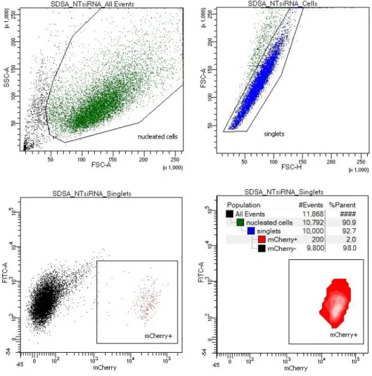
The SDSA assay design allows high throughput data analysis using flow cytometry based
on the cell size and fluorescence. Because U2OS cells adhere to the surface of the well, obtaining a single-cell suspension using trypsin-EDTA prior to analysis is necessary (see materials and
methods). During harvesting a number of cell dies or is still attached to their neighbors despite the treatment. Gating strategy (Fig. 4) facilitates cell debris and doublet discrimination to score single mCherry-positive cells exclusively.
Figure 4. Flow cytometry facilitates analysis of SDSA events by detecting mCherry-positive cells.
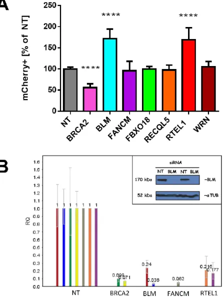
BRCA2 is essential for RAD51-mediated strand exchange and thus SDSA (Sharan et al., 1997). To validate the SDSA assay, I knocked down BRCA2 in the SDSA-U2OS cells which
yielded 50% reduction in SDSA (Fig. 5A). Residual SDSA in this experiment may have resulted from incomplete loss BRCA2 (Fig 5B)
Figure 5. BLM and RTEL1 are negative regulators of SDSA in human cells.
(A) Relative SDSA frequency in SDSA-U2OS cells upon siRNA treatment. X-axis: different siRNA treatments; Y-axis: percent of the cells exhibiting mCherry fluorescence; values: averaged results of 12 experiments with 41 data points (NT); averaged results of 9 experiments with 31 data points (BLM); averaged results of 5 experiments with 20 data points (RTEL1); averaged results of 3 experiments with 7 data points (FANCM and RECQL5); averaged results of 2 experiments with 6 data points (FBXO18 and WRN) error bars: standard deviation; **** p<0.0001
To determine which DNA helicases are involved in SDSA we used a candidate approach and selected a panel of genes consisting of: BLM, FANCM, FBXO18, RECQL5, RTEL1, and
WRN for siRNA knockdown (see materials and methods). These helicases have been either previously implicated in DNA DSBR via HR in human cells or identified as positive SDSA
regulators in model organisms (see introduction).
Surprisingly, BLM and RTEL1 helicase knockdowns resulted in a nearly two fold increase in the SDSA repair as measured by the percentage increase of the number of mCherry-positive
cells standardized to a non-targeting siRNA control (Fig. 5A). Knocking down FANCM, FBXO18, RECQL5, or WRN did not result in a significant change in SDSA.
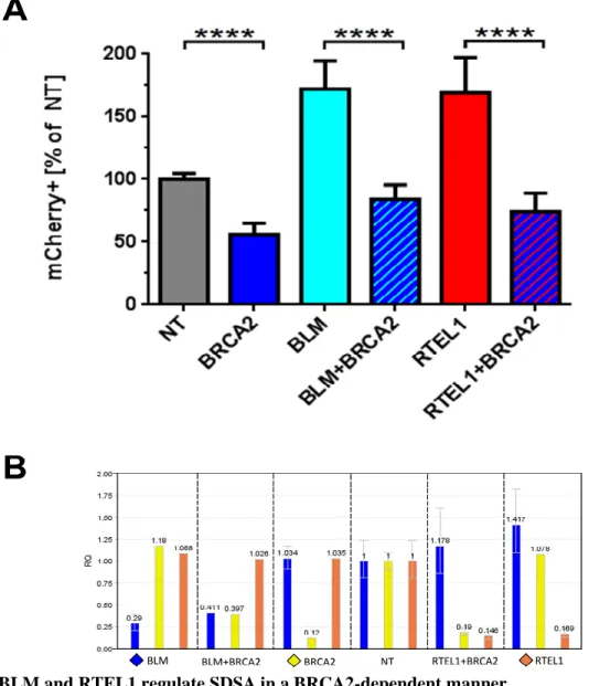
BLM and RTEL1 are BRCA2-dependent SDSA regulators
Both BLM and RTEL1 have multiple roles in genome maintenance. BLM, together with
Dna2, can catalyze extensive DSB end resection, though this function is redundant with Exo1 (Zhu et al., 2008). BLM with Topoisomerase 3 alpha, RMI1, and RMI2 can catalyze dHJ
dissolution (Singh et al., 2008; Wu and Hickson, 2003, 2006). RTEL1 was initially identified as a telomere length regulator and is responsible for T-loop disruption (Sarek et al., 2015).
Biochemical studies indicate that RTEL1is responsible for D-loop disassembly, (Youds et al.,
2010), but can unwind DNA secondary structures and promote replication fork progression (Vannier, 2013; Vannier and Sarek, 2014)
To assess whether BLM and RTEL1 act upstream or downstream from the homology search step during DSBR to regulate SDSA, I knocked down these helicases in combination with BRCA2, which is essential for homology search (Sharan et al., 1997). Consistent with this
down (Fig. 5A and 6A). When BRCA2 was knocked down simultaneously with BLM or RTEL1, decreases of similar magnitude (relative to the increased SDSA frequency of BLM and
RTEL1 single knockdowns) were observed. I conclude that BLM and RTEL1 impact SDSA through functions downstream of strand exchange into a homologous template.
Figure 6. BLM and RTEL1 regulate SDSA in a BRCA2-dependent manner.
(A) Relative SDSA frequency in SDSA-U2OS cells upon siRNA treatment. X-axis: different siRNA treatments; Y-axis: percent of the cells exhibiting mCherry fluorescence; values: averaged results of 5 experiments (4 for double KDs) with 20 data points per treatment (16 data points per treatment for double KDs); error bars: standard deviation;
Knocking down BLM or RTEL1 results in different repair outcomes
To further develop our SDSA assay and gain additional insights into the effects of
knocking down BLM and RTEL, I determined the structures of repair events produced in knockdown cells (Fig. 7). I analyzed 55 clones derived from single red-fluorescing cells,
including 23 from the NT control, 21 from BLM knockdown, ten from FANCM knockdown, and one from RTEL1 knockdown. All but one had the structure expected of SDSA (Fig. 2E). The remaining clone, which came from non-targeting (NT) siRNA treatment, had lost neoR and the
template spacer, and therefore may have arisen from SDSA followed by DSBR with crossover resolution. These results support our conclusion that cells with restored mCherry utilized SDSA
to repair the DSB, occasionally coupled with use of DSBR.
Figure 7. Structures of repair events in cells not expressing mCherry.
I also analyzed cells that failed to produce mCherry to determine whether some of these were produced by other repair processes. In the non-targeting control, all 45 lines examined
appeared to be identical to the initial construct (Fig. 7A). I titrated the viral concentration to a sublethal dose to obtain a maximal possible transfection efficiency, thus these events are likely
the result of a precise NHEJ.
The majority of clones from BLM or RTEL1 knockdown cells also had an intact I-SceI site; however, structures indicating other repair processes were observed in 11 of 34 clones from
BLM knockdown (P <0.0001 compared to NT) and 24 of 133 clones from RTEL1 knockdown cells (P = 0.0007). In four of BLM knockdown clones and 14 of the RTEL1 knockdown clones
the entire 3-kb copia spacer sequence was copied from the repair template into the upstream
mCherry (Fig. 7B and 7C). This extensive repair synthesis might occur through multiple cycles
of strand exchange, as is believed to occur in Drosophila gap repair by SDSA (McVey et al,
2004), or through a single, continuous synthesis event. Among the 17 examples in which the entire spacer was copied, one from each knockdown sample had lost neoR, a structure that is
most consistent with a dHJ being resolved to give a crossover (Fig. 7C). The other 15 may have arisen by long-tract SDSA or by dissolution or non-crossover resolution of a dHJ (Fig. 7B).
There were additional repair events from knockdown cells that also had evidence of
long-tract synthesis. Three events from BLM knockdown and seven from RTEL1 knockdown had a subset of the spacer copied into the upstream mCherry (Fig. 7D). These are most likely hybrid
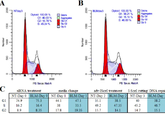
BLM helicase does not alter cell cycle phases
Double-strand break repair pathway choice is believed to be dependent on the phase of
the cell cycle; NHEJ preferred in G1 and HR favored in S/G2/M where a sister chromatid is available as a repair template. Detailed studies of the cell cycle progression coupled with DSBR
assays in human cells indicate that although HR is dominating over S-phase it decreases in G2/M (Mao, 2008). NHEJ levels, however, are high throughout the cell cycle with the maximum at G2/M. To assess whether BLM helicase knockdown affects the cell cycle and thus could account
for an increase in SDSA levels, I analyzed cell cycle profiles over the course of three days after BLM siRNA treatment and a non-targeting control (Fig. 8). The results indicate that BLM
knockdown does not affect cell cycle and thus the SDSA increase in BLM-deficient cells is due to the BLM processing recombination intermediates rather than a chance in cell cycle phases.
Figure 8. BLM helicase does not regulate the cell cycle phases.
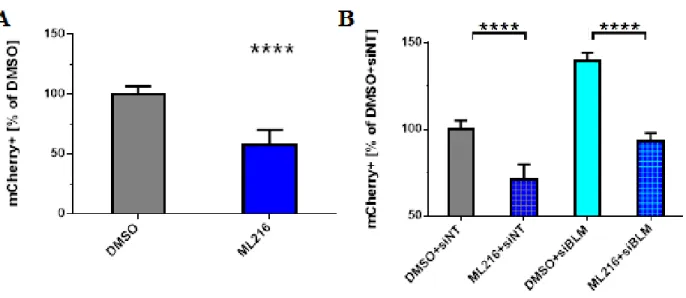
Small particle ML216, BLM inhibitor, affects SDSA
To gain further insights into the function of BLM helicase in SDSA, especially in regards of its inhibitory effects on SDSA revealed by the siRNA knockdown, I decided to test the effects
of ML216 on the DSBR repair in U2OS-SDSA cells. ML216 is a small particle discovered in a large screen for BLM inhibitors (Nguyen and Hickson, 2013). Molecularly ML216 is a DNA mimicking compound with two aromatic rings that prevents BLM helicase from interacting with
DNA. The authors of the paper characterized ML216 thoroughly using both biochemical and genetic approaches. 50 uM concentration of ML216 inhibits DNA binding and the helicase activity of both truncated and full-length BLM. Gel-based DNA unwinding assays reveled that
ML216 is inhibiting primarily BLM and at higher doses can also inhibit WRN helicase, but no inhibition was discovered for other RecQ helicases like RecQL1, RecQL5, and URVD. BLM
deficient cells exhibit high levels of SCEs (Wechsler et al., 2011) ML216 mimics this phenotype but to a lesser degree, however, it does not exacerbate the elevated SCE phenotype in the cells already devoid of BLM (Nguyen and Hickson, 2013). To assess the influence of ML216 on
SDSA in human cells, I treated the cells with the inhibitor at the same time when I-SceI-expressing virus was added. Surprisingly, ML216 treatment leads to a two-fold reduction of
SDSA frequency (Fig 9A) as oppose to a two-fold increase of SDSA in BLM siRNA knockdown (Fig 5A).
The striking contrast between ML216 and BLM siRNA phenotypes indicates that ML216
might have other, yet unknown, targets in the cell that are necessary for SDSA, or that ML216-bound and deactivated BLM is now binding and sequestering other DSB repair proteins
ML216 might have on the cell, I designed an experiment in which the cells are transfected with BLM siRNA prior to ML216 treatment. If ML216 acts on BLM helicase then knocking down
BLM should render ML216 ineffective and thus SDSA levels will go up. However, if ML216 has a different target, then the ML216 phenotype will supersede the one of BLMs, and the SDSA
levels will go down. The data of the ML216 treatment in conjunction with the siRNA
transfection (Fig. 9B) indicate that ML216 is likely targeting a different DNA DSBR protein target other than BLM helicase as the SDSA levels are reduced even in the cells treated with
BLM siRNA. It is worth noting that, although ML216 can inhibit WRN helicase, there must be other ML216 targets in the cell affecting SDSA since WRN knockdown does not affect SDSA
(Fig 5A).
Figure 9. BLM inhibitor, ML216, influences SDSA.
(A) Relative SDSA frequency in SDSA-U2OS cells upon ML216 treatment. X-axis: different drug treatments; Y-axis: percent of the cells exhibiting mCherry fluorescence; values: averaged results of 5 experiments with 14 data points per treatment; error bars: standard deviation; significance: ML216 vs.DMSO: ****; p< 0.0001.
(B) Relative SDSA frequency in SDSA-U2OS cells upon ML216 and siRNA treatment. X-axis: different drug and siRNA treatments; Y-axis: percent of the cells exhibiting mCherry fluorescence; values:
BLM helicase is a negative SDSA regulator in HeLa cells
To complement the research I have conducted in U2OS and to investigate whether SDSA
regulation is specific to U2OS or universal across all human cell, I repeated some of the
aforementioned experiments in HeLa cells. First, I stably integrated linearized pSDSA into HeLa
cells and upon infection with the I-SceI- expressing virus, I observed mCherry positive cells under the fluorescent microscope (Fig. 10A). Then, I tested the consequences of BLM
knockdown for SDSA regulation (Fig. 10B). BLM helicase in HeLa cells, just like in U2OS, is a
negative SDSA regulator, indicating that the roles of DNA helicases in all human cells are conserved.
Figure 10. BLM helicase is a negative SDSA regulator in HeLa cells.
Conclusion
In conclusion, I showed that human cells, as hypothesized, can utilize SDSA to repair
DSBs. I developed the first human cell SDSA assay, which is a tool designed specifically to detect SDSA in human cells by following mCherry fluorescence using flow cytometry
(Fig. 2 and Fig. 4). I also identified two helicases, BLM and RTEL1, which are SDSA regulators in human cells (Fig. 5). Unlike our initial hypothesis based on the results derived from yeast and fruit flies, BLM and RTEL1 do not promote SDSA, but rather are its negative regulators, which I
demonstrated by utilizing an siRNA knockdown approach targeting various helicases (Fig 5). I observed a nearly two fold increase (on average from 1.4% to 2.4%) in SDSA efficiency in the
cells devoid of BLM or RTEL1 (Fig 5). I also determined that these helicases, despite their multiple functions in the DSBR pathway and in the cell, act in a BRCA2-dependent manner (Fig. 6), which means that the SDSA regulation occurs post the homology search step in the
DSBR model (Fig 1).
To further investigate the contribution of BLM and RTEL1 to double-strand break repair,
I analyzed single-cell derived colonies of the siRNA treated repair events at the molecular level (Fig 2 and Fig. 7). All mCherry positive clones are SDSA events as determined by sequencing and resistance to I-SceI cutting (Fig. 2). mCherry negative clones from the BLM and RTEL1
siRNA treated cells exhibit various repair patterns (Fig. 7) indicating that they can be a result of SDSA, DSBR with dHJ resolution or dissolution, NHEJ, or a combination of the above; SDSA
followed by DSBR or end-joining.
Comprehensive molecular analysis indicates that the synthesis-tract length is extended in the helicase-deficient background which can account for observed SDSA increase. Regardless, I
samples. I conducted a series of SDSA assay experiments using BLM inhibitor – ML216, which unexpectedly revealed that ML216 treatment decreases SDSA efficiency in a BLM-independent
manner indicating that ML216 has another target in the cell. (Fig. 8). Lastly, I performed a detailed cell cycle analysis to reveal whether BLM helicase extends S-phase and thus promotes
HR, but I did not find any evidence to support it.
Together, the data from Chapter 2 confirm that SDSA is a major pathway of DSBR in human cells and that DNA helicases, such as BLM and RTEL1, regulate it. DNA helicases are
also thought to inhibit hyper-recombination by limiting the number of COs in mitotically dividing cells. A dramatic increase in HR in the cells with reduced BLM or RTEL1 levels
supports that notion. To attempt answering questions regarding the contribution of DNA helicases to CO regulation, I designed a fluorescence based CO-GC assay. The experimental approached successfully utilized in Chapter two has been instrumental in learning about SDSA
Materials and Methods
Construction of assay plasmids
The SDSA assay construct was based on pEF1a-mCherry-C1 vector (Clonetech #631972). A fragment of mCherry was removed by cutting with NheI and HindIII and inserting annealed
oligonucleotides containing an I-SceI recognition sequence and a part of the mCherry sequence. The product, pEF1a-mCherry-I, had 350 bp of mCherry deleted and the I-SceI sequence inserted. In parallel, 5’ and 3’ mCherry fragments, overlapping by 350 bp, were PCR-amplified and
cloned into a vector containing a fragment of the copia retrotransposon from D. melanogaster. A fragment of HPRT was cloned out of the DR-GFP construct. This entire module (5’ mCherry –
copia – 3’ mCherry – HPRT) was PCR-amplified and cloned into the pEF1a-mCherry-I to
produce pSDSA.
Generation of stably transfected cell lines
U2OS and HeLa cells were cultured under normal conditions (DMEM +10% FBS +
pen/strep) for 24h till they reached 80% confluency before transfection with the SDSA assay constructs using a Nucleofector™ 2b Device (Lonza #AAB-1001) and Cell Line Nucleofector®
Kit V (Lonza #VCA-1003). One week post-transfection appropriate antibiotics were added to
select for the cells with a stably-integrated construct. The SDSA assay construct contains a neoR
gene; cells receiving this construct were treated with 700 μg/ml G418 (Sigma # A1720) for one
DNA repair assay and flow cytometry
SDSA-U2OS cells were cultured in 10 cm dishes containing 10 ml of DMEM medium
with high glucose; Corning) until split onto 6-well plates at a concentration of 5x10⁴ cells per milliliter using 0.05% Trypsin 0.53% mM EDTA solution (Corning). Upon reaching ~60% confluency, the cell were treated with an siRNA reaction mixture (90 nmol of siRNA and 8 μl
lipofectamine 2000 reagent per well; Invitrogen). 24 hours after transfection the siRNA reaction mixture was replaced with the fresh culture medium. 12 hours later the cells were treated with 100 μl of I-SceI-expressing adenovirus (Anglana and Bacchetti, 1999) (previously titrated to a
non-lethal concentration). After another 24 hours the medium was replaced and thus the
adenovirus removed. 72 hours later the cells were harvested and resuspended in 1x PBS
(Corning) supplemented with 2% fetal bovine serum (FBS) and 5 mM EDTA for flow cytometry acquisition on a BD LSRFortessa, using 488nm and 561nm lasers to detect the mCherry
fluorescence.
Genomic DNA isolation
Cells were cultured in a 15 cm dish till they reached 100% confluency. Then they were rinsed gently with 1x PBS and harvested using 0.05% Trypsin 0.53 mM EDTA and pelleted by
centrifuging for three minutes at 2000 rpm. Cells were washed with PBS and transferred to 1.5ml microfuge tubes and spun for 10 sec to re-pellet. PBS was removed and cells were resuspended
in TSM + 0.5%NP-40 solution (140 mM NaCl; 10 mM Tris-HCl, pH 7.4; 1.5 mM MgCl2), then
incubated on ice for 2-3 minutes. After pelleting, cells were resuspended in 1 ml nuclei dropping buffer (0.075 M NaCl; 0.024 M EDTA, pH 8.0). The suspension was transferred to a 15 ml tubes
concentration = 0.2 mg/ml), and 0.5% of SDS. The cells were lysed overnight at 37°C. The next day an equal volume of phenol was added to lysed nuclei and mixed on an orbital shaker for two
hours followed by a five minute spin at 2000 rpm. The aqueous phase was transferred to a clean tube and an equal volume of chloroform was added and the mix was incubated for 30 mins on an
orbital shaker. After spinning, the aqueous phase was transferred to a new tube and 0.1 volumes of 3M sodium acetate was added, followed by one volume of isopropanol. The DNA was spooled out using a glass Pasteur pipette and resuspended overnight in 1 ml TE buffer (10 mM
Tris-HCl, pH 8.0; 1 mM EDTA, pH 8.0). The next day the DNA was precipitated using 0.5 volumes of 7.5M NH4OAc and two volumes of ethanol. DNA was spooled out and resuspended
in 0.5ml TE-4 buffer (10mM Tris-HCl, pH 8.0; 0.1mM EDTA, pH 8.0). Samples were stored in 4°C until analyzed.
PCR analysis of the repair events
DNA from the repair events was isolated according to the protocol above and used in a
PCR reaction to amplify a desired DNA fragment for sequencing or fragment length
characterization. 1.5ul DNA was added to each PCR mixture containing primer sets according to the Table 2, iProof polymerase (BioRad #424264), and a buffer. PCR amplification reaction
Table 2. PCR primers used in the SDSA assay study and their location
Western blot of BLM protein siRNA-treated cells
U2OS cells were transfected using BLM siRNA or NT siRNA (90nmol) and
lipofectamine 2000 (8 μl; Invitrogen) and harvested on the third day post-transfection using
0.05% Trypsin 0.53% mM EDTA solution (Corning). After washing with 1x PBS the cells were resuspended in a protein sample buffer (Tris-HCl; SDS; glycerol; bromophenol blue; 150mM DTT) and boiled. 20 μl of the protein sample was loaded on a 7.5% SDS-PAGE gel and the gel
was run for 1-2 hours at 100V. Protein was transferred to a PVDF membrane using a wet transfer method (1.5 h at 90 V in 4°C). The membrane was blocked in 5% milk PBS solution and
primer
sequence
amplicon size [bp]
1. neo_F
ggatgaggatcgtttcgcatg
2. neo_R
catagaaggcggcggtggaatcg
3. nheI_F
cgtgacgctagcgctaccgg
433 if intact I-SceI
4. hindiii_R
cgaagcttgagctcgagatc
783 if SDSA
5. pSDSAprom_F ggccaagatctgcacactgg
↓
6. SDSAseq1_R
cctcgcagcaaatgctggatc
1298
7. SDSAseq2_R
cagagaatcaactggctgac
1498
8. SDSAseq3_R
cgtgagagaagctctatggc
1707
9. SDSAseq4_R
ctgtgggcctattactccag
2640
10. SDSAseq5_R
gtcccacgcgtcgacaaggc
4112
incubated in PBS plus 0.1% Triton-X plus primary antibodies (rabbit anti-BLM [Abcam #2179] at 1:2000 and mouse anti-αTubulin [Sigma #T9026] at 1:8000) overnight at 4°C, rocking. The
membrane was then washed three times in PBS-T solution. HRP-conjugated secondary antibodies were added (goat anti-rabbit 1:5000; goal anti-mouse 1:100,000) and the blot was
incubated for 1 hour at room temperature. The membrane was washed three times in PBS-T solution and then incubated in an ECL solution (Thermo Fisher) for chemiluminescence for two minutes. The Western blot image was taken using BIO-RAD Molecular Imager (ChemiDoc
XRS+) or the X-ray film was developed using a developer.
qPCR evaluation of the siRNA knockdown efficiency
U2OS cells were transfected using desired siRNA (90 nmol) and lipofectamine 2000 (8 μl; Invitrogen) and harvested on the 3rd day post-transfection using 0.05% Trypsin 0.53% mM
EDTA solution (Corning). RNA was extracted using a manufacturer’s protocol for ReliaPrep RNA Cell Miniprep System (Promega). Purified RNA was used as a template to generate cDNA
library with QuantiTect Reverse Transcription Kit (Qiagen # 205310). The qPCR mix was contained gene-specific DNA primers, cDNA, and QuantiTect SYBR Green PCR kit (Qiagen #A 204141). Amplification and quantification was conducted on a RealTime PCR machine
(QuantStudio 6 flex Real Time PCR System).
Cell cycle analysis by DNA content using propidium iodide
U2OS-SDSA cells were washed by centrifugation (200 x g, 5 min, 4°C) two times in protein-free buffer Phosphate Buffered Saline without Ca+2 or Mg+2 (PBS). The cells were
Then the cells were vortexed gently, and then the cell suspension was added dropwise to 9 ml of 70% ethanol in a 15 ml polypropylene centrifuge tube (Falcon® Cat. No. [35]2097). Cell
precipitation was observed with a microscope to verify minimum cell clumping. The cells were stored at 4°C for 12 - 24 hours before the propidium iodide stating. The cells were centrifuged at
200 x g, 10 min, 4°C, and the pellet resuspended in 3 ml of cold PBS and transferred to Falcon® 12 X 75 mm (Cat. No. [35]2054) polystyrene tubes for staining Falcon® Cat. No. [35]2235. The cell were resuspended in 300 - 500 µl PI/Triton X-100 staining solution: 10 ml of 0. 1 %
(v/v) Triton X-100 (Sigma) in PBS add 2 mg DNase-free RNase A (Sigma) and 0.40 ml of 500 µg/ml PI (Roche # 11348639001). The cells were incubate 37°C for 15 minutes or for 30 min at
20°C, then stored at 4°C. The data was acquired on a BD LSRFortessa flow cytometer and analyzed using ModFit LT version 4.1 by Verity Software House.
Statistical data analysis
GraphPad Prism 6 software (version 6.07) was utilized to generate the figures presenting
1
CHAPTER 3CROSSOVERS AND GENE CONVERSIONS IN HUMAN CELLS
Introduction
Over a 100 years ago Thomas Morgan published his work describing chromosomes as the units of inheritance and proposed crossovers as a mechanism for unlinking genes and generating diversity during reproduction (Morgan et al., 1915). The first experimental evidence
however, came from the studies conducted by Barbara McClintock on crossing Zea mays and analyzing recombinant progeny (Creighton and McClintock, 1931). Over 50 years ago Robin
Holliday proposed a model in which the main recombination intermediate is a structure of two interconnected DNA helices that he called a Holliday junction (HJ) (Holliday, 1964). However, according to the most frequently cited model for HR, DSB processing generates not a single but
double HJ (dHJ) (Szostak et al., 1983), which needs to be cleaved for proper chromosomal segregation during cell division. HJs are cleaved by specialized endonucleases, called resolvases,
to generate either a reciprocal crossover (CO; Fig. 1E) or a noncrossover (NCO; Fig. 1E’). Either can be associated with gene conversion, in which the exchange of genetic material is unidirectional (Szostak et al., 1983). COs in meiotically dividing cells are programmed and
desired as they prevent chromosomal non-disjunction that could lead to chromosome number alterations like in Down syndrome. Genetic diversity is also a result of dHJ processing into COs.
2011) and chromosome rearrangements in cases of ectopic recombination between DNA
duplications (Liu et al., 2011). LOH has been implicated in carcinogenesis as it leads to the loss
of functional copies of tumor suppressor genes (Bignon, 2004; Knudson, 1971; Luo et al., 2000). Although the high level of SCEs is a cellular hallmark of BLM-deficient cells, crossovers
between sister chromatids do not explain propensity for mutations and cancer development in Bloom syndrome patients (Chaganti et al., 1974; Wechsler et al., 2011). These symptoms can be explained however if elevated levels of COs between homologous chromosomes leading to LOH
are present. Studies of LOH point out to BLM helicase as one of the important players preventing recombination (LaRocque et al., 2011). In the S/P assay developed by Jeannine
LaRocque in ES cells, LOH is measured after introducing a DSB in two allelic neoR genes residing on homologous chromosomes. The loss of polymorphisms between the two neoR alleles resulting from a CO conversion or a NCO conversion constitutes LOH. BLM-deficient cells
exhibit 12.9% of LOH while the control cells - less than 2.5%. Interestingly, in the control experiment all LOH events came from NCO conversion, while in the BLM-deficient cells the
majority came from the CO conversion.
In Chapter 2, I showed the development of the first SDSA assay in human cells and that DNA helicases such as BLM and RTEL1 are important SDSA regulators. The molecular analysis
data indicate that BLM and RTEL1, despite negatively controlling SDSA, still act as
recombination inhibitors (a dramatic elevation of HR in helicase knockdown cells). This raises
questions about the contribution of helicases to CO and GC regulation. This chapter is dedicated to the GC analysis conducted using DR-GFP assay and the development of a novel CO-GC assay in human cells using DR-GFP as a backbone.
Results
Gene conversion regulation in human cells using DR-GFP assay
DR-GFP, although initially developed in the Jasin laboratory (Pierce et al., 1999) in ES cells, quickly became a golden standard in studying HR and was adapted for human HEK293
and U2OS cells (Paliwal et al., 2014; Wang et al., 2011). In this assay a DSB is generated by introducing a source of I-SceI to cut a non-functional GFP gene (Fig. 11A). DSB can be repaired using a downstream template, iGFP, which will restore a functional GFP gene (Fig. 11B). The
restoration of a functional GFP can occur via SDSA and is often interpreted as such, however it can also occur via dHJ dissolution or resolution with the repair template on the homologous
chromosome. CO between the upstream GFP gene and the downstream template is also possible, although will not generate a functional GFP due to iGFP repair template missing the GFP 3’end sequence as indicated in Fig. 11B, and would lead to a deletion of the intervening sequence. The
same rationale applies to a possible SSA repair which would not result in the restoration of the functional GFP gene.
Figure 11. DR-GFP assay design.