Details of the Virus
Classified According to a. DNA or RNA
b. Enveloped or Non-Enveloped c. Single-stranded or double-stranded
Viruses contain only a few genes Reverse transcriptase
proteins to inhibit host synthesis to make coat proteins
Obligate parasites
•Smaller than bacteria
•Can not replicate without host - autonomous
replication in the host
•Composition
-nucleic acid
-protein coat
-envelope?
•Where did they come from?
Origin of Viruses
• Do not encode ribosomes or enzymes for
energy production
• Suggests they evolved after cells
• Evolved from small segments host cell DNA
or RNA
• had regions of homology with host cell DNA -
then could recombine and increased in
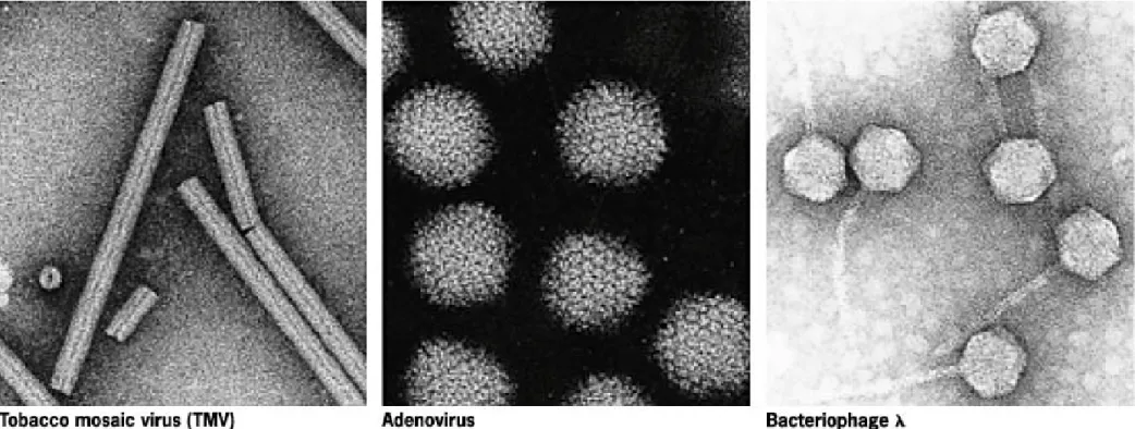
Fig 16.1 Electron micrographs showing the morphologies of a plant virus, an animal virus, and a bacterial virus.
© 2003 John Wiley and Sons Publishers
Credit: Courtesy of Robley Williams, University of California Berkeley and Harold Fisher,
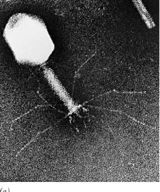
Fig 16.2a Electron micrograph showing the structure of bacteriophage T4.
© 2003 John Wiley and Sons Publishers
Credit: Courtesy of John Finch, Cambridge
Viruses
• Simplest of all organisms • proteins and nucleic acids
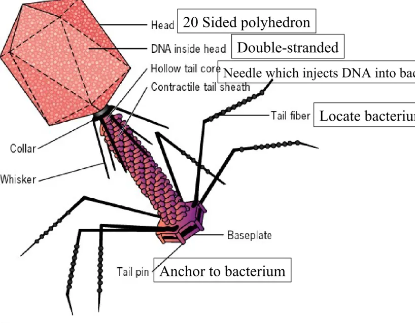
• T phages (T1-T7) - E. coli phage • Components (Figure 16.2)
– head: contains several proteins (20 sides) - DNA in head
– tail: has 2 hollow tubes (inner needle and outer sheath) – tail fibers: uses to find a bacterial host
Fig 16.2 Diagram showing the structure of bacteriophage T4.
© 2003 John Wiley and Sons Publishers
20 Sided polyhedron
Double-stranded
Locate bacterium
Anchor to bacterium
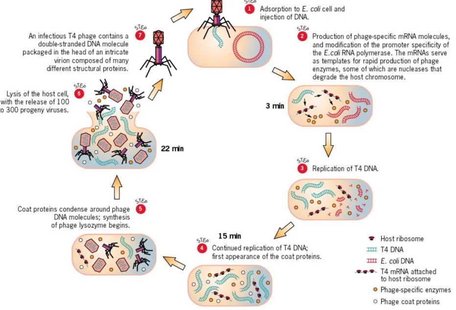
Fig 16.3 The life cycle of bacteriophage T4.
Select Details of Phage Lifecycle
A. Bacteriophage DNA enters bacterium
B. Viral proteins bind to host polymerase/inhibit host synthesis
these proteins also aid in host polymerase recognizing viral genes C. Host DNA is degraded by viral nucleases
D. Viral genes encode coat proteins
lysozyme (to break host wall for lysis)
Bacterial Defense
• Restriction endonucleases (restriction
enzymes) - help protect bacteria from
invasion by viruses
• can degrade C- and HMC-containing DNAs
• Viruses smart - add glucose to HMC
Mapping Phage Genome
• Normally, crossing organisms with different
alleles of a gene
• Don’t have this in viruses
• Only can been seen with electron
microscope
Phage Plaques
• Clear area on a lawn of bacteria- results
from lysis or killing of contiguous cells
• Host range - infect some bacterial strains
but not others
• Phage morphology factors:
X174
• Genes within genes - 5389 nt; should be
1795 amino acids, but there are 2300 amino
acids
• learned that they contain overlapping genes
(translated using different reading frames)
• See
Figures 16.17 and 16.18
- e.g., E gene
HIV
Human Immunodeficiency Virus
• Virus that causes AIDS (acquired immune
deficiency syndrome)
• 30 to 40 million infected
• Prolonged infection
• Primary effect of HIV infection: reduction in
T
Hcells
HIV
• Results of TH cell depletion:
– opportunistic infections (pneumonia) – tumors develop
• HIV mutates rapidly
• HIV is a retrovirus –genome RNA
• Member of lentivirus – “slow virus”
• Enveloped
• gp120 on virus binds to CD4 – T helper lymphocytes – macrophages
• gp41
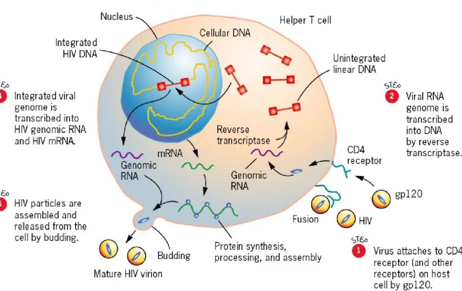
Life Cycle of HIV
FIGURE 16.20 (next slide)
1. virus attaches and penetrates host cells
2. Viral RNA converts to viral DNA (PROVIRUS)
3. Viral DNA integrates into human chromosome
4. Infected host cells produce new virus particles
5. New virus particles bud from host cell one-by-one, taking host cell's membrane along as envelope
Fig 16.20 Overview of the life cycle of HIV.
• Rnase H- step 5- degrades RNA in
DNA/RNA duplex
• integrase - enzyme that allows integration of
the virus into the host genome
• Long terminal repeats
– required for integration
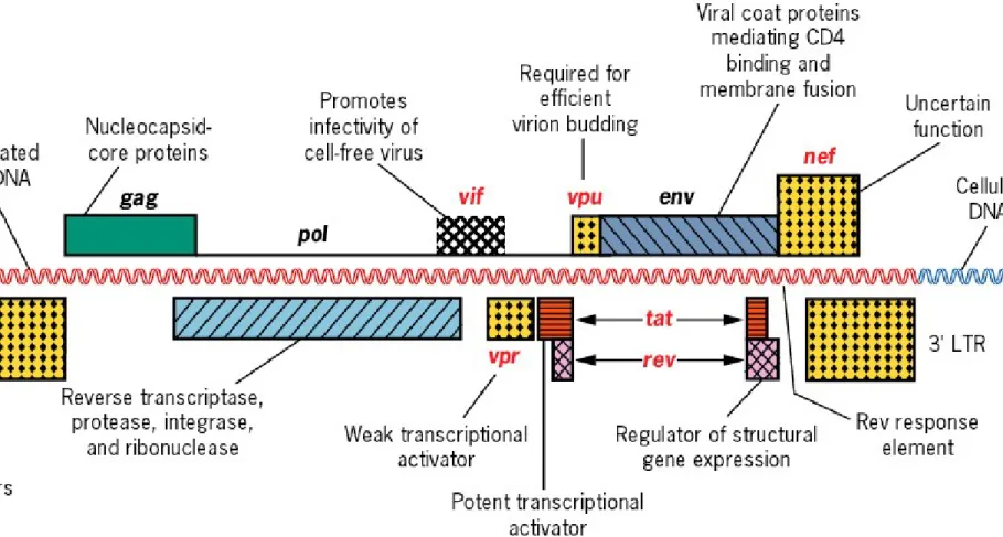
Fig 16.23 Map of the integrated HIV genome showing the location of regulatory genes and genes encoding important viral proteins.
Development of HIV Disease
Time Periods:
1. weeks 1-3: virus enters body, circulates, and makes
infected person contagious
2. weeks 1-8: acute viral syndrome
– short term
– mild or severe flu-like symptoms
Development of HIV Disease
3. 6 weeks - 6 months +: positive HIV antibody test
– seronegative: negative test
– seropositive: positive test for HIV
4. 2 yrs: onset of longer-lasting symptoms
5. 6 months-15 yrs: development of AIDS
yeast infections fungal pneumonia
Diagnosis of AIDS
• ELISA test
• Western Blot
HIV Therapy Strategies
• Boosting immune response insufficient
– once virus in T cells - humoral response ineffective
– cytotoxic T cells must kill infected cells, but viral antigens not displayed
• Interfere with:
– HIV attachment to Cells
– HIV integration into the host cell genome
– Virus replication
Possible HIV Therapies
• Preventing assembly of HIV virus – HIV protease inhibitor (Crixivan)
• Inhibit reverse transcription – AZT (azidothymidine) – Epivir, Retrovir
• Gene Therapy
– Antisense RNA
– Introduce into stem cells
• Preventing entry of HIV into uninfected cells gp120/viral envelope (bind with CD4)
– CD4 on the cell surface (bind with mAb)