Supporting Information
Lee
et al.
10.1073/pnas.0812801106
Fig. S1. Inhibition of HIF-1 transcriptional activity by anthracyclines. (A) HIF-1-dependent luciferase (Luc) activity was determined in Hep3B-c1 cells treated with 0, 0.2, or 1M idarubicin (IDA) or epirubicin (EPI) under 20% (open bars) or 1% (filled bars) O2for 24 h. Cells were lysed and analyzed for the ratio of firefly to RenillaLuc activity. (B) HEK293 cells were exposed to 20% (open bars) or 1% (filled bars) O2for 24 h in the presence of 0, 0.2, or 1M IDA or EPI. Total RNA was
isolated for determination of VEGF (Left) and GLUT1 (Lower) mRNA levels by quantitative real-time reverse-transcription (qRT) PCR. The mRNA levels were normalized to the levels of 18S rRNA in each sample, and each value was expressed relative to the levels in vehicle-treated cells exposed to 20% O2. Mean⫾SEM
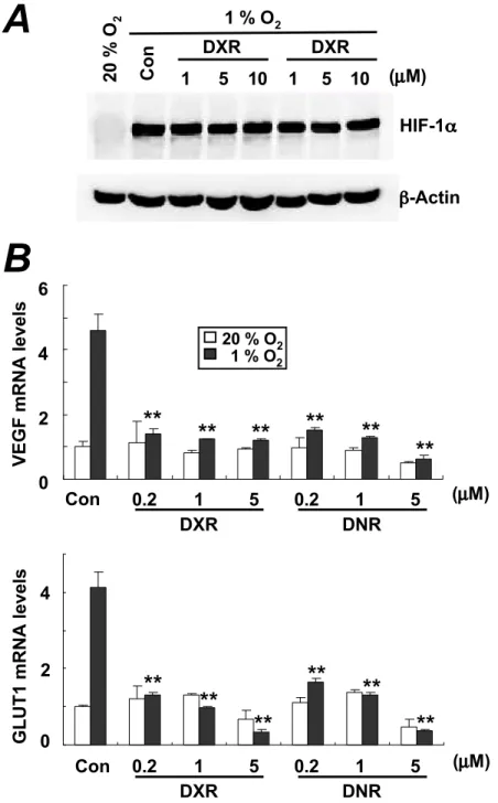
Fig. S2. Mechanism of action by which anthracyclines inhibit HIF-1 activity. (A) HIF-1␣protein levels were determined by immunoblot assays of lysates prepared from HEK293 cells that were cultured in the presence of 0, 1, 5, or 10M daunorubicin (DNR), doxorubicin (DXR), IDA, or EPI at 1% O2for 20 h. To verify equal
loading, the blot was stripped and reprobed with a-actin antibody. (B) HIF-1 transactivation domain function was analyzed by using GAL4/HIF-1␣fusion proteins. HEK293 cells were cotransfected with reporter plasmid pG5-E1b-Luc, which contains five tandem GAL4-binding sites upstream of a minimal E1b gene promoter and coding sequences for firefly Luc; pSV-Renilla, which contains coding sequences forRenillaLuc downstream of a basal SV40 promoter; and an expression vector encoding GAL-O [GAL4 DNA-binding domain (DBD) only], GAL-A (GAL4-DBD/HIF-1␣531– 826), GAL-C (GAL4-DBD/HIF-1␣653– 826), or GAL-H
(GAL4-DBD/HIF-1␣786 – 826). After 24 h, transfected cells were treated with 5M DNR, DXR, EPI, or IDA or vehicle (Con) and exposed to 1% (filled bars) or 20%
(open bars) O2for 24 h followed by cell lysis and determination of firefly andRenillaLuc activities. The mean⫾SEM ratio of firefly toRenillaLuc activity from
three independent transfections is shown (GAL act). (C) Dimerization of HIF-1␣and HIF-1was determined by using GST pulldown assay. Purified GST-HIF-111–510
fusion protein was incubated with lysates from HEK293 cells transfected with vector encoding FLAG-HIF-1␣in the presence or absence of 5M DNR, DXR, EPI, or IDA. Proteins were then pulled down with glutathione–Sepharose 4B beads and subjected to immunoblot (IB) assays by using anti-FLAG and anti-GST antibodies.
HIF-2
HIF-2
IgG
IgG
Input
Input
IP
IP
VEGF
Promoter
PDK1
Promoter
M
Con DXR DNR Con DXR DNR
M
gDNA
20% O
21% O
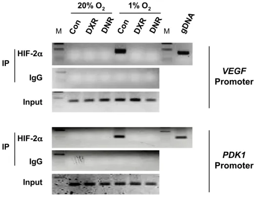
2Fig. S3. Effect of DXR and DNR on HIF-2␣DNA-binding activity. HEK293 cells were exposed to 1% or 20% O2in the presence of vehicle (Con) or 1 mM DXR
or DNR for 20 h. Input DNA was isolated from an aliquot of lysate before immunoprecipitation (IP), and lysates were then divided between anti-HIF-2␣and rabbit IgG for IP. PCR was performed by using the immunoprecipitates as template to amplifyVEGFandPDK1promoter sequences. PCR products were analyzed by 2% agarose gel electrophoresis and ethidium bromide staining. M, 200-bp size marker.
Fig. S4. Effect of DNR or DXR on HIF-1 activity in Hep3B-c1 cells. (A) HIF-1␣protein levels were determined by immunoblot assays of lysates prepared from Hep3B-c1 cells that were exposed to hypoxia (1% O2, 20 h) in the presence of vehicle control (Con) or 1, 5, or 10M DNR or DXR. (B) VEGF and GLUT1 mRNA
levels were determined by using qRT-PCR in Hep3B-c1 cells treated with vehicle (Con) or 0.2, 1, or 5M DNR or DXR under 1% (filled bars) or 20% (open bars) O2. Mean⫾SD (n⫽4) are shown.**,P⬍0.01 vs. control, Student’sttest.
Fig. S5. Effect of DNR and DXR treatment on HIF-1 activity in cultured PC-3 cells. (A) HIF-1␣levels were determined by immunoblot assay of lysates prepared from PC-3 cells that were exposed to 1% O2for 20 h in the absence (Con) or presence of 1, 5, or 10M DNR or DXR. (B) Hypoxia response element (HRE)-driven
Luc activity was determined in PC-3 cells. Cells were cotransfected with HRE-driven firefly Luc and controlRenillaLuc reporter plasmids and treated with DXR or DNR for 24 h at 20% (open bars) or 1% (filled bars) O2. Cells were lysed and analyzed for the ratio of firefly toRenillaLuc activity. (C) qRT-PCR assays were
performed to quantify VEGF and GLUT1 mRNA levels in PC-3 cells treated with 0.2, 1, or 5M DNR or DXR at 20% (open bars) or 1% (filled bars) O2for 24 h.
Table S1. Primers used for quantitative real-time reverse-transcription PCR
Gene Sequences
VEGF Fwd: CTTGCCTTGCTGCTCTAC
Rev: TGGCTTGAAGATGTACTCG
GLUT1 Fwd: CGGGCCAAGAGTGTGCTAAA
Rev: TGACGATACCGGAGCCAATG
HK1 Fwd: TGGAGTCCGAGGTTTATG
Rev: TTTGGATTGTTGGCAAGG
HK2 Fwd: CCAGTTCATTCACATCATCAG
Rev: CTTACACGAGGTCACATAGC
SDF1 Fwd: GTGGTCGTGCTGGTCCTC
Rev: CTCTGGCAACATGGCTTTCG
SCF Fwd: AAAGATTCCAGAGTCAGTGTC
Rev: TTCCAGTATAAGCTCCAAAAG
18 s rRNA Fwd: CGGCGACGACCCATTCGA AC
Rev: GAATCGAACCCTGATTCCCCGTC
Table S2. Primers used for the amplification of HIF-1

11–510 SequencesFwd: CGCGGATCCCGATGACATCAGATGATCCATCACTG Rev: ATAAGAATGCGGCCGCAGTTCTGTTTGCTGTTGCTGCTGCC
Table S3. Primers used for chromatin immunoprecipitation assay
Gene Region Sequences
VEGF ⫺531 to⫺374 Fwd: TCTTCGAGAGTGAGGACGTGTGT
Rev: AAGGCGGAGAGCCGGAC
PDK1 ⫺485 to⫺386 Fwd: CGCCCTGTCCTTGAGCC
Rev: CGGTATGGAGCGTCCCCT 550 to 646 Fwd: CGCGTTTGGATTCCGTG
Rev: CCAGTTATAATCTGCCTTCCCTATTATC