Original Research Article
Clinical profile and outcome of H1N1 influenza in children during
August 2016 to January 2017 at KIMS hospital in Bangalore,
Karnataka, India
Ramya H. S., N. Varsha Monica Reddy*, Kavya Shekar
INTRODUCTION
In June 11, 2009 World Health Organization (WHO) Director General Margaret Chan declared a pandemic alert level 6 due to the spread and outbreak of the Influenza A (H1N1) virus in at least two countries within
the WHO region.1 The epidemic was caused by the
swine-origin Influenza A (H1N1) virus (S-OIV) which was said to be made up of the HA gene derived from the 1918 swine flu virus and other genes from human, avian and Eurasian swine influenza viruses.2
ABSTRACT
Background: Influenza virus is a common respiratory pathogen. In April 2009, a new strain of Influenza virus A H1N1 was evolved and spread in several countries around the world and caused pandemic. After that the resurgence of H1N1 epidemic occurs almost every year and it has caused significant morbidity and mortality. Author present the clinical profile of suspected cases of swine flu infection among children attending our hospital.
Methods: This prospective study was conducted at KIMS hospital among 70 children aged 1month to 17 yrs admitted with pneumonia from August 2016 to January 2017 and who consented for the study were included. Institutional ethical clearance obtained. All cases were classified according to WHO ABC category and PRESS (Pediatric Respiratory Severity Score) was applied. Outcome of patients were calculated based on requirement for PICU care, ventilation and PRESS score 4-5. Suspected high risk cases, category B2, and category C cases were started on
antiviral Oseltamivir and throat swabs were sent for detection of H1N1 by rRT-PCR.
Results: Out of 70 pneumonia cases 44 cases improved with antibiotics and supportive therapy, 26 cases who belonged to category B2 and C and PRESS score 4-5 (severe) were screened for H1N1 influenza and 26 cases were started with empirical antiviral drug oseltamivir.10 out of 26 cases (38.4%) were positive for H1N1 influenza. In our study, fever, cough, breathlessness, and chest x-ray abnormalities were the most common clinical features in both suspected and confirmed cases of H1N1 influenza. Out of 10 cases 2 succumbed (20%). Both the cases had prolonged hospital stay.
Conclusions: During monsoon, high degree of suspicion is required in patients presenting with fever, cough, cold and hurried breathing with supportive lab investigations and early administration of antiviral drug provides better outcome, reduces the length of hospital stay and mortality.
Keywords: Clinical profile, H1N1 influenza, Oseltamivir
Department ofPediatrics, Kempegowda Institute of Medical Sciences, Bangalore, Karnataka, India
Received: 14 December 2017
Received: 02 March 2018
Accepted: 07 March 2018
*Correspondence:
Dr. N. Varsha Monica Reddy, E-mail: varshareddy29@gmail.com
Copyright: © the author(s), publisher and licensee Medip Academy. This is an open-access article distributed under the terms of the Creative Commons Attribution Non-Commercial License, which permits unrestricted non-commercial use, distribution, and reproduction in any medium, provided the original work is properly cited.
Genetic characterization suggested that introduction of this virus into humans occurred as a single event, followed by multiple human to human transmission. On 11th of June 2009, WHO declared the outbreak a pandemic.3
The clinical presentation of H1N1 infection varies from a usually mild influenza like illness to rapidly worsening pneumonia with ARDS.3,4 Some countries have reported
a greater number of severe illnessess and deaths; whereas others have reported only mild influenza like illness. Some studies have shown that one third of patients remain asymptomatic, one third has a short febrile illness and one third require hospitalization.4
This study aimed to describe the clinical profile and outcome of pediatric patients with reverse transcription-polymerase chain reaction (RT-PCR)-confirmed Influenza A (H1N1) from August 2016 to January 2017 at KIMS hospital, Bangalore.
METHODS
This prospective study was conducted at KIMS hospital among 70 children aged 1month to 17 yrs admitted with pneumonia from August 2016 to January 2017 and who consented for the study were included. Institutional ethical clearance obtained.
An observational clinical study was conducted at Kempegowda Institute of Medical Sciences Hospital, located in Bangalore, Karnataka between the period of August 2016 to January 2017.
Study population
children aged 1month to 17 yrs admitted with suspected pneumonia in pediatric intensive care and wards.
Inclusion criteria
All children aged 1 month to 17 yrs with suspected pneumonia admitted in pediatric intensive care and wards who consented for the study.
Exclusion criteria
Infants less than 1 month and 18yrs above.
Stastical analysis
Microsoft Excel spread sheet was used for data collection and the data analyzed using SPSS version 24. Microsoft word and excel have been used to generate graphs, tables etc.
In this study, 70 cases suspected to have pneumonia from the august 2016 to January 2017 were included. All cases were classified according to WHO ABC category and PRESS (Pediatric Respiratory Severity Score) was
applied. Outcome of patients were calculated based on requirement for PICU care, ventilation and PRESS score 4-5.
Out of 70 cases, 57 cases belonged to category A of which 44 cases improved with antibiotics and got discharged, the remaining 13 cases from cat A were also started with antiviral due to worsening of symptoms. The other 13 cases out of remaining 26 were highly suspected belongs to category B2(6cases), and category C(7cases) were treated with antiviral drug and throat swab was sent for detection of H1N1 by rRT-PCR for all the remaining 26 cases.
All positive cases were continued with antiviral and supportive treatment and negative cases were further diagnosed and appropriately managed.
RESULTS
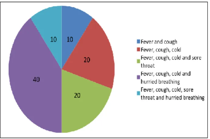
A total number of suspected pneumonia cases admitted during the study period was 70. Out of which, 9(12.9%) were children <1yr age, 30(42.9%) were children in 1-5 years age group, 19(27.1%) in 6-10 years age group and 12(17.1%) among 11-17years age group. Majority 37(52.9%) were females and 33(47.1%) were males. Among 70 cases, patients were categorized into ABC and 57(81.4%) were cat A, 6(8.6%) were cat B and 7(10%) were cat C. Fever and cough were equally present in all the cases (100%), 41(58.6%) had cold, 12(17.1%) had sore throat and 13(18.6%) had hurried breathing. History of contact with H1N1 was present in family in 3 cases and all recovered with prophylactic antiviral drugs. 27 out of 70 cases were admitted in PICU and 12 cases required ventilation (6 were on NIV and 6 were on mechanical ventilation).
Figure 1: Percentage of clinical features.
Table 1: Age sex clinical features and others distribution and percentage of patients.
Features N(%)
Age in category in years
<1 9 (12.9)
>1 to 5 30 (42.9)
6-10 19(27.1)
>10 12(17.1)
Gender
Male 33(47.1)
Female 37(52.9)
Clinical features
Fever 70(100)
Cough 70(100)
Cold 41(58.6)
Sore throat 12(17.1)
Hurried breathing 12(18.6)
Risk factors
History of contact 4(5.7)
No immunization 2(2.9)
Nutritional status 2(2.9)
Sick child 8(11.4)
Family history 3(4.3)
Category
A 57(81.4)
B 6(8.6)
C 7(10)
Investigations
Chest X ray
Bilateral 35(50)
Unilateral 35(50)
H1N1 swab taken 26(37.1)
Result positive 10(14.3)
TLC (<4000) 6(8.6)
Treatment related
Tamiflu given 26(37.1)
PICU care 27(38.6)
NIV 6(8.6)
Mechanical ventilation 6(8.6)
Outcome
Good 43(61.4)
Bad 27(38.6)
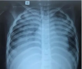
Chest X-ray of H1N1 influenza showing B/L involvement (Figure 2).
Author collected data on five components using the PRESS, namely, respiratory rate, wheezing, accessory muscle use, SpO2, and feeding difficulties (Table 2). Accessory muscle use was defined as visible retraction of one or more of the sternomastoid/suprasternal, intercostal, and subcostal muscles. Wheezing was defined by auscultation performed by experienced pediatricians. SpO2 was evaluated as above or below 95%. Feeding difficulties were assessed using information provided by the parents. Each component was given 0 or 1 point and the PRESS total score was classified as mild (0-1 points),
moderate (2-3 points), or severe (4-5 points). Respiratory rate was evaluated based on the American Heart Association guidelines 6 (Table 2).
Figure 2: CXR of H1N1 positive case showing B/L involvement and extensive pneumonia.
Table 2: Pediatric respiratory severity score.
Score
component Operational definition Scoring
Respiratory rate
Respiratory rate at rest, on
room air* 0 or 1
Wheezing High pitch expiratory sound
heard by auscultation 0 or 1 Accessory
muscle use
Any visible use of
accessory muscles 0 or 1
SpO2 Oxygen saturation <95% on
room air 0 or 1
Feeding difficulties
Refusing feedings 0 or 1 Sum of five components
PRESS score 0-1: mild 2-3: moderate 4-5:
severe 0-5
Criteria of tachypnea* RR
Month
<12 60< 1
>12, <35 40< 1 >36, <156 <30 1
156< <20 1
*Respiratory rate evaluated according to American Heart Association guideline, PRESS- Pediatric Respiratory Severity Score, RR- Respiratory rate
Table 3: Details of H1N1 positive case (N=10).
Features Mean(SD)
Illness duration 3.37(1)
PRESS score 2.67(1.2)
Hospital stay 0.61(1.6)
Table 4: Details of those patients not started on treatment (N=44).
Features N(%)
Tuberculosis 10(22.7)
WALRI 10(22.7)
Croup 6 (13.6)
Bronchiolitis 10(22.7)
Bacterial pneumonia 8(18.1)
Those cases which improved with antibiotics were classified according to diagnosis of which TB were 10 cases (22.7%), WALRI were 10 (22.7%), croup was 6 (13.6%), bronchiolitis 10 (22.7%) and bacterial pneumonia 8 (18.1%).
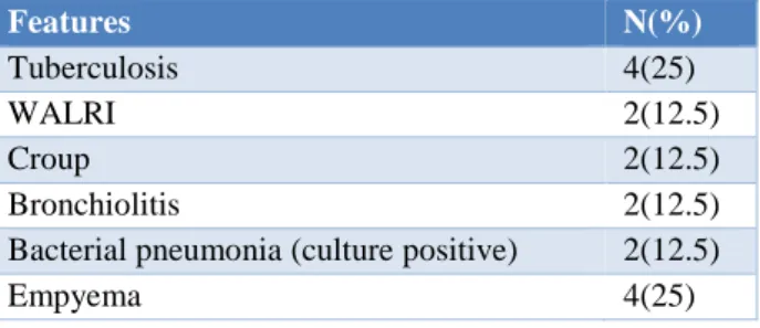
Table 5: Details of swab negative suspects of H1N1 (N=6).
Features N(%)
Tuberculosis 4(25)
WALRI 2(12.5)
Croup 2(12.5)
Bronchiolitis 2(12.5)
Bacterial pneumonia (culture positive) 2(12.5)
Empyema 4(25)
Of those cases which were negative for H1N1 were diagnosed to be Tuberculosis 4 cases (25%), WALRI 2 cases (12.5%), croup 2 (12.5%), bronchiolitis 2 (12.5%), bacterial pneumonia (culture positive) 2 (12.5%) and empyema 4 (25%).
DISCUSSION
The 2009 pandemic of H1N1 influenza rapidly spread globally, causing significant mortality and morbidity. It was first global pandemic since 1968. H1N1 is a flu virus and spreads between people in the same way that seasonal flu viruses spread, i.e., through droplets or fomites. Incubation period is around 2-7 days. Symptoms of H1N1 are flu-like symptoms, fever, cough, coryza, headache, myalgia, and joint pain. Less common symptoms are vomiting, diarrhea, conjunctivitis, and parotitis. In this study, author observed that fever; cough, breathlessness, and coryza were the most common clinical features in both suspected and confirmed cases of H1N1 influenza.
Respiratory tract infections in childhood can readily lead to respiratory distress and sometimes to severe dyspnea, which requires further examinations and hospitalization. An objective bedside assessment of the respiratory symptoms would enable such examinations and treatment to be commenced quickly, to avoid the condition worsening. In the present study, we used a simple scoring system PRESS score based on respiratory symptoms for
assessing the need for further examinations and hospitalization.5
The majority were in the age group of 1-5 years and 47.1% were males, contrary to other previous studies.6-10
Similar findings were reported by Das et al, Mehta et al, Prakash, and Kumar et al.7-10 In similar to the many
studies which reported higher admission rate in females than males.11-15
Among the positive cases, h/o contact in family was present in 3 cases emphasizes the widespread prevalence of H1N1.
All children with positive swab for H1N1 were treated with oseltamivir and majority did not require mechanical ventilation. Of the cases which were negative for H1N1 were classified into other bacterial infections and treated accordingly. Suspected cases were treated with antiviral drugs and had good outcome. Hence early suspicion and treatment with antiviral oseltamivir reduces the morbidity and mortality. Out of 70 cases, two children (2.8%) succumbed to the illness. The remaining cases recovered.
CONCLUSION
During monsoon, high degree of suspicion is required in patients presenting with fever, cough, cold and hurried breathing with supportive lab investigations and early administration of antiviral drug provides better outcome, reduces the length of hospital stay and mortality.
ACKNOWLEDGEMENTS
Authors would like to thank Dr. Srinivasa S., for his constant help to undertake this study.
Funding: No funding sources Conflict of interest: None declared
Ethical approval: The study was approved by the Institutional Ethics Committee
REFERENCES
1. Department of Health. Influenza A H1N1 update: Statement of Secretary Francisco T. Duque III, MD on the WHO Declaration of Pandemic Alert Level 6. Available at http://www. DOH.gov.com. August 2009.
2. Chang LY, Shih SR, Shao PL, Huang DT, Huang LM. Novel swine-origin influenza virus A (H1N1): the first pandemic of the 21st century. Journal of the Formosan Medical Association. 2009 Jul 1;108(7):526-32.
Jocob JT, Mahesh M. Pandemic Influenza in India. Indian pediatrics. 2010;47:25-31.
3. International Society of Infectious Diseases. Influenza Pandemic (H1N1) 2009 (20): Peru, 33%
http://www.promedmail.org/pls/otn/f?p=2400:1202: 739. Accessed 17 November 2009.
4. Yumiko M, Kazuko S, Asako N. Pediatric Respiratory Severity Score (PRESS) for Respiratory Tract Infections in Children. Austin Virol and Retrovirology. 2015;2(1):1009.
5. ECC Committee, Subcommittees and Task Forces of the American Heart Association. 2005 American Heart Association Guidelines for Cardiopulmonary Resuscitation and Emergency Cardiovascular Care. Circulation. 2005;112:151-203.
6. Das RR, Sami A, Lodha R, Jain R, Broor S, Kaushik S, et al. Clinical profile and outcome of swine flu in Indian children. Indian Pediatr. 2011;48:373-8.
7. Mehta VK, Sharma P, Guleria RC, Ganju SA, Singh D, Kanga A. Clinico-epidemiological profile, pandemic influenza H1N1/2009 and seasonal influenza, August 2009-March 2013, Himachal Pradesh, India. IJCM. 2016;41(1):69-71.
8. Prakash G. Epidemiological and clinical profile of patients with swine flu (Influenza A, H1N1) attending Guru Govind Singh Government Hospital, Jamnagar, India. J Res Med Dent Sci. 2013;1(1):1-6.
9. Kumar SB, Kumar SK, Rau AT, Somashekhar AR. Clinical profile of “pandemic swine flu (H1N1)” in children. J Paediatr Sci. 2010;4:e40.
10. Mandal PK, Sardar JC, Bhandari B. Clinical profile of H1N1 influenza: A hospital based
epidemiological study in Kolkata, India. SJPH. 2013;8(1):21-4.
11. Van‘t Klooster TM, Wielders CC, Donker T, Isken L, Meijer A, Van den Wijngaard CC, et al. Surveillance of hospitalisations for 2009 pandemic influenza A (H1N1) in the Netherlands, 5 June-31 December 2009. Euro Surveill. 2010 Jan 14;15(2):19461.
12. Fielding JE, Higgins N, Gregory JE, Grant KA, Catton MG, Bergeri I, et al. Pandemic H1N1 influenza surveillance in Victoria, Australia, April-September, 2009. Eurosurveillance. 2009 Oct 22;14(42):19368.
13. Tulloch F, Correa R, Guerrero G, Samaniego R, Garcia M, Pascale JM, et al. Profile of the first cases hospitalized due to influenza A (H1N1) in Panama City, Panama. May-June 2009. J Infect Dev Ctries. 2009;3:811-6.
14. New South Wales Public Health Network. Progression and impact of the first winter wave of the 2009 pandemic H1N1 influenza in New South Wales, Australia. Euro-surveillance. 2009 Oct 22;14(42):19365.