34
Effects of Parenteral Amino Acid Administration on
the Postoperative Nutritional Status and Wound Healing
of Protein-Malnourished Rats
Akira Wada, Chiaki Sonoda, Yuya Makino, Yuki Hama, Akihiro Nagahama and Daisuke Harada
Naruto Research Institute, Research and Development Center, Otsuka Pharmaceutical Factory, Inc., Naruto, Tokushima 772–8601, Japan
(Received January 12, 2017)
Summary In Japan, parenteral nutrition (PN) solutions are frequently administered
to patients in the postoperative short-term period. In these cases, amino acid-containing peripheral parenteral nutrition (PPN) solutions, amino acid-free maintenance solutions or combinations of the two are used. However, consensus regarding the most beneficial solu-tion for these patients is lacking. Here, we examined the nutrisolu-tional status and wound heal-ing outcomes in protein-malnourished rats receivheal-ing postoperative administrations of PPN solution, maintenance solution or combinations of the two solutions. Protein malnutrition was induced in Sprague-Dawley rats by feeding an AIN-93G-based low-protein diet (5% casein) for 2 wk. After laparotomy, dorsal skin incision, and placement of a jugular vein catheter, the rats were divided into 3 groups. Each group was administered 113 kcal/kg/d, with group A receiving maintenance solutions without amino acid, group B receiving PPN with 1.5% amino acid, and group C receiving PPN with 3% amino acid. After 5 d post-oper-ative administration, we measured the tensile strength of the wound area, skeletal muscle weights, and nutritional parameters. Significantly higher plasma nutritional parameters and gastrocnemius and extensor digitorum longus (EDL) muscle weights were observed in groups B and C than in group A. Group C exhibited significantly elevated tensile strength of the wound area along with up-regulation of type I collagen mRNA expression compared to group A. These findings demonstrate the nutritional status and wound healing benefits of short-term postoperative administration of PPN solutions containing amino acids in pro-tein-malnourished rats.
Key Words PPN solution, parenteral nutrition, amino acids, wound healing, protein
mal-nutrition
In Japan, peripheral parenteral nutrition (PPN) is generally used for patients in the short-term period after surgery (1, 2), and during the transition to oral intake or enteral nutrition (EN) therapy. Most commercial PPN solutions contain 3% amino acids, 7.5% dextrose, and electrolytes. However, for short-term postoperative fluid therapy such as 3–5 d, maintenance solutions contain-ing 2.5–10% dextrose and electrolytes without amino acids are sometimes used solely instead of the PPN solu-tions or combinasolu-tions of the two.
During the perioperative period, malnutrition can influence patient outcomes such as morbidity, mortality, length of stay, infection rates, and wound healing (3, 4). Garwe et al. (5) examined geriatric trauma patients who were admitted to the emergency department and found that 65% had protein-energy malnutrition (PEM). In addition, these patients had higher risk of in-hospital complications. Furthermore, other previous studies have determined that malnutrition during the periop-erative period was a risk factor for delay in wound heal-ing (6, 7). A study investigatheal-ing early administration of
EN during the postoperative period showed improved wound healing in these patients (8). However, there are some cases where patients who cannot use the gastro-intestinal tract are unable to receive EN. Even if EN is feasible, it is difficult to satisfy their energy requirements by EN alone. Therefore, parenteral nutrition (PN) is indi-cated in these situations. However, there are few stud-ies that have investigated changes in wound healing with administration of PPN solutions immediately after surgery.
Even for a short term, the effects of amino acids in the PPN solutions should be commonly recognized among clinicians. Thus, the current study examined the nutritional status and wound healing outcomes in protein-malnourished rats receiving postoperative administrations of PPN solution, maintenance solution or combinations of the two.
MATERIALS AND METHODS
Animals. Male 5-wk-old Sprague-Dawley rats were purchased from Charles River Japan (Yokohama, Japan). All rats were housed under constant tempera-ture (2363˚C) and humidity (55615%) conditions
ing a 12 h light/dark cycle. This study was approved by the Committee for the Care and Use of Laboratory Ani-mals of the Otsuka Pharmaceutical Factory, Inc., Japan (Authorization number: OPFCAE-14-421).
Experimental design and operative procedures. After 12 d of adaptation, rats were fed an AIN-93G-based low-protein diet (5% casein) or standard AIN-93G (20% casein) diet for 2 wk. The rats fed the low-protein diet (protein-malnourished rats) were divided into three groups, namely, A (n512), B (n510), and C (n511). Rats fed the standard AIN-93G diet were used as the control group D (n56). All the animals of each group were fasted overnight at the end of 2 wk of feeding. Next, all rats underwent a 4-cm midline laparotomy and 4-cm dorsal skin incision while under sodium pen-tobarbital anesthesia. Rats in groups A–C received a silicon catheter that was placed in the jugular vein of each animal. After the operation, all rats were placed in individual metabolic cages for 5 d. Immediately after the surgery, rats in groups A–C were administered different nutrient solutions, and were deprived of both food and water. The control rats in group D were provided with ad libitum access to the standard AIN-93G diet and water. This group was set as the target baseline as it mimicked the clinical setting of patients who do not have PEM before surgery and can eat normal meals sufficiently after surgery.
Parenteral nutrient solutions. A commercial mainte-nance solution containing 10% dextrose and electro-lytes (Physio35; Otsuka Pharmaceutical Factory, Inc., Tokushima, Japan) and a commercial PPN solution con-taining 3% amino acids, 7.5% dextrose and electrolytes (BFLUID; Otsuka Pharmaceutical Factory, Inc.) were used. Rats in group A received Physio35 (0% amino acids), whereas rats in group B received a mixed solution that contained equal volumes of Physio35 and BFLUID (1.5% amino acids); rats in group C received BFLUID (3% amino acids). Table 1 lists the composition of each nutrient solution and the daily dosage. Rats in group A–C were provided 113 kcal/kg/d of each nutrient solution. This dosage corresponds to about 2,000 mL/d (840 kcal/d) of commercial PPN solution that is
cur-rently used in persons weighing 50 kg. This dosage was based on a previous study about PPN-infused rats, which indicated that the calorie intake of 120 kcal/kg/d for rats corresponds to calorie intake of 1,000 kcal/d in a person weighing 60 kg (9). Table 2 lists the amino acid composition of PPN solution (BFLUID).
Measurements of nutritional parameters. Body weights were measured prior to surgery (day 0) and at the end of the study (day 5). On day 5, rats were anesthetized using intravenous injection of sodium thiopental. After the collection and processing of blood from an abdomi-nal vein, plasma samples were stored at 280˚C until analysis. Plasma levels of albumin, total protein, and blood urea nitrogen were measured using the Hitachi 7180 Biochemistry Automatic Analyzer (Hitachi High-Technologies Corp., Tokyo, Japan). The rapid-turnover proteins, prealbumin (transthyretin) and transferrin, were analyzed using ELISA kits (Abnova Corporation, Taipei, Taiwan and Aviva Systems Biology Corporation, San Diego, CA) according to the manufacturer’s proto-col. In all animals, the gastrocnemius, soleus, and EDL muscles were excised and weighed.
Wound healing assay. During the surgery, the dor-sal skin incision site was closed using a surgical stapler. Five days after the initial surgery, the dorsal skin inci-sion area was harvested, and the surgical staples were removed. The tensile strength of the skin wound was measured using a Digital Force Gauge ZP-500N (Imada Inc., Aichi, Japan) to evaluate wound healing.
Quantitative real-time PCR. After measurement of the tensile strength of the dorsal skin incision site, samples that contained the incised and closed sites were
Table 1. Composition of parenteral nutrient solutions (/1,000 mL) and daily dosage.
A B C
Composition
Dextrose (g) 100 87.5 75
Amino acid (g) 0 15 30
NPC/N 0 149 64
Total energy (kcal) 400 410 420 Daily dosage
Volume (mL/kg/d) 284 277 270 Total energy (kcal/kg/d) 113 113 113 Dextrose (g/kg/d) 28.3 24.2 20.3 Amino acid (g/kg/d) 0 4.1 8.1
NPC/N: non-protein calories/nitrogen ratio.
Table 2. Amino acid composition of PPN solution (BFLUID).
Amino acids g/1,000 mL
l-Leucine 4.20
l-Isoleucine 2.40
l-Valine 2.40
l-Lysine 3.15
l-Threonine 1.71
l-Tryptophan 0.60
l-Methionine 1.17
l-Cysteine 0.30
l-Phenylalanine 2.10
l-Tyrosine 0.15
l-Arginine 3.15
l-Histidine 1.50
l-Alanine 2.40
l-Proline 1.50
l-Serine 0.90
Glycine 1.77
l-Aspartic acid 0.30 l-Glutamic acid 0.30 Total amino acids 30.0 BCAA content rate 30% w/w
frozen in liquid nitrogen and stored at 280˚C until RNA extraction. Total RNA was prepared using the PureLink® RNA mini kit (Life Technologies, Carlsbad, CA). Extracted total RNA was reverse transcribed into cDNA using ReverTra Ace® qPCR RT Master Mix with genomic DNA remover (Toyobo Co., Ltd., Osaka, Japan). Quantitative real-time PCR was performed using the 7500 Fast real-time PCR system (Applied Biosystems, Foster City, CA) with the Thunderbird SYBR qPCR mix (Toyobo). The primer sets used included Gapdh (for-ward: 5′-GGCACAGTCAAGGCTGAGAATG-3′, Reverse: 5′-ATGGTGGTGAAGACGCCAGTA-3′), Col1a1 (for-ward: 5′-CTGCATCAGGGTTTCAGAGCAC-3′, Reverse: 5′-TCCACATGCTTTATTCCAGCAATC-3′), and Col3a1 (forward: 5′-CGGAAGCACTGGTGGACAGA-3′, reverse: 5′-GAAGGCCAGCTGTACATCAAGGA-3′) (Takara Bio Inc., Otsu, Japan). The mRNA expression was quanti-fied using the comparative DCT method, with Gapdh as the reference gene. The mRNA expression levels were expressed relative to the mean expression observed in group A.
Plasma amino acid analysis. Plasma samples were diluted with water and deproteinized using acetonitrile. Subsequently, all the samples were centrifuged and the supernatants were collected. The amino acid concentra-tions in the supernatants were analyzed by LC/MS/MS using the Acquity UPLC I Class with Xevo TQ-S system (Waters Corporation, Milford, MA).
Statistical analysis. All data represent means6
stan-dard deviation (SD). The statistical significance among all of the groups compared with group D was assessed using one-way ANOVA followed by Dunnett’s test. The statistical significance among the three nutrient solu-tion groups was assessed using one-way ANOVA fol-lowed by the Tukey-Kramer test. All statistical tests were performed using the EXSUS Version 7.7.1 software (CAC Croit Corporation, Tokyo, Japan). Values of p,0.05 were considered statistically significant.
RESULTS Nutritional parameters
Table 3 lists the nutritional parameters, which include the body weight and plasma biochemical parameters. After receiving the low-protein diet for 2 wk, the body weight of rats of groups A, B, and C fell significantly (initial body weight) compared to the weight of group D rats. After the 5 d infusion period for each of the nutrient solutions, the final body weight of group C rats and the body weight gain for the group B and C rats increased significantly compared to those of group A rats. Levels of albumin and total protein in group A were significantly lower compared to those of group D; in addition, the levels in the group B and C rats were sig-nificantly higher than that observed for the rats in group A. The blood urea nitrogen levels gradually increased from group A to group D, which reflects the infused or ingested amino acid dose in each group. Evaluations of the plasma rapid-turnover protein levels showed that
Table 3. Nutritional parameters.
A B C D
Body weight
Initial body weight (day 0) (g) 267.9617.1* 268.5614.6* 270.0615.7* 311.0630.9 Final body weight (day 5) (g) 251.2616.6* 267.1617.9* 274.2622.8*# 329.3628.5
Body weight gain (g/5 d) 216.765.6* 21.465.4*# 4.269.8*# 18.367.1
Plasma biochemical parameters
Albumin (g/dL) 2.0860.18* 2.3460.21# 2.3560.19# 2.5460.19
Total protein (g/dL) 4.1460.22* 4.9560.30# 4.8760.31# 5.1160.25
Prealbumin (mg/dL) 41.79610.9* 58.60617.4* 70.63632.8# 108.18668.2
Transferrin (mg/dL) 100.50636.1 176.81670.4# 169.61665.4# 144.07619.5
Blood urea nitrogen (mg/dL) 1.9360.57* 5.2661.51*# 11.0962.06*#† 13.8762.04
Results represent means6SD. *indicates p,0.05 vs. group D among all experiment groups by Dunnett’s test. # indicates p,0.05 vs. group A and † indicates p,0.05 vs. group B among three nutrient solution groups by the Tukey-Kramer test. (A: n512, B: n510, C: n511, D: n56)
Table 4. Skeletal muscle weight.
A B C D
Gastrocnemius (g) 1.56860.084* 1.73260.079*# 1.69860.166*# 2.21960.258
Soleus (g) 0.10760.009* 0.11060.011* 0.11360.011* 0.15060.016 EDL (g) 0.12360.007* 0.13560.009*# 0.13460.011*# 0.14960.016
EDL: extensor digitorum longus.
Results represent means6SD. *indicates p,0.05 vs. group D among all experiment groups by Dunnett’s test. # indicates p,0.05 vs. group A among three nutrient solution groups by the Tukey-Kramer test. (A: n512, B: n510, C: n511, D: n56)
the concentrations of prealbumin in group A were sig-nificantly lower than that observed in the group D rats. Prealbumin levels were significantly higher in group C rats than in group A rats. Transferrin levels were signifi-cantly higher in the group B and C rats than in group A rats.
Skeletal muscle weight
After 2 wk on the low-protein diet and 5 d adminis-tration of the nutrient solutions, there was a significant reduction in skeletal muscle weights for the gastrocne-mius, soleus, and extensor digitorum longus (EDL) mus-cles of the rats in groups A, B, and C compared to those in the group D rats. In addition, a significant increase in the weights of the gastrocnemius and EDL muscles were observed in the group B and C rats compared to those of the group A rats (Table 4).
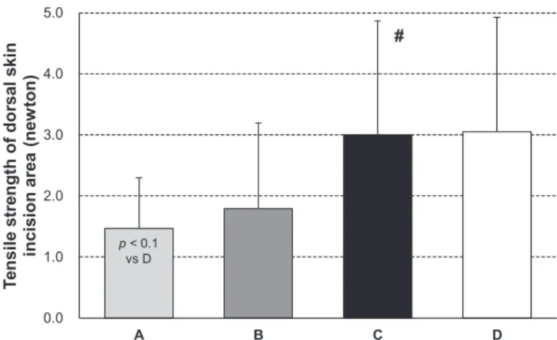
Tensile strength of the dorsal skin incision site (wound heal-ing assay)
We performed a wound healing assay to evaluate the effect of each of the nutrient solutions on wound heal-ing in protein-malnourished rats. As seen in Fig. 1, the tensile strength of the dorsal skin wound area in group C rats was almost identical to that observed for group D rats, whereas it was significantly elevated compared to those in group A rats.
mRNA expression levels of type I and type III collagen We analyzed the mRNA expression of wound healing markers (type I and type III collagen) in the dorsal skin incision area to investigate the potential mechanisms of wound healing. As seen in Fig. 2A, there was a sig-nificant up-regulation of the type I collagen (Col1a1) mRNA expression in group C compared to that in group A. Figure 2B shows that there was a tendency for up-reg-ulation of the type III collagen (Col3a1) mRNA expres-sion in group C compared to that of group A (p,0.1). Profile of plasma amino acid levels
Table 5 presents the plasma amino acid profiles. Com-pared to the other groups, group A exhibited signifi-cantly increased levels of plasma glutamine, glutamic acid, and alanine. After administration of the nutrient solutions in groups A–C, we observed infused amino acid concentration-dependent increases in the plasma arginine, isoleucine, leucine, lysine, phenylalanine, tryptophan, and valine concentrations. These amino acids were all included in the PPN solution used in this study.
Fig. 1. Using the tensile strength of the dorsal skin incision area to evaluate wound healing. Results are presented as the means6SD. # indicates p,0.05 compared to group A among the three nutrient solution groups (Tukey-Kramer test) (A: n512, B: n510, C: n511, D: n56).
Fig. 2. mRNA expression levels of type I collagen (Col1a1) (A) and the type III collagen (Col3a1) (B) in the dorsal skin incision area. Results are presented as the means6SD. *indicates p,0.05 compared to group D among all of the experimental groups (Dunnett’s test). # indicates p,0.05 compared to group A among
the three nutrient solution groups (Tukey-Kramer test) (A: n512, B: n510, C: n511, D: n56).
DISCUSSION
PEM is one of the factors that can delay wound heal-ing (6, 7). In contrast, appropriate nutritional support improves nutritional status and wound healing. The present study examined the effect of immediate short-term postoperative administrations of PPN solution with amino acids or maintenance solution without amino acids on nutritional status and wound healing in protein-malnourished rats.
Our initial findings showed that the feeding of a low-protein diet for 2 wk induced a significant reduction of the overall body weight. We examined the effect of administering each solution immediately after surgery in these protein-malnourished animals. At the end of the study, our findings demonstrated that rats admin-istered PPN solutions containing 1.5% or 3% amino acids exhibited improved nutritional status, such as a body weight gain, and increase in the plasma levels of albumin and total protein. Moreover, the levels of the rapid-turnover proteins such as prealbumin and trans-ferrin increased in these groups. Previous studies have shown that plasma rapid-turnover proteins have a short half-life and thus are more precise nutritional markers than albumin during the acute phase (10, 11). Indeed, Jiménez Jiménez et al. (12) examined the clinical use of PPN solutions and reported that a 5 d infusion of PPN solutions immediately after surgery improved nitrogen balance and levels of prealbumin, retinol-binding
pro-tein, and 3-methylhistidine compared to the baseline values of the surgical patients.
Our study also showed that a 5 d administration of PPN solutions containing 1.5% and 3% amino acids increased the weights of the gastrocnemius and EDL muscles of rats. Studies in humans and animals show that branched-chain amino acids (BCAA), especially leucine, stimulate muscle protein synthesis (13, 14). And Sugawara et al. (15) reported the inhibitory effect of leucine on muscle protein degradation in low-protein diet-fed (5% casein) rats. They showed that addition of leucine to low protein diet suppressed the decrease in gastrocnemius muscle weight and the release of 3-methylhistidine from EDL muscle. Since the PPN solution used in this study was BFLUID, which is a BCAA- (30% w/w BCAA of total amino acids) and leu-cine (4.2 g/1,000 mL)-enriched solution (Table 2), the observed increase in the muscle weight might be related to the their content of the solution. Moreover, clinical reports indicated that BCAA provides precursors for glu-tamine and alanine synthesis during catabolic states, in addition to enriching the nutritional support and pre-serving the nitrogen balance of surgical patients (16). Sun et al. (17) observed that administration of BCAA-enriched total parenteral nutrition (TPN) solutions improved the nutritional status and reduced the postop-erative complications in malnourished surgical patients with gastrointestinal cancer.
Our study also evaluated the tensile strength of the
Table 5. Profile of plasma amino acid levels (nmol/mL).
A B C D
Alanine 515.2678.5* 282.1653.0# 267.9634.5# 261.3656.1
Arginine 157.4643.6 303.5624.6*# 367.3682.1*#† 200.2630.0
Asparagine 35.964.0* 22.865.2# 19.263.0*# 27.765.0
Aspartic acid 10.563.0* 7.560.8# 7.561.0# 6.860.7
Citrulline 27.964.6* 32.366.2* 34.964.6*# 52.764.2
Cysteine 18.962.6 22.761.9*# 22.662.9*# 17.262.7
Glutamic acid 43.1610.6* 29.268.2# 26.066.0# 28.563.7
Glutamine 7,396.66467.9* 5,757.46374.7# 5,350.56321.8# 5,764.46359.4
Glycine 186.1627.7 227.2629.2# 233.6631.2# 200.1630.5
Histidine 95.468.7 91.866.2 127.96116.0 67.169.4 Hydroxyproline 18.062.8 18.362.3 19.463.7 17.964.4 Isoleucine 47.166.3 60.969.5 88.5651.5# 77.865.0
Leucine 82.4610.7* 114.2618.1 165.1660.8#† 128.2611.4
Lysine 548.3680.8* 763.86110.8# 969.46214.6#† 820.56167.4
Methionine 26.865.5* 43.966.3*# 49.264.6*# 20.363.8
Ornithine 150.5671.0 134.7625.1 429.56638.6 99.5624.3 Phenylalanine 48.667.0 77.0612.2 96.4646.6*# 54.467.1
Proline 157.1614.8* 159.8619.5* 187.7651.3* 262.9692.0 Serine 335.0656.8* 208. 5631.4*# 178.0620.7*# 85.7621.1
Threonine 140.1628.8 419.5670.4*# 343.06110.1*# 108.1635.2
Tryptophan 52.3616.2* 96.6611.7# 118.1619.7*#† 79.167.8
Tyrosine 29.467.4 40.369.5# 41.263.9# 34.366.8
Valine 95.2612.4* 132.6621.1* 211.0670.8#† 183.6616.8
Results represent means6SD. *indicates p,0.05 vs. group D among all experiment groups by Dunnett’s test. # indicates p,0.05 vs. group A and † indicates p,0.05 vs. group B among three nutrient solution groups by the Tukey-Kramer test. (A: n512, B: n510, C: n511, D: n56)
dorsal skin wound area and demonstrated that PPN solu-tions containing 3% amino acids promoted wound heal-ing. To the best of our knowledge, this is the first experi-mental animal study to demonstrate that postoperative administration of PPN solutions not only improves the nutritional status but also the wound healing outcomes. In another recent study involving animal experiments, Gündog˘du et al. (18) reported that preoperative oral nutritional intervention improved anastomotic wound healing in calorically-restricted rats. In our current study, we found that the postoperative administration of PPN solutions containing 3% amino acids promoted wound healing in protein-malnourished rats. Thus, the administration of PPN solutions containing amino acids immediately after surgery would be efficacious for wound healing. Furthermore, the significant increase in type I collagen and a tendency for increase in type III collagen mRNA expression indicate that these factors might play a part in the mechanism that underlies the promotion of wound healing. Type I and type III collagens have been shown to play crucial roles in wound healing (19). Oishi et al. (20) reported that 8 d of a protein-free diet induced down-regulation of the type I and type III col-lagen mRNA expression in the dorsal skin of rats. This result suggests that protein malnutrition can deteriorate skin conditions. The wound healing process consists of 3 consecutive phases, namely, the inflammatory phase, proliferative phase, and remodeling phase. Type III col-lagen is produced in the proliferative phase, and thereaf-ter, collagen replacement from type III to type I occurs in the remodeling phase (21). Thus, the differences in the collagen mRNA expression levels observed between groups C and D may have contributed to this process. Moreover, it should be noted that the tensile strength of the wound area for these groups was similar. How-ever, with respect to collagen mRNA expression levels, the wound healing of group D rats with a normal nutri-tional state was faster than that of the group C rats in which type III collagen was replaced by type I collagen.
Our analysis of plasma amino acids revealed changes in the levels of various amino acids. Glutamine, glu-tamic acid, and alanine concentrations were especially elevated in the group that was administered the main-tenance solutions without amino acids. Since muscle tissues contain large amounts of these amino acids (22), the significant reduction in muscle weight indi-cates that amino acids from the degraded muscle were released into the blood. Moreover, we also observed that the concentrations of the plasma arginine, isoleucine, leucine, valine, lysine, phenylalanine, and tryptophan were higher in the group that was administered the PPN solutions containing 3% amino acids. Murakami et al. (23) reported that the administration of a combination of BCAAs and glutamine stimulated dermal collagen protein synthesis in protein-malnourished rats. In addi-tion, Zandifar et al. (24) reported that arginine admin-istration improved wound healing in diabetic rats. Over-all, these previous findings are partially consistent with our results and speculation that the elevation of these plasma amino acid levels is associated with promotion
of wound healing.
However, our current study is not without limitations. First, we estimated type I and type III collagen mRNA expressions only 5 d after the post-operative administra-tion of PPN to investigate the mechanisms underlying the promotion of wound healing. However, the wound healing process is very complex (21), and thus, other factors may be involved in wound healing and other time points may be required. Second, the energy intake between the groups administered with the nutrient solutions (A–C), which were given equal amounts of energy, and the control group rats, who could consume experimental diet ad libitum, might have varied. How-ever, since our objective was to investigate the effects of the nutrient solutions on nutritional status and wound healing, we believe that the conditions used to evaluate the postoperative administration of nutrient solutions with or without amino acids are appropriate. Third, we used young rats in this study. However, the majority of patients with PEM are elderly individuals (5). Therefore, further studies using aged rats are required to better mimic the clinical conditions of elderly patients with PEM.
In conclusion, our results suggest that even short-term postoperative administration of PPN solutions con-taining amino acids increases skeletal muscle weight, improves nutritional status, and promotes wound heal-ing. Therefore, amino acids in the PPN solutions are efficacious, and the use of PPN solutions within a clini-cal setting might be a valuable asset in the recovery of protein-malnourished patients.
Acknowledgments
We would like to thank Mitsuo Nakayama, Toshimi Ohno, Takao Imanaka, Takeshi Iwamoto, Akiyoshi Kuroda and Takashi Kuwahara (Otsuka Pharmaceu-tical Factory, Inc.) for their helpful comments and discussions.
Authorship
AW and DH contributed to the conception and design of the study. AW, CS, YM, YH, and AN contributed to the acquisition of data, analysis, and interpretation of the data. AW drafted the article. AW and DH critically revised the manuscript. All authors gave final approval for the final manuscript.
REFERENCES
1) Yamazaki E, Horikawa M, Fukushima R. 2011. Vitamin
C supplementation in patients receiving peripheral par-enteral nutrition after gastrointestinal surgery. Nutrition
27: 435–439.
2) Mimura Y, Yamakawa M, Maeda J, Tateno I, Araki S,
Fujita T, Sugizaki K, Furuya K, Oohara T. 1997. Efficacy of amino acid infusion for improving protein metabo-lism after surgery: a prospective randomized study in patients undergoing subtotal gastrectomy. J Am Coll Surg
185: 163–171.
3) Ali Abdelhamid Y, Chapman MJ, Deane AM. 2016.
Peri-operative nutrition. Anaesthesia 71: 9–18.
Prog-nostic impact of disease-related malnutrition. Clin Nutr
27: 5–15.
5) Garwe T, Albrecht RM, Stoner JA, Mitchell S, Motghare
P. 2016. Hypoalbuminemia at admission is associated with increased incidence of in-hospital complications in geriatric trauma patients. Am J Surg 212: 109–115.
6) Mäkelä JT, Kiviniemi H, Juvonen T, Laitinen S. 1995.
Factors influencing wound dehiscence after midline lap-arotomy. Am J Surg 170: 387–390.
7) Anderson K, Hamm RL. 2014. Factors that impair
wound healing. J Am Coll Clin Wound Spec 4: 84–91.
8) Schroeder D, Gillanders L, Mahr K, Hill GL. 1991. Effects
of immediate postoperative enteral nutrition on body composition, muscle function, and wound healing. JPEN
J Parenter Enteral Nutr 15: 376–383.
9) Nakayama M, Motoki T, Kuwahata T, Onodera R. 2002.
The optimal nitrogen proportion to non-protein calories in normal rats receiving hypocaloric parenteral nutri-tion. Nutr Res 22: 1091–1099.
10) Robinson MK, Trujillo EB, Mogensen KM, Rounds J,
McManus K, Jacobs DO. 2003. Improving nutritional screening of hospitalized patients: the role of prealbu-min. JPEN J Parenter Enteral Nutr 27: 389–395.
11) Raguso CA, Dupertuis YM, Pichard C. 2003. The role of
visceral proteins in the nutritional assessment of inten-sive care unit patients. Curr Opin Clin Nutr Metab Care 6: 211–216.
12) Jiménez Jiménez FJ, Leyba CO, Jiménez Jiménez LM,
Valdecasas MS, Montero JG. 1995. Study of hypocaloric peripheral parenteral nutrition in postoperative patients (European project). Clin Nutr 14: 88–96.
13) Fujita S, Volpi E. 2006. Amino acids and muscle loss
with aging. J Nutr 136: 277S–280S.
14) Leenders M, van Loon LJ. 2011. Leucine as a
pharma-conutrient to prevent and treat sarcopenia and type 2 diabetes. Nutr Rev 69: 675–689.
15) Sugawara T, Ito Y, Nishizawa N, Nagasawa T. 2007.
Sup-plementation with dietary leucine to a protein-deficient diet suppresses myofibrillar protein degradation in rats. J
Nutr Sci Vitaminol 53: 552–555.
16) Choudry HA, Pan M, Karinch AM, Souba WW. 2006.
Branched-chain amino acid-enriched nutritional sup-port in surgical and cancer patients. J Nutr 136: 314S–318S.
17) Sun LC, Shih YL, Lu CY, Hsieh JS, Chuang JF, Chen FM,
Ma CJ, Wang JY. 2008. Randomized, controlled study of branched chain amino acid-enriched total parenteral nutrition in malnourished patients with gastrointestinal cancer undergoing surgery. Am Surg 74: 237–242.
18) Gündog˘du RH, Yas¸ar U, Ersoy PE, Ergül E, Is¸ıkog˘lu S,
Erhan A. 2015. Effects of preoperative nutritional sup-port on colonic anastomotic healing in malnourished rats. Ulus Cerrahi Derg 31: 113–117.
19) Campos AC, Groth AK, Branco AB. 2008. Assessment
and nutritional aspects of wound healing. Curr Opin Clin
Nutr Metab Care 11: 281–288.
20) Oishi Y, Fu Z, Ohnuki Y, Kato H, Noguchi T. 2002. Effects
of protein deprivation on alpha1(I) and alpha1(III) col-lagen and its degrading system in rat skin. Biosci
Bio-technol Biochem 66: 117–126.
21) Reinke JM, Sorg H. 2012. Wound repair and
regenera-tion. Eur Surg Res 49: 35–43.
22) Holecek M, Sispera L. 2016. Effects of arginine
supple-mentation on amino acid profiles in blood and tissues in fed and overnight-fasted rats. Nutrients 8: 206–216.
23) Murakami H, Shimbo K, Takino Y, Kobayashi H. 2013.
Combination of BCAAs and glutamine enhances dermal collagen protein synthesis in protein-malnourished rats.
Amino Acids 44: 969–976.
24) Zandifar A, Seifabadi S, Zandifar E, Beheshti SS, Aslani
A, Javanmard SH. 2015. Comparison of the effect of topical versus systemic L-arginine on wound healing in acute incisional diabetic rat model. J Res Med Sci 20: 233–238.