M E T H O D O LO G I E S
Alveolar antral artery management during sinus
elevation: A case report of a novel approach with review of
the literature
Nicola Alberto Valente1, Konstantinos Harogiannis2, Sebastiano Andreana2
1Department of Periodontics and Endodontics, School of Dental Medicine, State University of New York at Buffalo, Buffalo, New York, United States, 2Department of Restorative Dentistry, School of Dental Medicine, State University of New York at Buffalo, Buffalo, New York, United States
Abstract
Maxillary sinus elevation is a widely used and relatively safe and predictable technique. Frequently, when aberrant anatomical conditions of the sinus are present, the handling of such a procedure might become more complex, and subjected to possible intra- and post-operatory complications. One of the most challenging anatomical conditions to manage is the alveolar antral artery (AAA), when it is unusually wide in diameter and passes through the area of the osteotomy with a complete intraosseous course. In the literature, many treatment options have been proposed for the surgical management of such an anatomical condition. The aim of this case report is to describe the clinical handling of an AAA with a piezosurgical approach, including the artery in the bony window design, but avoiding its displacement and possible tearing that might cause severe intra-operatory bleeding and post-operatory complications.
Keywords: Piezoelectric surgery, sinus augmentation, sinus blood supply Correspondence
Dr. Nicola Alberto Valente, Department of Periodontics and Endodontics, School of Dental Medicine, State University of New York at Buffalo, 250 Squire Hall, 3435 Main Street, 14201, Buffalo, New York, United States. Phone: (716) 939‑5979, E‑mail: [email protected]
Received 27 February 2015; Accepted 28 April 2015
doi: 10.15713/ins.ijcdmr.67
How to cite the article:
Nicola Alberto Valente, Konstantinos Harogiannis, Sebastiano Andreana, “Alveolar antral artery management during sinus elevation: A case report of a novel approach with review of the literature,” Int J Contemp Dent Med Rev, vol.2015, Article ID: 100215, 2015. doi: 10.15713/ins.ijcdmr.67
Introduction
The maxillary sinus floor elevation technique with a lateral approach was first described by Tatum in 1976[1] and first published by Boyne and James in 1980.[2]
The technique has been further developed and modified over the years and its indications and directions have been refined
by several authors. We distinguish now between a one stage procedure, with simultaneous insertion of the implants, and a two-stage procedure, when the residual bone height is <4 mm, but the basic procedure has essentially remained the same as was
firstly described.[3]
Despite the widespread use of this surgical technique and its relative safety there are several variables and possible complications that must be taken into consideration when planning and during the surgical procedure itself. One of these is the blood supply and vascularization of the sinus cavity and Schneider membrane and, in particular, the lateral maxillary wall that is of crucial importance as a source of the blood supply for
our graft material and because the accidental severing of a vessel
during the antrostomy can be, in some case, a significant
intra-operatory complication.
The blood supply of the maxillary sinus is provided by three branches of the maxillary artery (MA): The greater palatine artery, the infraorbital artery (IOA), and the posterior superior alveolar artery (PSAA).[4,5]
Usually, the PSAA and the IOA form anastomoses inside and outside the bony lateral antral wall that supply the Schneiderian membrane and the epiperiosteal vestibular tissues. According to the literature, an intraosseous anastomosis is constantly present while an extraosseous anastomosis is present in about 44% of the cases.[6,7] Of particular importance is the intraosseous
anastomosis, which is also called alveolar antral artery (AAA).
It was first described in 1934 and it passes through the area
second premolar to second molar area in 100% of cases and,
in such an area, the AAA was strictly close to the Schneiderian membrane and partially encased in the lateral sinus wall in all specimens.[9]
Despite severing of the AAA, it’s not life-threatening. It could dramatically complicate the procedure, damage this bony vessel can cause intense bleeding, obscuring the vision, and may lead to perforation of the Schneiderian membrane, which prolongs the operation and assessment of the sinus
membrane reflection.[10] In more than 10% of cases, there is a
risk of bleeding because of an artery with a diameter of more
than 0.5 mm. In a patient with an artery with a diameter of more than 0.5 mm (1-2 mm), the probability of a high risk of hemorrhage is about 57%.[11] These findings must be taken into
consideration when preparing the bony window to prevent an accidental severing of the AAA.
Furthermore, the presence and integrity of the AAA might be crucial during surgery in order to prevent local bone necrosis and optimize the healing of the graft material, which could be impaired if the vessel is transacted.[7]
Different methods have been suggested to prevent the
complication caused by the resection and bleeding of an AAA with a wide diameter. Of major importance is the use of the piezoelectric surgical handpiece that selectively cuts hard tissues and it’s harmful to the soft tissues, leaving delicate structures like nerves and vessels undamaged.[12] If the course
of the AAA is only partially intraosseous with the vessel being in contact with the Schneider membrane, it can be isolated and
reflected together with the sinus membrane itself. When we are
dealing with a completely intraosseous course, the AAA can be isolated, using traditional rotating burs, through a double window technique in which the design of the window takes the form of a double window with the top and bottom separated by the long course of the AAA which is left covered in length by the maxillary sinus lateral bone wall.[13] Finally, the more drastic
solution is the ligation of the vessel, with the disadvantage of depriving the sinus from the blood supply assured by the AAA [14]
On the basis of these assumptions, the use of a computed tomography (CT) scan before proceeding with sinus lift procedures is strongly recommended in order to carefully evaluate any aberrant anatomical factor of the sinus such as an unusually wide AAA or with a complete intraosseous course, but also to detect patencies of the ostium, often responsible of persistent infections, and any preexisting sinus disease. All of these issues must be taken into account before commencing a sinus lift/grafting procedure. If the dental clinician is unfamiliar with reading a CT of the paranasal sinuses, a radiologist should be consulted to review the scan.[15] Moreover, the evaluation of
the CT scan allows for the evaluation of the bucco-palatal width
of the sinus cavity to be filled, which is important in terms of
bone regeneration and healing time.[16]
In this case report, we describe a new approach to the sinus elevation in a patient with an AAA with an unusually wide diameter.
Case Report
A 68-year-old Caucasian male patient presented with a completely
edentulous upper maxilla. The medical history revealed type 2 diabetes and hypertension. The patient was taking metoprolol
(25 mg) twice per day, metformin (500 mg) twice per day, rosuvastatin (80 mg) once per day, esomeprazole (40 mg) once per day, and aspirin (85 mg) once per day.
The patient was wearing a full upper denture. According
to the patient’s request of having a fixed upper prosthesis, the
decision was made for an upper bridge supported by six implants. An immediate placement of the posterior implants was rendered
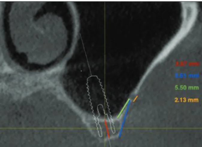
impossible by the severe resorption of the ridges with insufficient vertical distance between the crest and the floor of the sinus that was of 3.87 mm, as measured in the cone-beam computed
tomography (CBCT), thus preventing also a sinus elevation procedure with contextual implant placement. Decision was taken for a two-staged technique with sinus elevation and delayed implant placement.
We here report the surgical elevation of the left maxillary sinus.
The CBCT showed a bilateral AAA with a diameter,
measured in the CBCT, of about 2.13 mm, an AAA-alveolar crest, and AAA-sinus floor distances, respectively of 8.86 mm and 5.50 mm, in the area planned for sinus elevation (second premolar-first molar) [Figure 1].
Due to the presence of such a delicate and wide (in diameter) vascular structure in the area subjected to the surgical procedure, the decision was made for the use of the piezoelectric handpiece.
A full thickness flap was elevated and, due to the large
dimension, the AAA was already visible through the cortical bone of the lateral sinus wall [Figure 2]. The design of the osseous
window could not avoid to include the course of the artery. The osteotomy was performed with the aid of the piezoelectric bur and the bone was gently removed around the portions of AAA passing through the line of the window’s design. The mobility of the bony piece was assessed and the Schneider
membrane was elevated, avoiding tearing of the artery. A perforation of the membrane was accidentally created with the piezoelectric bur and later covered with a collagen membrane (Collatape) [Figure 3].
The bony block, resulting from the creation of the window, was not removed or pulled away with the Schneider membrane but rather rotated upwards, with the course of the AAA being the fulcrum of such a rotation, thus avoiding any stretching of the vessel that would have resulted in a laceration with intense bleeding.
The sinus cavity was then filled with 3 cc irradiated allogenic
cancellous bone and marrow particulate (Rocky Mountain
Tissue Bank, 2993 S. Peoria St. Ste 390, Urora, CO 80014)
mixed with platelet-rich plasma prepared from the patient’s own
blood. The window was covered with an additional 20 × 28 mm collagene membrane (Collatape) and the flap was then sutured with 3-0 cytoplast interrupted sutures [Figures 4 and 5].
Systemic antibiotics, amoxicillin (875 mg) twice per day for a total of 10 days, starting from the day before the
surgery, was prescribed. Local application of chlorhexidine
gluconate 0.12% twice per day was recommended. Suture removal was done 14 days following surgery.
The healing was uneventful, with complete closure of the wound and no discomfort reported by the patient.
The case has been monitored for 3 months with no adverse events, with high patient satisfaction, and stability of the graft as shown in the panoramic X-ray [Figure 6].
Discussion
A patient with a severely resorbed maxillary ridge was treated with a sinus elevation technique. The decision was made for a two-stage surgery, avoiding immediate implant placement and for the use of piezosurgery. Despite the
Figure 2: Alveoar antral artery visible through the cortical bone
Figure 3: Bony window created with no resection of the alveoar
antral artery
Figure 4: Sinus cavity filled with bone graft
Figure 5: Collagen membrane closing the lateral window
use of a piezoelectric handpiece might results in a longer surgery time when adopted for other procedures, it was demonstrated to be advantageous for sinus elevation and in procedures involving delicate structures such as arteries and nerves.[12,17,18]
In the case hereby reported the diameter of the artery was
2.13 mm, wider than the mean diameters reported by many
authors. Both the distance of the artery from the alveolar crest
(8.86 mm) and from the sinus floor (5.50 mm) were ways below
the mean values reported in the literature [Table 1].
As shown in the Table, mean values for AAA diameters
range from 0.8 mm and 1.59 mm. Mean values for distance
between AAA and alveolar crest and AAA and sinus floor
range from 16.88 mm to 23.56 mm and from 6.57 mm to 9.70 mm.
The AAA to sinus floor distance of 5.50 mm here reported
is consistent with only one study in literature where the mean
values for the second premolar area and first molar area (the area
subjected to surgery in the case here reported) were respectively
5.80 mm and 5.90 mm.
The anatomical findings of this case report are consistent with
the data reported by Park et al.,[26] Ilgüy et al.[23] and Rysz et al.,[27]
that show that the distance between the AAA and the alveolar crest is always consistently shorter in edentulous patients when compared with dentate patients.
Conclusions
The patient (subject) of this case report has been successfully treated with a sinus elevation procedure, showing no signs of
bone loss, soft tissues inflammatory activity, or any other negative
occurrence as of 3 months after the surgery. According to the clinical outcomes of this particular case, the preservation of the AAA intraosseous course, avoiding dislocation with rotation of the window osseous block using a piezosurgical approach, can be a suitable and recommendable treatment option in cases of unusually lower course of the AAA in the area of the window opening.
References
1. Tatum H Jr. Maxillary and sinus implant reconstructions. Dent Clin North Am 1986;30:207-29.
2. Boyne PJ, James RA. Grafting of the maxillary sinus floor with autogenous marrow and bone. J Oral Surg 1980;38:613-6. 3. Misch CE. Maxillary sinus augmentation for endosteal implants:
Organized alternative treatment plans. Int J Oral Implantol 1987;4:49-58.
4. Flanagan D. Arterial supply of maxillary sinus and potential for bleeding complication during lateral approach sinus elevation. Implant Dent 2005;14:336-8.
5. Van den Bergh JP, Ten Bruggenkate CM, Disch FJ, Tuinzing DB. Anatomical aspects of sinus floor elevations. Clin Oral Implants Res 2000;11:256-65.
6. Solar P, Geyerhofer U, Traxler H, Windisch A, Ulm C, Watzek G. Blood supply to the maxillary sinus relevant to sinus floor elevation procedures. Clin Oral Implants Res 1999;10:34-44. 7. Traxler H, Windisch A, Geyerhofer U, Surd R, Solar P, Firbas W.
Arterial blood supply of the maxillary sinus. Clin Anat 1999;12:417-21.
8. Strong C. The innervation and vascular supply of the antrum: (Section of laryngology). Proc R Soc Med 1934;27:745-51. 9. Rosano G, Taschieri S, Gaudy JF, Weinstein T, Del Fabbro M.
Maxillary sinus vascular anatomy and its relation to sinus lift surgery. Clin Oral Implants Res 2011;22:711-5.
10. Chanavaz M. Sinus grafting related to implantology. Statistical analysis of 15 years of surgical experience (1979-1994). J Oral Implantol 1996;22:119-30.
11. Ella B, Sédarat C, Noble Rda C, Normand E, Lauverjat Y, Siberchicot F, et al. Vascular connections of the lateral wall of the sinus: Surgical effect in sinus augmentation. Int J Oral Maxillofac Implants 2008;23:1047-52.
12. Vercellotti T, De Paoli S, Nevins M. The piezoelectric bony window osteotomy and sinus membrane elevation: Introduction of a new technique for simplification of the sinus augmentation procedure. Int J Periodontics Restorative Dent 2001;21:561-7.
13. Maridati P, Stoffella E, Speroni S, Cicciu M, Maiorana C. Alveolar antral artery isolation during sinus lift procedure with the double window technique. Open Dent J 2014;8:95-103. 14. Testori T, Rosano G, Taschieri S, Del Fabbro M. Ligation
Table 1: Summary of anatomical findings in literature
Study Number
of sinuses Mean diameter (mm) Mean distance from crest (mm) Mean distance from sinus floor (mm)
Solar et al., 1999[6] 18 1.59 18.90-19.60
(minimum-maximum) No data
Ella et al., 2008[11] 134 1.2 No data No data
Hur et al., 2009[19] 42 0.8 23.56 9.70
Kim et al., 2011[20] 400 1.52 17.13 No data
Güncü et al., 2001[21] 252 1.3 18.00 No data
Kang et al., 2013[22] 150 1.18 17.03 8.25
Ilgüy et al., 2013[23] 135 0.94 16.88 No data
Apostolakis and Bissoon, 2014[24] 256 1.1 No data 6.47
of an unusually large vessel during maxillary sinus floor augmentation. A case report. Eur J Oral Implantol 2010;3:255-8. 15. Almaghrabi BA, Hatton MN, Andreana S, Hoeplinger MA.
Treatment of severe sinus infection after sinus lift procedure: A case report. Implant Dent 2011;20:430-3.
16. Kutkut AM, Andreana S, Kim HL, Monaco E. Clinical recommendation for treatment planning of sinus augmentation procedures by using presurgical CAT scan images: A preliminary report. Implant Dent 2011;20:413-7.
17. Valente NA, Raffaelli L, Manicone PF, D’Addona A. Influence of the piezosurgery in the intra and post-operative course: Preliminary results. Dent Cadmos 2010;78:79-88.
18. Bovi M. Mobilization of the inferior alveolar nerve with simultaneous implant insertion: A new technique. Case report. Int J Periodontics Restorative Dent 2005;25:375-83.
19. Hur MS, Kim JK, Hu KS, Bae HE, Park HS, Kim HJ. Clinical implications of the topography and distribution of the posterior superior alveolar artery. J Craniofac Surg 2009;20:551-4. 20. Kim JH, Ryu JS, Kim KD, Hwang SH, Moon HS. A radiographic
study of the posterior superior alveolar artery. Implant Dent 2011;20:306-10.
21. Güncü GN, Yildirim YD, Wang HL, Tözüm TF. Location of posterior superior alveolar artery and evaluation of maxillary sinus anatomy with computerized tomography: A clinical study.
Clin Oral Implants Res 2011;22:1164-7.
22. Kang SJ, Shin SI, Herr Y, Kwon YH, Kim GT, Chung JH. Anatomical structures in the maxillary sinus related to lateral sinus elevation: A cone beam computed tomographic analysis. Clin Oral Implants Res 2013;24 Suppl A100:75-81.
23. Ilgüy D, Ilgüy M, Dolekoglu S, Fisekcioglu E. Evaluation of the posterior superior alveolar artery and the maxillary sinus with CBCT. Braz Oral Res 2013;27:431-7.
24. Apostolakis D, Bissoon AK. Radiographic evaluation of the superior alveolar canal: Measurements of its diameter and of its position in relation to the maxillary sinus floor: A cone beam computerized tomography study. Clin Oral Implants Res 2014;25:553-9.
25. Watanabe T, Shiota M, Gao S, Imakita C, Tachikawa N, Kasugai S. Verification of posterior superior alveolar artery distribution in lateral wall of maxillary sinus by location and defect pattern. Quintessence Int 2014;45:673-8.
26. Park WH, Choi SY, Kim CS. Study on the position of the posterior superior alveolar artery in relation to the performance of the maxillary sinus bone graft procedure in a Korean population. J Korean Assoc Oral Maxillofac Surg 2012;38:71-7. 27. Rysz M, Ciszek B, Rogowska M, Krajewski R. Arteries of the