Mixed-Type Melasma Treated with Low Fluence Q-Switched
Nd-YAG 1064 nm Laser: A Case Report
Aninda Fitri Nugrahani, Sinta Murlistyarini
Department of Dermatology and Venereology, DR. Saiful Anwar Hospital, Malang, East Java Faculty of Medicine, Universitas Brawijaya, Malang, East Java
ABSTRACT
Background: Melasma is an acquired, chro-nic hypermelanosis condition. Melasma is more common in women of all races and occur espe-cially on the face. The pathogenesis of melasma is very complex and the treatment is still a challenge. The purpose of this study was to report mixed-type melasma treated with low fluence Q-switched Nd-YAG 1064 nm laser. Case Presentation: A case of melasma in 54 year old woman was reported. Dermatological examination showed presence of brownish macules and patches in the centro facial area with symmetrical distribution. Examination with wood lamp showed mixed type. Patients have received topical therapy but there were no improvement.
Results: The patient was then treated with low fluence Q-switched Nd-YAG 1064 nm laser for three sessions with an interval of 2 weeks. At 6
weeks of treatment the modified MASI (mMASI) value was reduced from 8.4 to 4.6 and the VAS value was increased from 2 to 8. Conclusion: The depth of the pigment deter-mined the response to therapy. In the mixed type melasma the response for therapy is only partial. The low-fluence Q-switched Nd-YAG 1064 nm laser can penetrate deeper into the dermis and damage melanin in a short time.
Keywords: mixed typed melasma, low fluence, Q-switched Nd-YAG laser
Correspondence:
Aninda Fitri Nugrahani. Department of Derma-tology and Venereology, DR. Saiful Anwar Hospital, Malang, East Java/ Faculty of Medi-cine, Universitas Brawijaya, Malang, East Java. Email: aninda16fitri@gmail.com
Cite this as:
Nugrahani AF, Murlistyarini S (2020). Mixed-Type Melasma Treated with Low Fluence Q-Switched Nd-YAG 1064 nm Laser: A Case Report. Indones J Med. 05(02): 95-101. https://doi.org/10.26911/theijmed.20-20.05.02.01.
Indonesian Journal of Medicine is licensed under a Creative Commons Attribution-NonCommercial-ShareAlike 4.0 International License.
BACKGROUND
Melasma is an acquired, chronic hyper-melanosis condition that can cause aesthe-tic disorders and decrease the patients’ qua-lity of life. Melasma is common in women of reproductive age of all races and is characterized by symmetrical, homogenous and irregular brown macules in areas exposed to sunlight, especially the face (Tamega et al, 2013). Epidemiological data in the Cosmetic Dermatology Clinic of Der-matology and Venereology Department in Dr. Cipto Mangunkusumo Hospital, Jakarta in 2011 showed the number of melasma
patients was 18.1% of the total 3,763 visits, with a distribution of 98.4% was female patients and the remaining 1.6% was male (Melyawati et al, 2014).
chemical peeling, and various laser therapy (Choi et al, 2015). Melasma often does not improve with chemical peeling therapy or topical therapies such as hydroquinone, retinoid, azelaic acid or kojic acid due to resistance to these agents and rapid recur-rence in the formation of melasma lesions (Sim et al, 2015). Since 2006, the use of low-fluence 1064-nm Q-switched Neody-mium: Yttrium-Aluminum-Garnet (QS Nd-YAG), a technique known as "laser toning", became popular for melasma therapy. Although said to be effective, this Nd-YAG QS laser can cause hypopigmentation in some patients(Kim et al, 2013)
In this case report a 54-year-old woman with mixed-type melasma will be treated with a 1064-nm low-fluence Q-switched Neodymium-Yttrium-Aluminum-Garnet laser.
CASE PRESENTATION
A 54-year-old woman came to the Derma-tology and Venereology Clinic of Dr. Saiful Anwar Hospital Malang, with complaint of dark spots on his face. These spots have appeared since 1 year ago. Spots initially appear in the nose area, then spread to the right and left cheeks and forehead. The spots do not feel itchy, hot, or painful. Before the spots appear the patient denies the presence of acne or irritation in the area. One month before going to the hos-pital the patient visited his son who lived in Kalimantan and the patient felt his spots darken.
Patient is a housewife. Besides doing household activity, she also actively partici-pating in the recitation around the house. The patient said that she had routinely used cream from a beauty clinic >5 years. About 4 months ago the patient changed her beauty clinic and got a morning and night cream. The creams are used for 2.5 months. The patient said there was no change in the
spots. The past month, she has not used any treatment cream. Patients said she never wear sunscreen routinely every day.
The patient has 2 children. Between the first and the second child, she used implant contraceptive for 5 years. After the birth of the second child, she underwent tubectomy. The patient said that she had never taken hormonal drugs such as birth control pills or other drugs on a regular basis. The patient said that neither the patient's mother nor the patient's brother had the same complaints as the patient.
RESULTS
On physical examination the patient's blood pressure was obtained 120/80 mmHg, with a pulse of 68x/ min, a respiratory rate of 20x / min, and a temperature of 36.7oC. On dermatological examination on the face at centrofacial area, patches and macules of dark brown color with firm boundaries and irregular shapes are found. On examination with Wood's lamp, it was found that there were some spots with firm boundaries and other spots with unclear boundaries.
Figure 1 depicted dermatological examination. Figure 1 showed dark brown patches and macules with firm borders and irregular shapes at the centro facial part of the face.
Figure 2 depicted wood lamp exami-nation. Figure 2 obtained some spots with firm boundaries and other spots with indefinite boundaries.
Figure 1 A, B, C. Dermatological examination.
Figure 2 A, B, C. Wood lamp examination
Before laser therapy, the patient is given topical anesthesia with 2.5% lidocaine and 2.5% prilocaine for 30-45 minutes. After that topical anesthesia was cleaned with sterile gauze moistened with 0.9% NaCl. Patients received QS: Nd-YAG laser therapy with a wavelength of 1064 nm, spot size of
8 mm, fluence 2.0 J / cm2, and 3 times fit the entire face. The end point of this thera-py is the presence of erythema on the skin and warm palpable skin. After therapy, a cold compress with 0.9% NaCl is applied to reduce the patient's burning sensation. Laser therapy is performed every 2 weeks.
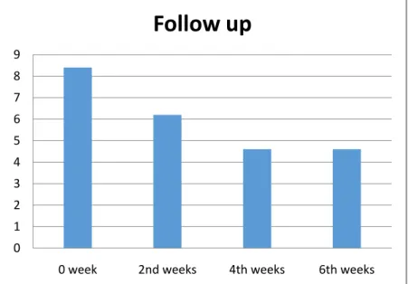
Figure 4. The improvement of mMASI value
At the time of initial examination, the modified MASI (mMASI) score was 8.4. Two weeks after the first laser, the mMASI score decreased by 2.2, so the score on the
second week became 6.2. In the fourth week the score dropped again to 4.6 and in the sixth week the mMASI score remained 4.6 (Figure 4).
Figure 5. The improvement of VAS values
0 1 2 3 4 5 6 7 8 9
0 week 2nd weeks 4th weeks 6th weeks
Follow up
0 2 4 6 8 10
0 week 2nd weeks 4th weeks 6th weeks
VAS value
To evaluate patient satisfaction, Visual Analogue Scale (VAS) is used in the range 0-10, where 0 is no improvement and 10 is a very satisfying improvement. Eva-luation was carried out at weeks 0, 2, 4, 6, and 8. VAS values obtained were 2, 5, 6, and 8. There were no significant side effects in patients (Figure 5).
DISCUSSION
Melasma is a chronic condition of hyper-melanosis obtained on the skin, which is characterized by brown macules with irre-gular shapes and symmetrically distributed in areas of the body exposed to sunlight, especially the face(Handel et al, 2014). This disorder occurs most commonly between the ages of 30 and 40 years. Family history affects the occurrence of melasma in people with Fitzpatrick skin types II and III, whereas in darker skin types family history has no effect. Melasma is more common in female with a ratio of 9: 1 compared to male. In Southeast Asia the prevalence in adult women reaches 40% and in adult men 20% (Cestari et al, 2017).
Several studies have shown that there are increasing in both melanocytosis and melanogenesis in melasma patients. Some factors that can be triggers include sun exposure, use of oral contraceptives, thy-roid disease, pregnancy, and medications (Sarkar et al, 2014). Study conducted by KrupaShankar et al in India shows that the most dominant trigger factor for melasma is sun exposure(KrupaShankar et al, 2014).
Facial melasma can be divided into 3 patterns based on the affected face area, namely centro facial pattern, malar pattern, and mandible pattern. In the centro facial pattern the affected area is the forehead, cheeks, upper lip, nose, and chin. Whereas in malar pattern the lesions are only found on the cheeks and nose, and in the pattern of the mandible the area involved is the
ramus of the mandible(Rigopoulos et al, 2017). While based on the depth of the pig-ment, the type of melasma is divided into three, namely the dermal type, epidermal type, and mixed type. On Wood's lamp exa-mination, dermal-type melasma is clearer and darker, while in the dermal type less fluorescence can be captured. The depth of the pigmentation also affects melasma therapy. In epidermal type, the response to therapy is better than the dermal type. And in mixed types, the response produced is only partial (Sarkar et al, 2014). In this case report found melasma in 54 years old woman. Patients never use sunscreen. Der-matological examination revealed brownish macules in the centro facial area and Wood's lamp examination showed mixed type.
The treatment of melasma consists of medical and non-medical therapy. In medi-cal management can be given topimedi-cal drugs, systemic drugs, and minimally and invasive treatment such as lasers and light and che-mical peeling. Non-medical management can be topical such as skin care products, decorative cosmetics, or skin lightening cosmetics, and can also be systemic for example consumption of antioxidants (Wasitaadmaja et al, 2018).
sub-termolytic laser therapy is <5 J / cm2 (Wattanakrai et al, 2010). Low fluence thera-py using a wavelength of 1064 nm (QS: Nd-YAG) can penetrate deeper into the dermis and a little affects the epidermis (Trivedi et al, 2017).
The selection of the right beam dia-meter or spot size is important in the treat-ment of pigtreat-mented lesions. Large spot sizes are safer for the epidermis and are pre-ferred for dark skin types (Small et al, 2016). Spot sizes between 7-10 mm produce maximum penetration to the dermis tissue. Deeper penetration will not be achieved if the spot size is above 10-12 mm(Nouri, 2014).
Patients were given a 1064 nm Q-switched Nd-YAG laser therapy with 2 J / cm2 fluence, 8 mm spot size, and 3 passes. 3 laser sessions with a 2-week interval showed improvement in the lesion marked by a decrease in the mMASI score and an increase in the VAS score. There were no laser side effects in patients, either hyper-pigmentation, hypohyper-pigmentation, or the presence of scars.
AUTHOR CONTRIBUTION
Both authors developed the concept for this manuscript and edited multiple drafts of the manuscript.
FUNDING AND SPONSORSHIP
There was no sponsorship in this study.
CONFLICT OF INTEREST
There is no conflict of interest in this study.
ACKNOWLEDGEMENT
The authors were fully responsible for all content and editorial decisions and received no financial support or other form of compensation related to the development of this manuscript.
REFERENCE
Arora P, Sarkar R, Garg VK, Arya L (2012). Lasers for treatment of melasma and
post-inflammatory
hyperpigmenta-tion. Journal of cutaneous and aesthe-tic surgery, 5(2): 93. doi: 10.4103/09-74-2077.99436
Cestari T, Peruzzo J, Giongo N (2017). Defi-nition, incidence, and etiology of melasma in brown skin. In Melasma and Vitiligo in Brown Skin. Springer, New Delhi.doi: 10.1007/978-81-322-3664-1_3
Choi CP, Yim SM, Seo SH, Ahn HH, Kye YC, Choi JE (2015). Retrospective analysis of melasma treatment using a dual mode of low-fluence Q-switched and long-pulse Nd: YAG laser vs. low-fluence Q-switched Nd: YAG laser mono-therapy. Journal of Cosmetic and Laser Therapy, 17(1): 28. doi: 10.3109/14764172.2014.957217
Handel AC, Miot LDB, Miot HA (2014). Melasma: a clinical and epidemiologi-cal review. Anais brasileiros de der-matologia, 89(5): 771782. doi: 10.15-90/abd1806-4841.20143063
Kim JE, Chang SE, Yeo UC, Haw S, Kim IH (2013). Histopathological study of the treatment of melasma lesions using a low‐fluence Q‐switched 1064‐nm neo-dymium: yttrium–aluminium–garnet laser. Clinical and Experimental Der-matology: Clinical dermatology, 38 (2): 167171. doi: 10.1111/j.1365-223-0.2012.04473.x
Melyawati SS, Bernadette I, Legiawati L (2014). Perkembangan terbaru etio-patogenesis melasma. Media Derma-to-Venereologica Indonesiana, 41(3): 133-138. Retrieved from http://-www.perdoski.or.id/doc/mdvi/fulltex t/33/220/13_Tinjauan_Pustaka.pdf. Moubasher AE, Youssef EM, Abou-Taleb
DA (2014). Q-switched Nd: YAG laser versus trichloroacetic acid peeling in the treatment of melasma among Egyptian patients. Dermatologic Sur-gery,40(8): 874882.doi: 10.1097/D-SS.0000000000000065
Nouri K (2014). Handbook of Laser in Dermatology. London: Springer. Rigopoulos D, Katoulis AC (2017).
Hyper-pigmentation. CRC Press. Retrieved from https://www.crcpress.com/Hy- perpigmentation/Rigopoulos-Katou-lis/p/book/9781498740173
Sarkar R, Arora P, Garg VK, Sonthalia S, Gokhale N (2014). Melasma update. Indian dermatology online journal, 5(4): 426.doi: 10.4103/2229-5178.14-2484
Sim JH, Park YL, Lee JS, Lee SY, Choi WB, Kim HJ, Lee JH (2014). Treatment of melasma by low-fluence 1064 nm Q-switched Nd: YAG laser.Journal of
Dermatological Treatment,25(3):
212217. doi: 10.3109/09546634.20-12.735639
Small R, Hoang DA (2016). Practical Guide to Laser Procedures. Philadelphia: Walters Kluwer
Tamega ADA, Miot LDB,Bonfietti C,Gige TC, Marques MEA,Miot HA (2013). Clinical patterns and epidemiological characteristics of facial melasma in Brazilian women.Journal of the Eu-ropean Academy of Dermatology and Venereology,27(2): 151156. Retrie-ved
fromhttps://doi.org/10.1111/j.1468-3083.2011.04430.x
Trivedi MK, Yang FC, Cho BK (2017). A review of laser and light therapy in melasma. International journal of
women's dermatology, 3(1): 1120.
doi: 10.1016/j.ijwd.2017.01.004
Wasitaadmaja SM,Norawati L (2018). Pedoman Diagnosis dan Tata Laksana Melasma di Indonesia. Jakarta: Badan Penerbit FKUI