Revealing the secret life of pre-implantation embryos
by time-lapse monitoring: A review
Azita Faramarzi1, 2 Ph.D., Mohammad Ali Khalili1 Ph.D., Giulietta Micara3 Ph.D., Azam Agha-Rahimi1 Ph.D.
1.Research and Clinical Center for Infertility, Yazd Reproductive Sciences Institute, Shahid Sadoughi University of Medical Sciences, Yazd, Iran.
2.Department of Anatomical Sciences and Biology, Faculty of Medicine, Kermanshah University of Medical Sciences, Kermanshah, Iran.
3.Department of Obstetrics and Gynecology, UOC OGC03 Infertility and IVF University of Rome La Sapienza, Viale Regina Elena 324-00161 Rome
Corresponding Author: Mohammad Ali Khalili, Research and Clinical Center for Infertility, Yazd Reproductive Sciences Institute, Bouali Ave., Safaeyeh, Yazd, Iran.
Email: khalili59@hotmail.com Tel: (+98) 35 38247085-6
Received: 5 October 2016
Revised: 5 January 2017
Accepted: 20 February 2017
Abstract
High implantation success following in vitro fertilization cycles are achieved via the transfer of embryos with the highest developmental competence. Multiple pregnancies as a result of the transfer of several embryos per cycle accompany with various complication. Thus, single-embryo transfer (SET) is the preferred practice in assisted reproductive technique (ART) treatment. In order to improve the pregnancy rate for SET, embryologists need reliable biomarkers to aid their selection of embryos with the highest developmental potential. Time-lapse technology is a noninvasive alternative conventional microscopic assessment. It provides uninterrupted and continues the survey of embryo development to transfer day. Today, there are four time-lapse systems that are commercially available for ART centers. In world and Iran, the first time lapse babies were born in 2010 and 2015, respectively, conceived by SET. Here, we review the use of time-lapse monitoring in the observation of embryogenesis as well as its role in SET. Although, the findings from our review support common use of time-lapse monitoring in ART centers; but, future large studies assessing this system in well-designed trials are necessary.
Key words: Embryo selection, Kinetic, Assisted reproductive technology, Time-lapse.
Introduction
n vitro fertilization (IVF) as one of assisted reproductive technique (ART) methods is used in infertility centers for conceiving infertile couples. In vitro culture of human embryo technique has improved considerably in recent years. New incubators and medium have been bettered
which provide embryo development to
blastocyst (1). In spite of this, best embryo selection for transfer is the main challenge in ART clinics. The failure to suitably know the
embryos with highest developmental
competence can lead to failed ART cycles. Grading categorical based on morphology is the preferred method for recognizing embryo developmental potential, despite promising other methods (2).
However, embryologists still depend on
daily discrete assessments of embryo
development based on cell count on cleavage days 2 or 3, the extent of blastulation as well as the quality of the inner cell mass or trophectoderm in blastocysts on day 5, the
degree of fragmentation, the rate of
nucleation, and symmetry in cleavage
embryos (1). Unfortunately, conventional
morphological techniques for evaluating
embryo development have been preliminary and have not yet increased desirable results of implantation and pregnancy (3). TLM has first used three decades ago for the study of the developmental progression of bovine embryos (4, 5). Recent interest in TLM for
assessment of clinical embryos was
engendered following numerous studies
investigating the potential benefit of multi-day scoring of embryos for selection of the most robust embryo(s) for ET (6).
Despite conflicting findings, it is assumed that compared with static observations, frequently captured images will provide
substantial information regarding the
association between morphological
development and embryo viability and a more
developmental kinetics (1). Motato et al
showed that main advancement in the selection of the best embryos with TLM is recognition of the embryos with high
I
implantation competence (7). However, the comparison of TLM with conventional embryo incubation was reviewed by Cochrane in 2015, comparing clinical pregnancy, live birth, miscarriage, and stillbirth rates. The data showed that there were no significant differences in any of these clinical outcomes (8). Some data extraction was thus done on 13 summarized papers in table I.
In this review, an overview of the current
literature concerning the use of this
technology in regards to the best embryo
selection with a possible increase of
implantation rate in ART cycles is discussed. A literature search in PubMed, Scopus, Ovid MEDLINE, Web of Science, and Science Citation Index was done to identify studies that evaluated the embryo selection based on morphokinetics by TLM system.
Why should be chosen the best embryo?
In spite of ASRM guidelines to reduce the number of embryos transferred, many centers still transfer 2-3 embryos despite the increase in the risk of multiple pregnancies (9). Multiple pregnancies are unfavorable due to the poor neonatal outcome, maternal complications, long-term developmental problems and high costs. Multiple pregnancies are associated with increased rates of stillbirths, neonatal
deaths, and infant mortality. Multiple
gestations have an increased morbidity rate due to complications associated with the increased risk of prematurity, low birth weight. Multiples also have a compromised long-term outcome, including an increased risk of long-term medical and developmental problems, such as learning problems, cerebral palsy, as well as adult health risks.
Pregnancy-induced maternal
complications, particularly hypertensive
disorders, are more common in association with multiple pregnancies (10). It also entails
an increased risk for intrapartum
complications as a consequence of uterine atony and malpresentation, resulting in high
incidences of cesarean sections and
postpartum hemorrhage. In addition, the
financial consequences of multiple
pregnancies are substantial for both parents and healthcare providers (9).
SET is now considered as the preferred practice in IVF cycles to reduce the risk of adverse outcomes associated with multiple gestations. However, in order to improve the pregnancy outcomes for SET, embryologists
depends on biomarkers to aid their selection
of the embryos with the highest
developmental potential (11). In the SET program, that includes the transfer of a single fresh embryo in selected women with good prognosis (<35 yrs of age), pursued by one or more frozen-warmed embryo transfer cycle(s)
as required, has diminished multiple
pregnancy rates, while keeping passable live birth rates (12). Over the past decade, SET utilization has been an increment in assisted conception facilities. However, there are challenges to increase use of SET as standard ET program. It involves fine embryo selection, patient education and provider, and successful cryopreservation (13).
Best embryo selection procedure
As SET becomes increasingly applied in many ART practices, the challenge of identifying the embryo with the highest developmental competence becomes crucial. Therefore, there have been investigations in search of additional viability markers to supplement current criteria for selection, such
as aneuploidy screening, O2 respiration,
metabolic profiling and gene expression analysis (14). Although, these methods are surely promising, but grading systems based on embryo morphology still remain the preferred method for assessing embryonic competence (15).
However, these morphological
assessments are generally limited to once a day checking, since repeated removal of embryos from the incubator for observation resulted in undesired temperature and pH fluctuations in the culture dish. Embryo development is a dynamic event and static observations of their growth can be limiting in their ability to discern differences between embryos at similar cell stages (16, 17). Blastocyst transfer is one of the successful approaches for SET, but it may increase neonatal complications, such as of preterm birth, low birth weight, respiratory diagnoses and low APGAR score, owing to the extended
culture condition (18, 19). Therefore,
identifying cleaving embryos that will develop to the blastocyst stage is necessary (20). The early prediction that which embryo may reach to the blastocyst before day 3 of the development will let an earlier transfer to be done. Hence, the need for longer culture to blastocyst is well eliminated, which is possible by using TLM (21).
Modern embryo survey: morphokinetics (using TLM)
The new era for embryo quality evaluation
takes a different method: dynamic
morphokinetics. It is the result of the application of TLM in the ART centers which combines morphokinetics and morphology of embryo development. The precise timing of specific events, such as pronuclear formation, syngamy appearance, early cleavage events, cell cycle intervals, the synchronicity of cell division and initiation of blastulation are indicators of an embryo’s developmental potential. The ability to continuously monitor an embryo’s progression certainly aids in selecting the best embryo/s for uterine transfer (16, 17).
Correlation of morphokinetics and aneuploidy in embryos
Absence or presence of an additional chromosome which causes to divergence from 46 chromosomes in humans is defined as aneuploidy. Embryo aneuploidy results in
implantation failure and increases in
miscarriage rates (22). The conventional procedures to distinguish aneuploidy in human embryos are preimplantation genetic screening (PGS) and preimplantation genetic diagnosis (PGD), which are expensive technologies and not routinely accessible. Also, biopsy of one or more cells of embryos is needed for analysis (23).
TLM by using certain morphokinetic parameters may distinguish aneuploidy Thus, TLM can be used to select the euploid embryos noninvasively as an alternative to PGS (24). Recently, Chawla and associates assessed the morphokinetic parameters such as timings of the extrusion second PB, pronuclei appearance and fading, division timing and second and third cleavages duration of 460 embryos in order to discriminate between euploid and aneuploid
embryos. Their results showed that
morphokinetic parameters differed
significantly for euploid and aneuploid
embryos (25).
In contrast to this, some studies showed contradictory results, with regards to knowing kinetic parameters that are classified as euploid or aneuploid embryos (26). Although TLM could be a potential selection instrument for women who are not a candidate for PGS, there is still a need for more studies to be
done before replacing PGS and/or PGD with TLM (27).
Time-lapse systems models
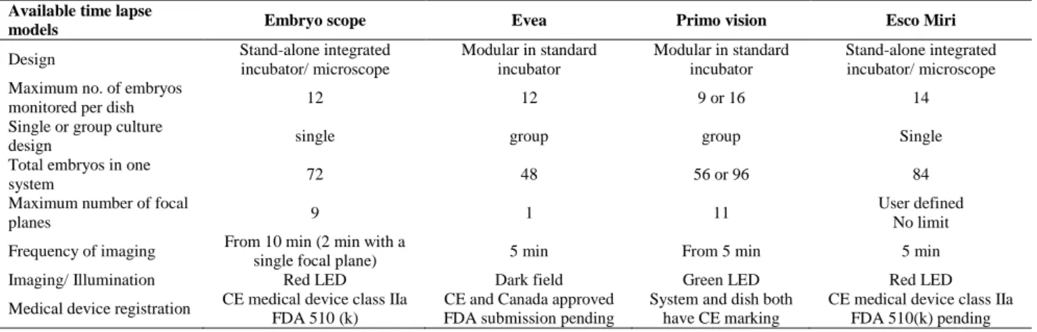
At present, there are four TLM systems which are used in the embryology laboratory, namely Primo Vision, EmbryoScope, Ecso Miri and Eeva systems (Table II). They all require the use of a digital inverted microscope that acquires images of the embryos at preset intervals which are integrated to create live images. The Embryo Scope and Ecso Miri are compact, a self-contained incubator with a built-in camera. While, both Eeva and Primo Vision systems comprise a camera that is placed in a traditional incubator (9, 11, 14, 15, 28).
Each of the systems uses a different light source and differs in the way the embryos are brought into the field of view (no movement of the embryo culture dish vs. constant movement of the dish). The EmbryoScope, Esco Miri and Primo Vision systems use bright field technology that allows the assessment of both kinetic parameters and morphology of the embryos. While, the dark field technology used with Eeva allows the assessment of kinetic parameters, but provides limited information on the morphological features.
Although, all systems use an oil overlay on culture microdrops, but differ in the way the embryos are cultured: the Eeva dish and the microwell group culture dish for Primo Vision provide group culturing, in which 12 micro-wells (Eeva) and 9-16 micro-wells (Primo Vision) share a common 50-120 μl volume of medium. In contrast, the EmbryoScope provides an individual culture set-up, in which the culture dish has 12 wells, each holding 20-25 μl of culture medium (1, 9, 28).
The benefits and safety of time-lapse technology
The first advantage of TLM is that the embryos are retained in a low disturbed environment during development, as they are not subjected to alteration in gas, pH, or temperature changes, or to the movements that are done daily for embryo assessment under conventional conditions. The second
advantage correlated to the extra
developmental morphokinetic that are
obtained as compared with conventional assessment at different time points. Human embryos display discontinuously morphologic features that are commonly used during
conventional morphological grading system (1, 8).
Key morphokinetics parameters (Table III) from TLM for prognostication of blastocyst formation, aneuploidy and implantation have been surveyed during recent years (25, 29, 30-35). Multiple studies have reported that
apply of determined morphokinetic
parameters is correlated with betters
prediction of embryo selection. Furthermore, increased implantation and pregnancy rates have been shown which has encouraged various IVF clinics to purchase TLM system (8). In spite of increased studies showing models and algorithms for selecting the best embryos, it is presumably which timing of development is distinguished by the health of embryo. Though, further culture condition variables, intrinsic patient characteristics, as well as the type of ovarian stimulation protocol and use of ICSI, could play important roles (1).
Also, TLM has improved the knowledge on the mechanism of fertilization and early
preimplantation embryo development.
Beneficial data for embryo selection is obtained by TLM to pursue the dynamic pattern of embryo development. In addition,
TLM lets observation of specific time point to be related to the capacity of embryo development and implantation. Moreover, TLM prepares a united monitoring including a safe culture environment that determines critical event of embryo development (36, 37). Before applying TLM in clinical centers, the safety of it is an important issue to be considered. TLM entails serial light exposure. It has been reported that vast exposure to light may be deleterious to the embryo, and particularly that exposure of wavelength light ought to be minimized (15). However, it has been reported there was no detrimental effect of obtaining images of a microscope on the development of human embryos (38).
Down intensity red light (635 nm) from a single light-emitting diode with low illumination times of 30 ms per image to short embryo exposure to light and to elude harming short wavelength light is used in Embryo Scope. Also, evaluations were made on conventional microscope applied in IVF centers. The time of light exposure in the time-lapse system during 3 days for a total of 1420 images was 57s, compared with a higher light exposure time of 167s reported for a conventional IVF system (38).
Table I. Association of TLM parameters with human embryo development in 15 eligible studies retrieved from electronic database search and reference list review
Authors Embryos no./ Patients no.
Start of imaging/ the time
between image acquisition Comments Cetinkaya et al
(2015) (43) 3,354 /626 time of insemination/ 20 min
Cleavage relative timings were better indicators of blastocyst formation and quality compared to absolute time-points
Storr et al
(2015) (30) 380/108 time of ICSI/ 7 to 20 min
Eight significant predictive parameters of a top quality blastocyst were known: s3, t6, t7, t8, tM, tSB, tB and tEB *
Siristatidis et al
(2015) (44) Not reported/239 Time of ICSI/ 10 min
Early embryo morphokinetics parameters were associated with the subfertile patients characteristics
Almagor et al
(2015) (45) Not reported/253 Time of ICSI/ Not reported
Irregular cleavage embryos that are prevalent in younger women may have implantation potential and live birth.
Motato et al
(2015) (7) 7,483/not reported Time of ICSI/ 15 min
Morphokinetics parameters including tM t8-t5 interval, tEB could predict blastocyst formation and implantation **
Sunddvall et al
(2015) (46) 1388/249 Time of entry/ 20 min Developmental timings in PCOS were not linked to live birth *** Wdowiak et al
(2015) (47) Not reported/165 Time of ICSI/ 10 min
Higher SDF levels could be slow down morphokinetic parameters, and might be decreasing of pregnancy rate ****
Wu et al
(2016) (48) 212/109 After PNA/ Not reported
The use of morphokinetic parameters to select embryo improved implantation and live birth rates
Adamson et al
(2016) (49) Not reported/319 After PNA/ Not reported
The use of combined conventional morphology and morphokinetics survey improved implantation rate
Mizobe et al
(2016) (50). 791/164 Time of entry/ Not reported
Blastocyst transfers that derived from faster first and second cleavage embryos, improved pregnancy rate
Goodman et al
(2016) (51) Not reported/235 Time of entry/ 10 min
The use of morphokinetics was not improving ART outcomes, significantly. Although, it associated with blastocyst implantation rates
Liu et al
(2016) (29) Not reported/265 Time of insemination/ 10 min
Qualitative and quantitative de-selection proposed model predicted implantation
Nogales et al
(2017) (52) 485/112 Time of ICSI/ 15 min
Chromosome aneuploidy affects embryo morphokinetics. TLM was useful to know discarded embryos
* S3: third synchronization, t6: time to 6 cells, t7: time to 7 cells, t8: time to 8 cells, tM: time of morula formation, tSB: time to sign of blastulation, tB: time to blastocyst, tEB: time to expanded blastocyst
**: tM: time of morula formation, t8-t5: interval 5 cells to 8 cells, tEB: time to expanded blastocyst ***: PCOS: poly cystic ovarian syndrome ****: SDF: Sperm DNA fragmentation
Table II. Comparisons of Time lapse Systems Currently Available. Available time lapse
models Embryo scope Evea Primo vision Esco Miri
Design Stand-alone integrated
incubator/ microscope
Modular in standard incubator
Modular in standard incubator
Stand-alone integrated incubator/ microscope Maximum no. of embryos
monitored per dish 12 12 9 or 16 14
Single or group culture
design single group group Single
Total embryos in one
system 72 48 56 or 96 84
Maximum number of focal
planes 9 1 11
User defined No limit
Frequency of imaging From 10 min (2 min with a
single focal plane) 5 min From 5 min 5 min
Imaging/ Illumination Red LED Dark field Green LED Red LED
Medical device registration CE medical device class IIa FDA 510 (k)
CE and Canada approved FDA submission pending
System and dish both have CE marking
CE medical device class IIa FDA 510(k) pending
Table III. Definition of morphokinetics parameters of embryos
CS2-4 The Cleavage Synchronicity from the 2- to 4-cell CS4-8 The Cleavage Synchronicity from 4- to 8-cell
DR The DNA Replication time ratio was defined and calculated by the formula= (t3-t2) / (t5-t3). T5-PNF Time from pronuclear fading to 5-cell stage
S2 or P3 duration of 3-cell stage
DC direct cleavage or where either 2- or 4-cell stage was less than 5 hours
RC reverse cleavage or where either daughter cells fused after cleavage division or the blastomere failed to divide after karyokinesis, ICCP intercellular contact points
P2 duration of 2-cell stage Pn-t1 Time of pronuclei formation NEBD Nuclear envelope break down t2 Time of cleavage to a 2-cell embryo t3 Time of cleavage to a 3-cell embryo t4 Time of cleavage to a 4-cell embryo t5 Time of cleavage to a 5-cell embryo t6 Time of cleavage to a 6-cell embryo t7 Time of cleavage to a 7-cell embryo t8 Time of cleavage to a 8-cell embryo tM Time to full compaction or Morula tSB Time to the first signs of blastulation tB Time to full blastocyst
tEB Time to expanded blastocyst tHB Time to hatching blastocyst
s1 Time between NEBD and subsequent division to 2 cells
s3 Time between division to 5 cells and subsequent division to 8 cells t4 interval Time between division to 4 cells and subsequent division to 5 cells t5-t2 Time between division to 2 cells and subsequent division to 5 cells CC2 Duration of the second cell cycle
CC3 Duration of the third cell cycle S3 time between division from 5 to 8cells tPNf Time to pronuclear fading or syngamy
Limitations of TLM
Currently, the high expenses of TLM do not let their implementation in many ART centers (39). Although, there are several studies presenting algorithms that may help in the selection of the best embryo, although the timing of early embryo development is mainly determined by the embryo. However, other factors such as the type of insemination, culture condition, type of ovarian stimulation and intrinsic women properties could play a role (1, 40). Also, one of the drawbacks of TLM is that it does not allow rolling of the embryos, causing limited visual observation,
especially when a high level of fragmentation exists or blastomeres overlapping other blastomeres (38).
First time-lapse babies in the world and Iran
In the year 2010, the analysis of time-lapse records was used to choose a single blastocyst for transfer, which resulted in a singleton pregnancy and first baby conceived with TLM IVF born in Hungary (41). The first Iranian live birth using TLM to select best embryos for transfer was reported in August 2015. A case with tubal factor infertility was
admitted to IVF program with
normozoospermia. After ovarian hyperstimulation, 6 cumulus oocyte complex (COCs) were retrieved and inseminated with 25,000 progressive sperms/ oocyte.
Five zygotes were placed individually into the micro wells of equilibrated embryo scope dish for a digital TL microscope (Primo Vision, Vitrolife Co, Sweden) observation, and incubated at 37oC, 6% CO2, O2 5% and N2
89%. The following early kinetic markers were
assessed: time to 2nd polar body (PB)
extrusion, pronuclei (PN) appearance, PN fading or syngamy (tPNf), time to 2 cells (c) (t2), 3c (t3), 4c (t4), 5c (t5), 6c (t6), 7c (t7), and 8c (t8). Durations of the second cell cycle (cc2; t3-t2) and the time to complete
synchronous divisions s2 (t4-t3) were
calculated. Cleavage anomaly was monitored: direct cleavage (single blastomere divided from 1 to 3 cells). The presence of multinucleation, vaculation, and fragmentation were also recorded on day 3, SET took place based on kinetic parameters of the embryos. Clinical pregnancy was confirmed 7 wk after SET (42).
Conclusion
In general, the practice of multiple embryo transferring not only increase the implantation rates, but also increase the multiple gestations associated with many complications. The ideal state would be high implantation potential SET. In recent years, many efforts have been done to finding suitable approaches to identify the best embryo. Despite of promising other
methods, embryo selection based on
morphology remains preferred method.
However, conventional morphology
assessment is subjective and provides limited and discrete data. Recently, the emerging
embryo TLM tools have enabled full
observation of embryo development. Every change in embryo morphology, from extrusion of the second polar body to the complete
blastocyst hatching can be recorded,
monitored and assessed.
In addition, all irregularities and
abnormalities of embryo development can be observed, which only monitored by TLM. The
application of time lapse microscopy
increases embryologist knowledge on embryo morphology and development. Also, it is an effective way for culturing and assessing embryos with minimum disturbing of optimal
embryo condition. The data may be used for better selection of embryos for SET, in order to prevent multiple gestations. Continuing monitoring with the use of TLM system lets a more exact identity of embryos that follow likely chromosomally normal.
Moreover, significant events could be assessed retrospectively at any time before embryo selection for ET. Finally, TLM technology in ART has the great benefit to be a non-invasive method, enable embryo development in very stable condition and correct embryologist decision on selection of embryos for transfer or cryopreservation. Although, the majority of publications have shown optimism regarding the successful application of TL technology in a SET program. Nevertheless, we aspire that large well designed RCTs will define the safety and efficacy of TLM for SET.
Conflict of interest
There is no conflict of interests of each author.
References
1. Racowsky C, Kovacs P, Martins WP. A critical appraisal of time-lapse imaging for embryo selection: where are we and where do we need to go? J Assist Reprod Genet 2105; 32: 1025-1030. 2. Faramarzi A, Khalili MA, Ashourzadeh S. Oocyte
morphology and embryo morphokinetics in ICSI program: is there a relationship? Zygote 2017; 25: 190-196.
3. Meldrum DR. Introduction: nongenetic markers of oocyte and embryo competence. Fertil Steril 2015; 103: 301-302.
4. Massip A, Mulnard J. Time-lapse cinematographic analysis of hatching of normal and frozen-thawed cow blastocysts. J Reprod Fertil 1980; 58: 475-478. 5. Massip A, Mulnard J, Vanderzwalmen P, Hanzen C,
Ectors F. The behaviour of cow blastocyst in vitro: cinematographic and morphometric analysis. J Anat 1982; 134: 399-405.
6. Finn A, Scott L, O’Leary T, Davies D, Hill J. Sequential embryo scoring as a predictor of aneuploidy in poor-prognosis patients. Reprod Biomed Online 2010; 21: 381-390.
7. Motato Y, de los Santos MJ, Escriba MJ, Ruiz BA, Remohí J, Meseguer M. Morphokinetic analysis and embryonic prediction for blastocyst formation through an integrated time-lapse system. Fertil Steril 2016; 105: 376-384.
8. Armstrong S, Arroll N, Cree LM, Jordan V, Farquhar C. Time-lapse systems for embryo incubation and assessment in assisted reproduction. Cochrane Database Syst Rev 2015; 2: Cd011320.
9. Desai N, Ploskonka S, Goodman LR, Austin C, Goldberg J, Falcone T. Analysis of embryo morphokinetics, multinucleation and cleavage anomalies using continuous time-lapse monitoring in blastocyst transfer cycles. Reprod Biol Endocrinol 2014; 12: 54.
10. Keel BA, May JV, De Jonge CJ. Handbook of the assisted reproduction laboratory. CRC Press; 2000. 11. Chen AA, Tan L, Suraj V, Pera RR, Shen S.
Biomarkers identified with time-lapse imaging: discovery, validation, and practical application. Fertil Steril 2013; 99: 1035-1043.
12. Grady R, Alavi N, Vale R, Khandwala M, McDonald SD. Elective single embryo transfer and perinatal outcomes: a systematic review and meta-analysis. Fertil Steril 2012; 97: 324-331.
13. Practice Committee of the American Society for Reproductive Medicine. Multiple gestation associated with infertility therapy: an American Society for Reproductive Medicine Practice Committee opinion. Fertil Steril 2012; 97: 825-834. 14. Seli E, Robert C, Sirard MA. OMICS in assisted
reproduction: possibilities and pitfalls. Mol Hum Reprod 2010; 16: 513-530.
15. Kirkegaard K, Agerholm IE, Ingerslev HJ. Time-lapse monitoring as a tool for clinical embryo assessment. Hum Reprod 2012; 27: 1277-1285. 16. Chamayou S, Patrizio P, Storaci G, Tomaselli V,
Alecci C, Ragolia C, et al. The use of morphokinetic parameters to select all embryos with full capacity to implant. J Assist Reprod Genet 2013, 30: 703-710. 17. Conaghan J, Chen AA, Willman SP, Ivani K,
Chenette PE, Boostanfar R, et al. Improving embryo selection using a computer-automated time-lapse image analysis test plus day 3 morphology: results from a prospective multicenter trial. Fertil Steril 2013; 100: 412-419.
18. Dar S, Lazer T, Shah PS, Librach CL. Neonatal outcomes among singleton births after blastocyst versus cleavage stage embryo transfer: a systematic review and meta-analysis. Hum Repord Update 2014; 0: 1-10.
19. Källén B, Finnström O, Lindam A, Nilsson E, Nygren, KG, Olausson PO. Blastocyst versus cleavage stage transfer in in vitro fertilization: differences in neonatal outcome? Fertil Steril 2010; 94: 1680-1683.
20. Liu Y, Chapple V, Roberts P, Matson P. Prevalence, consequence, and significance of reverse cleavage by human embryos viewed with the use of the Embryoscope time-lapse video system. Fertil Steril 2014; 102: 1295-1300.
21. Milewski R, Kuc P, Kuczynska A, Stankiewicz B, Lukaszuk K, Kuczynski W. A predictive model for blastocyst formation based on morphokinetic parameters in time-lapse monitoring of embryo development. J Assist Reprod Genet 2015; 32: 571-579.
22. Fragouli E, Alfarawati S, Spath K, Jaroudi S, Sarasa J, Enciso M, et al. The origin and impact of embryonic aneuploidy. Hum Genet 2013; 132: 1001-1013.
23. Campbell A, Fishel S, Bowman N, Duffy S, Sedler M, Thornton S. Retrospective analysis of outcomes after IVF using ananeuploidy risk model derived from time-lapse imaging without PGS. Reprod Biomed Online 2013; 27: 140-146.
24. Daughtry BL, Chavez SL. Chromosomal instability in mammalian pre-implantation embryos: potential causes, detection methods, and clinical consequences. Cell Tissue Res 2016; 363: 201-225.
25. Chawla M, Fakih M, Shunnar A, Bayram A, Hellani A, Perumal V, et al. Morphokinetic analysis of cleavage stage embryos and its relationship to aneuploidy in a retrospective time-lapse imaging study. J Assist Reprod Genet 2015; 32: 69-75. 26. Kramer YG, Kofinas JD, Melzer K, Noyes N,
McCaffrey C,Buldo-Licciardi J, et al. Assessing morphokinetic parameters via time lapse microscopy (TLM) to predict euploidy: are aneuploidy risk classification models universal? J Assist Reprod Genet 2014; 31: 1231-1242.
27. Gardner DK, Meseguer M, Rubio C, Treff NR. Diagnosis of human preimplantation embryo viability. Hum Reprod Update 2015; 21; 727-747. 28. Campbell A, Fishel S. Atlas of Time Lapse
Embryology. CRC Press; 2015.
29. Liu Y, Chapple V, Feenan K, Roberts P, Matson P. Time-lapse deselection model for human day 3 in vitro fertilization embryos: the combination of qualitative and quantitative measures of embryo growth. Fertil Steril 2016; 105: 656-662.
30. Storr A, Venetis CA, Cooke S, Susetio D, Kilani S, Ledger W. Morphokinetic parameters using time-lapse technology and day 5 embryo quality: a prospective cohort study. J Assist Reprod Genet 2015; 32: 1151-1160.
31. Chavez SL, Loewke KE, Han J, Moussavi F, Colls P, Munne S, et al. Dynamic blastomere behavior reflects human embryo ploidy by the four-cell stage. Nat Commun 2012; 3: 1251.
32. Paternot G, Debrock S, De Neubourg D, D’Hooghe TM, Spiessens C. Semi-automated morphometric analysis of human embryos can reveal correlations between total embryo volume and clinical pregnancy. Hum Reprod 2013; 28: 627-633. 33. VerMilyea MD, Tan L, Anthony JT, Conaghan J,
Ivani K, Gvakharia M & et al. Computer-automated time-lapse analysis results correlate with embryo implantation and clinical pregnancy: a blinded, multi-centre study. Reprod BioMed Online 2014; 29: 729-736.
34. Kirkegaard K, Campbell A, Agerholm I, Bentin-Ley U, Gabrielsen A, Kirk J, et al. Limitations of a time-lapse blastocyst prediction model: a large multicentre outcome analysis. Reprod BioMed Online 2014; 29: 156-158.
35. Cruz M, Gadea B, Garrido N, Pedersen KS, Martínez M, Pérez-Cano I & et al. Embryo quality, blastocyst and ongoing pregnancy rates in oocyte donation patients whose embryos were monitored by time-lapse imaging. J Assist Reprod Genet 2011; 28: 569-573.
36. Yalçınkaya E, Ergin EG, Çalışkan E, Öztel Z, Özay A, Özörnek H, et al. Reproducibility of a time lapse embryo selection model based on morphokinetic data in a sequential culture media setting. J Turk Ger Gynecol Assoc 2014; 15: 156-160.
37. Molina I, Martínez JV, Pertusa JF, Balasch S, Iniesta I, Pellicer A. Assessment of the implantation of day-2 human embryos by morphometric nonsubjective parameters. Fertil Steril 2014; 102: 1022-1028.
38. Meseguer M, Herrero J, Tejera A, Hilligsøe KM, Ramsing NB, Remohí J. The use of morphokinetics as a predictor of embryo implantation. Hum Reprod 2011; 26: 2658-2671.
39. Kaser DJ, Racowsky C. Clinical outcomes following selection of human preimplantation embryos with time-lapse monitoring: a systematic review. Hum Reprod Update 2014; 20: 617-631.
40. Herrero J, Meseguer M. Selection of high potential embryos using time-lapse imaging: the era of morphokinetics. Fertil Steril 2013; 99: 1030-1034.
41. Pribenszky C, Mátyás S, Kovács P, Losonczi E, Za´dori J, Vajta G. Pregnancy achieved by transfer of a single blastocyst selected by time-lapse monitoring. Reprod Biomed Online 2010; 21: 533-536.
42. Faramarzi A, Khalili MA, Soleimani M. First successful pregnancies following embryo selection using Time-lapse technology in Iran: Case report. Iran J Reprod Med 2015; 13: 253-258.
43. Cetinkaya M, Pirkevi C, Yelke H, Colakoglu YK, Atayurt Z, Kahraman S. Relative kinetic expressions defining cleavage synchronicity are better predictors of blastocyst formation and quality than absolute time points. J Assist Reprod Genet 2015; 32: 27-35. 44. Siristatidis C, Komitopoulou MA, Makris A,
Sialakouma A, Botzaki M, Mastorakos G, et al. Morphokinetic parameters of early embryo development via time lapse monitoring and their effect on embryo selection and ICSI outcomes: a prospective cohort study. J Assist Reprod Genet 2015; 32: 563-570
45. Almagor, Or Y, Fieldust S, Shoham1 Z. Irregular cleavage of early preimplantation human embryos:
characteristics of patients and pregnancy outcomes. J Assist Reprod Genet 2015; 32: 1811-1815. 46. Sundvall L, Kirkegaard K, Ingerslev HJ, Knudsen
UB. Unaltered timing of embryo development in women with polycystic ovarian syndrome (PCOS): a time-lapse study. J Assist Reprod Genet 2015; 32: 1031-1042.
47. Wdowiak A, Bakalczuk S, Bakalczuk G. The effect of sperm DNA fragmentation on the dynamics of the embryonic development in intra cytoplasmic injection. Reprod Biol 2015; 15: 94-100.
48. Wu L, Han W, Zhang X, Wang J, Liu W, Xiong S, et al. A retrospective analysis of morphokinetic parameters according to the implantation outcome of IVF treatment. Eur J Obstet Gynecol Reprod Biol 2016; 197: 186-190.
49. Adamson GD, Abusief ME, Palao L, Witmer J, Palao L, Gvakharia M. Improved implantation rates of day 3 embryo transfers with the use of an automated time-lapse-enabled test to aid in embryo selection. Fertil Steril 2016; 105: 369-375.
50. Mizobe Y, Oya N, Iwakiri R, Yoshida N, Sato Y, Miyoshi K, et al. Effects of early cleavage patterns of human embryos on subsequent in vitro development and implantation. Fertil Steril 2016; 106: 348-353.
51. Goodman LR, Goldberg J, Falcone T, Austin C, Desai N. Does the addition of time-lapse morphokinetics in the selection of embryos for transfer improve pregnancy rates? A randomized controlled trial. Fertil Steril 2016; 105: 275-285. 52. Nogales MDC, Bronet F, Basile N, Martínez EM,
Liñán A, Rodrigo L, et al. Type of chromosome abnormality affects embryo morphology dynamics. Fertil Steril 2017; 107: 229-235.