Vol. 6, Number 1, Winter / Spring 2016/65-74
Isolation of biologically active Actinomycetes from
untouched soils: a case study from Karaj district,
Iran
Ensieh Salehghamari*; Maryam Najafi
Department of Cell and Molecular Science, School of Biological Science, Kharazmi University, Moffateh Str., Tehran, Iran.
Received: November 29, 2015; Accepted: June 1, 2016
Actinomycetes are a source of a broad variety of secondary metabolites with diverse biological activities, such as antifungi, antibiotics and antitumorals; many of which have been developed for clinical use. In this study, 34 actinomycetes from untouched soils were isolated from Alborz Province-Iran. Evaluation of antifungal and antibacterial activities of these isolates, demonstrated the capability of the isolates to inhibit the growth of fungi and bacteria using agar well diffusion method. Moreover, the ethyl acetate and ethanol extracts of an isolate were also tested against Staphylococcus aureus (MRSA) and their inhibited zones were measured. Among the studied isolates 53% were active against at least one of the seven tested pathogens and 32% of actinomycetes were active against tested pathogenic fungi. Some of the actinomycetal isolates had shown strong antifungal and antibacterial activity which promises a good source of novel antimicrobial agents. As a case, isolate act-3 was selected for its high antimicrobial activity against MRSA. These results suggested that actinomycetes from Alborz province have a good potential for the production of biologically active compounds.
Keywords: Alborz province; Antimicrobial activity; Methicillin resistant; Staphylococcus aureus
Introduction
Microbial Natural Products (MNPs) are increasingly required for diverse industrial applications. One of the main targets is focused on the discovery of new antimicrobial compound, to face antibiotic resistant pathogens (1-2). Infections caused by fungi and multidrug-resistant (MDR) bacteria continue to be a worldwide problem. Currently society faces a rapidly growing resistance among pathogens that cause infection in the nosocomial environment (3-4). One of the most relevant multidrug resistant bacteria is
particularly antimicrobial producers and are aerobic, Gram positive bacteria comprising a group of branching microorganism (10-11). Many studies have been conducted in the search for novel actinomycetes (12-15).
Actinomycetes are present in various ecological habitats such as soil, water, lake, sewage and marine environment (5, 12, 16). Although soils have been screened by the pharmaceutical industry for about 50 years, only a small fraction of the surface of the earth has been screened, and only a small fraction of actinomycetes taxa has been identified (17).
The soils of Alborz province in Iran have rarely been explored for microbial metabolites. Hence, there is an immense possibility to identify new actinomycetes to discover novel bioactive compounds. In line with our previous investigations on biodiversity of actinomycetes in Alborz province and their natural products, we chose various geographical points Western to former study sites (18). After antimicrobial tests and selection of the best anti MRSA producer strain, its polarity is determined by two different solvent extractions. Finally the 16s rDNA gene for this strain is sequenced and its phylogenetic placement is studied.
Materials and methods
Soil sampling
A total of eight samples were collected from soil depth of 5-15 cm in Karaj city, Alborz province, Iran (location: 35°48' N, 51°00' E) during spring in May 2015. In our previous work (18) we had collected the samples from western part of Alborz province 5 to 15 cm depth, without any sorting. The samples were stored in sterilized 500-ml plastic zip-lock bags and transported to the laboratory (≤ 1 h) and stored overnight at 4°C until use.
Isolation of soil actinomycetes
All the soil samples were subjected to pre-heat treatment prior to serial dilution. The treatment was performed by incubating the soil samples at 100°C for one hour (15). One gram of soil sample was mixed in 10 ml of 0.9% sodium chloride solution, and mixed thoroughly in a shaker for 30 min at 150 rpm. The suspension was serially diluted up to 10-8 dilution. An
amount of 0.1 ml of the 10-4, 10−6, and 10-8 dilutions was spread plated on ISP-2 agar plates and incubated at 28°C for 7 days (7). Nystatin (50 mg/ ml) and nalidixic acid (10 mg/ml) were added for the inhibition of fungi and bacteria, respectively, during initial selection. After the incubation period, the plates were examined for the presence of actinomycetes colony. The suspected colonies were picked up and purified on ISP-2 media until pure cultures were obtained. The isolates were maintained on plates for short-term storage, and long-term strain collections were set up in medium supplemented with 30 % glycerol at −20°C (18).
Morphological characterization
Actinomycetes colonies were recognized on the basis of morphological characteristics by light microscopy according to the recommendations of International
Streptomyces Project (ISP) (19).
Indicator microorganisms
Methicillin resistant Staphylococcus aureus ATCC 33591, Klebsiella pneumoniae ATCC 10031,
Escherichia coli ATCC 29998, Serratia marcescens
ATCC 13880, Pseudomonas aeruginosa ATCC 9027,
Aspergillusniger ATCC 16404 and Candida albicans
ATCC 10231 were used as indicator strains. They were obtained from Persian Type Culture Collection (PTCC).
Preparation of inoculum
Bacterial inoculums were prepared by growing cells in Nutreint Broth (N.B.) for 24 h at 37°C. These cell suspensions were diluted with sterile N.B. to provide initial cell counts of about 10-4 CFU/ml. Aspergillus niger was grown on Sabouraud Dextrose Agar (SDA) slants at 28°C for 10 days. The spores were collected. Yeast was grown on Sabouraud Dextrose Broth (SDB) at 28°C for 48 h; size of inoculums was 106 CFU/ml (1, 20).
Screening for antimicrobial activity by modified spektra-plak method
and anti-candidal assay, Mueller Hinton Agar (MHA) plates were inoculated with actinomycetes cultures by a small circle of actinomycete isolates in the center of the petri dish and incubated at 28◦C for 4 days. The inoculums were cultured with swab in MHA around cultured actinomycetes.
For antifungal assay, Starch-casein agar plates were inoculated by actinomycete isolates around the plate and A. niger in the center at the same time. Antagonism was detected by formation of inhibition zone and measured by the determination of the size of the inhibition zone.
Secondary screening for actinomycete antimicrobial activity
Actinomycetes were inoculated in ISP-2 medium and incubated at 28°C, 220 rpm for 5 d. Cells were harvested by centrifugation at 4000 rpm for 10 minutes and the supernatant was collected into a fresh tube, filterated by nitrocellulose filter paper (0.4 µm) and tested for antimicrobial activity by the agar well diffusion method (21). The test pathogenic microbes were inoculated on MHA plate and wells of 6 mm diameter were prepared. In each of the plates, wells were filled with 100μl of clear supernatant of actinomycetes and the plates were incubated at 37°C for 48 h. All experiments were performed in triplicates.
Extraction of antimicrobial metabolites
The total culture filtrate of selected actinomycete isolate act-3 (650 ml) was used for solvent extraction with ethyl acetate and ethanol equally. The ratio of filtrate and solvent (1:1 v/v) was taken in a separating funnel and shaken vigorously. The organic layer was collected and the solvent was evaporated using vacuum rotary evaporator. MRSA as a test organism was used to test antimicrobial activity of the extract as was explained above. The solvents were included in the assays as negative controls. The plates were incubated for 24 h at 37°C.
DNA extraction, 16S rDNA amplification and phylogenetic analysis
The isolate act-3 was grown for 4 days at 28°C with
agitation in 100 ml flasks containing 10 ml of ISP-2 medium. Biomass was harvested by centrifugation at 4000 rpm for 10 min and washed twice with 0.9% sodium chloride. About 200 mg of pellet was used for DNA extraction as described by Kieser et al. (22). The
16S rDNA was amplified using PCR with Taq DNA
polymerase and primers 9F (5´
AAGAGTTTGATCATGGCTCAG 3´) and 1542R (5´ AGGAGGTGATCCAACCGCA 3´). The reaction mix (25 μl) contained 1μl of genomic DNA, 25 μl of Taq Master Mix (2X), 0.4 mm of each primer and 4% DMSO. The reaction was started with an initial denaturation at 96 ⁰C for 300 second followed by 30
cycles of denaturation at 96 ⁰C for 30 second, annealing at 54.5 ⁰C for 30 second and extension at 72 ⁰C for 90 second, with a final extension at 72 ⁰C for 300 second.
The PCR products were analyzed by agarose gel electrophoresis and submitted for purification and sequencing to Macrogen Inc. (Seoul, Korea).
The identification of phylogenetic neighbors and the calculation of pairwise 16S rDNA sequence similarities were achieved using the eztaxon server (http://www.ezbiocloud.net/eztaxon). Sequences were aligned using CLUSTAL X software (version 2.0, Conway Institute, USA), and a phylogenetic tree was constructed by the neighbor-joining method using MEGA software (version 5.0, Biodesign Institute, USA). The bootstrap was calculated from 1,000 replicates to assess the reliable level to the nodes of the tree.
Results
Screening and morphology
Out of 34 isolates, the majority (n = 13, 38.2%) were isolated from woody root, followed by other roots (n =10, 29.4%), 5 cm depth of soil (n = 6, 17.6%) and 15 cm depth of soil (n = 5, 14.7%).
Antimicrobial test
They also screened primarily against bacteria and fungi (Fig. 1). Among 34 isolates, 4 (11.8 %) isolates showed inhibition against P. aeruginosa, 9 (26.5%) against MRSA, 2 (5.9 %) isolates against E. coli and one (3%) isolate against K. pneumonia and Se. marcescens. Out of 34 isolates, merely 32 % isolates displayed potential antifungal activity in preliminary screening. The maximum antifungal activity was exhibited by 6 isolates against C. albicans (23.1 %), 5 isolates showed activity against A. niger (19 %). This result revealed that the Actinobacteria associated with the soils of Alborz Province possess the potential ability to produce diversely reactive secondary metabolites against pathogens. Isolates act-1, act-3, act-6 and act-8 exhibited the highest and broad
spectrum of antimicrobial activities and are considered as the most promising isolates for further attention. Act-6 showed activity against MRSA (34 mm), C. albicans (16 mm), K. pneumoniae (24 mm) and P. aeroginosa (18 mm), antimicrobial activity of act-3 was also found to be high against MRSA (24 mm), act-1 showed high antimicrobial activity against MRSA (22 mm) and C. albicans (21 mm), and act-8 had acute activities against P. aeruginosa (25 mm), MRSA(20 mm) and A. niger (22 mm) (Figs. 2, 3).
Secondary screening
As MRSA is considered to be one of the most important pathogenic bacteria, it was used as bioassay microorganism in this research. Four isolates including act-1, 3, 6 and 8 showed the most antibacterial activity against MRSA (Fig. 3). They were taken to study for secondary screening. Act-1 and mostly act-3 were the best anti-MRSA metabolite producer. As it is shown in Figure 4, they had the highest antibacterial activity against MRSA (22 mm).
Figure 1. Antimicrobial activity of 34 isolated actinomycetes against human pathogens. The tested pathogens were Staphylococcus aureus (MRSA), Klebsiella pneumoniae, Escherichia coli, Serratia marcescens,
Figure 2. Primary screening of antibacterial activity for isolated actinomycetes against bacterial pathogens, using modified spektra-plak method.
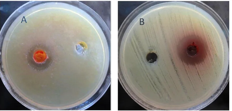
Figure 4. Antimicrobial activity of act-1 (A), 3, 6 and 8 (B) crude extracts against MRSAgrown on Muller Hinton Agar plates, using well-diffusion agar method.
Extraction
Based on the secondary screening results, act-3 was taken to study the extraction and evaluation of its antimicrobial property. The ethyl acetate and ethanol
extracts of act-3 was tested against MRSA. The Inhibition zone of ethyl acetate and ethanol extracts were 18 and 22 mm (Fig. 5).
Bacterial identification and phylogenetic analysis
Genomic DNA of the act-3 isolate was isolated and
16S rDNA gene was PCR amplified with specific forward and reverse primers. Based on the 16S rDNA
gene sequence, isolate act-3 shared the highest levels of sequence similarity (100%) with the species
Streptomyces setonii NBRC 13085T (AB184300). DNA sequence was deposited in GenBank under accession number: KU161102. Phylogenetic trees based on the 16S rDNA sequences constructed with the NJ method (Fig. 6).
Figure 6. Phylogenetic tree, based on the Neighbor joining algorithm, of the 16S rDNA sequences, showing the position of isolate act-3 and closely related species of the genus Streptomyces. Bootstrap values are indicated at branch-points. Mycobacterium
llatzerense MG13T was used as the outgroup. Bar = 1% sequence divergence.
Discussion
Many researchers have isolated actinomycetes from different natural sources like soil (18, 23-24), sediments (25), marine areas (26-27), plants (28-29), termite guts (30) which are capable of producing new bioactive metabolits for the treatment of infections (31-32).
In this research, actinomycetes were isolated, mainly from the untouched area of four different regions of Alborz province- Iran. Sampling was performed on different soil types like near plant’s root or different depth. The results indicate a dynamic population diversity of bacteria (Table 1). Most of the isolates were screened from rhizosphere. Similar results were obtained by other authors who showed that Actinomycetes are abundantly present in the rhizosphere due to their antagonistic and competitive characteristics toward other colonizers such as soil
microorganisms. The importance of these species and their complex interactions with plants and other organisms from different rhizospheric soils has also been revealed (33).
From 8 soil samples collected, 34 isolates of actinomycetes were isolated in ISP2 medium, which seems to be the most specific and sensitive medium for these bacteria, since it contains glycerol as a carbon source that most actinomycetes use. These results were anticipated because earlier studies have shown the importance of the ingredients of ISP2 under which the actinomycetes were cultivated (34).
The isolated bacteria were tested for their capacity to produce antimicrobial metabolites with efficacy against Gram-positive and Gram-negative bacteria and fungi, and in particular antibiotics that inhibit growth of one or more of the tested common human pathogens. The observed antimicrobial activity Streptomyces yanii NBRC 14669T
(AB006159)
Streptomyces atratus NRRL B-16927T
(DQ026638)
Streptomyces pulveraceus LMG 20322T
(AJ781377)
Streptomyces anulatus NRRL B-2000T
(DQ026637)
Streptomyces setonii NBRC 13085T (AB184300)
act3
Streptomyces longisporoflavus NBRC 12886T
(AB184220)
Streptomyces aureus NBRC 100912T
(AB249976)
Streptomyces graminilatus JL-6T
(HQ268006)
Streptomyces pratens BK 138T
(FR692098)
Streptomyces herbaceus BK 119T
(FR692091)
Mycobacterium llatzerense MG13T
profiles of the actinomycetes confirmed the importance of screening for antibiotics against specific human pathogens, in addition to the search for novel antimicrobial agents. Gram-negative and positive pathogens are a major problem in the nosocomial environment (35) and therefore many screening works focus on searching novel antibiotics against these pathogens.
We detected significant antimicrobial activity in isolated actinomycetes against Gram positive and Gram negative bacteria and also fungi. The most important aspect of our work was to establish novel bacterial sources for antibiotic discovery.
The presence of antibiotic activity against tested bacterial and fungal pathogens guaranteed further study to search for potential antimicrobial compounds. For example isolate act-6 showed significant antibiotic activity against MRSA(34 mm).
In our study, 9 of the 34 isolates that were identified as the antibiotic producers inhibited the Gram-positive MRSA and 6 inhibited C. albicans, whilst a significantly lower number of isolates inhibited the growth of Gram-negative pathogens. Similar results were reported in other antibacterial studies (18, 36) which might be due to the nature of outer membrane of Gram negative strains which does not permit antimicrobial agent penetration.
Only four of the 34 antibiotic-producing isolates could inhibit the growth of P. aeruginosa, which is the major pathogen in cystic fibrosis lungs (37). In comparison, one of the 34 isolates inhibited growth of
K. pneumoniae, which is also a very dangerous pathogen recently found in major outbreaks in hospitals.
It is, therefore, particularly interesting to note that act-3 inhibited exclusively the growth of MRSA but not any of the other pathogens. It has been underlined that specific antimicrobial agents that act against a certain pathogen may have advantages over traditional broad-spectrum agents, whereby resistance development and side effects could potentially be reduced (38). Ethyl acetate and ethanol extracts of act-3 showed high antibiotic activity against MRSA.
Figure 6 showed that the yield of ethanol extract was higher than the yield of ethyl acetate extract. This difference could be attributed to the selectivity of extraction. Ethanol is a polar solvent known to extract a wide range of molecules including sugar, glycoside and weakly polar compounds. Whereas, ethyl acetate is an intermediary polar solvent that extracts preferentially more hydrophobic compounds like aglycone and long carbon chain ones. Furthermore, ethanol is considered as a low toxic solvent, thus its use is preferable to minimize impact on the environment (39).
Act-6 presented the widest antibacterial spectrum, exhibiting inhibitory activity against several Gram-positive and Gram-negative and fungi species such as MRSA, K. pneumonia and C. albicans.
As screening of soil samples has not been performed extensively in Iran, we hope the isolates from this study may provide us with rare and novel industrial antibiotics or metabolites, which might be more effective than the existing ones, to cure diseases.
Acknowledgement
The authors gratefully acknowledge university of Kharazmi for supporting this study.
1. Duraipandiyan, V.A.H., Sasi, V.I.H., Islam, M., Valanarasu, S. and Ignacimuthu S. (2010) Antimicrobial properties of actinomycetes from the soil of Himalaya, J. Mycol. Med., 20, 15-20.
2. Payne, D.J., Gwynn, M.N., Holmes, D.J. and Pompliano, D.L. (2007) Drugs for bad bugs: confronting the challenges of antibacterial discovery. Nat. Rev. Drug. Discov., 6, 29-40.
4. Giske, C.G., Monnet, D.L., Cars, O. and Carmeli, Y. (2008) Clinical and economic impact of common multidrug-resistant gram-negative bacilli. Antimicrob. Agents Chemoter., 52, 813–821.
5. Claverias, F.P., Undabarrena, A., Gonzalez, M., Seeger, M. and Camara, B. (2015) Culturable diversity and antimicrobial activity of Actinobacteria from marine sediments in Valparaísobay, Chile. Front. Microb., 6, 737.
6. Chambers, H.F. and DeLeo, F.R. (2009) Waves of resistance: Staphylococcus aureus in the antibiotic era.
Nat. Rev. Microbiol.,7, 629-641.
7. Ramesh, S. and Mathivanan, N. (2009) Screening of marine actinomycetes isolated from the Bay of Bengal, India for antimicrobial activity and industrial enzymes. World. J. Microb. Biot., 25, 2103–2111. 8. Goodfellow, M. and Williams, S.T. (1983) Ecology of actinomycetes. Annu. Rev. Microbiol., 98, 119–142. 9. Prabavathy, V.R., Mathivanan, N., Murugesan, K. (2006) Control of blast and sheath blight diseases of rice
using antifungal metabolites produced by Streptomyces sp. PM5. Biol. Co., 39, 313-319.
10. Claessen, D., Rozen, D.E., Kuipers, O.P., Sogaard-Andersen, L. and van Wezel, G.P. (2014) Bacterial solutions to multicellularity; a tale of biofilms, filaments and fruiting bodies. Nat. Rev. Microbiol., 12, 115-124.
11. Hopwood, D.A. (2007) Streptomyces in nature and medicine: The antibiotic makers. New York: Oxford University Press.
12. Rakshith, D. and Satish, S. (2016) Antimicrobial properties of endophytic actinomycetes isolated from
Combretum latifolium Blume, a medicinal shrub from Western Ghats of India. Front. Bio., 10, 528-536. 13. Passari, A.K., Mishra, V.K., Saikia, R., Gupta, V.K., and Singh, B.P. (2015) Isolation, abundance and
phylogenetic affiliation of endophytic actinomycetes associated with medicinal plants and screening for their in vitro antimicrobial biosynthetic potential. Front. Microbiol., 6, 273.
14. Patel, J.D., Parmar, M., Patel, P., Rohit, P., Taviyad, R., Ansari, P., Bhattacharya, B.V., yas, D., Kumar, V., Sahay, N.S. and Pawan, K. (2014) Dynamism of antimicrobial activity of actinomycetes- A case study from undisturbed microbial niche. Adv. Microbiol., 4, 324-334.
15. Ganesan, P., Reegan, A.D., David, R.H.A., Gandhi, M.R., Paulraj, M.G., Al-Dhabi, N.A. and Ignacimuthu, S. (2016) Antimicrobial activity of some actinomycetes from Western Ghats of Tamil Nadu, India.
Alexandria J. Med. In Press.
16. Goodfellow, M. and Fiedler, H.P. (2010) A guide to successful bioprospecting: informed by actinobacterial systematics. Avan. Leeuw. J. Microb.,37, 189–216.
17. Baltz, R.H. (2007) Antimicrobials from actinomycetes: Back to the future. Microbe., 2, 125-131.
18. Salehghamari, E., Soleimani, M., Tafacori, V. (2015) Antibacterial activity of some actinomycetes isolated from soils of Alborz province, Iran. Prog. Biol. Sci.,5, 159-167.
19. Shirling, E.B., Göttlieb, D. (1966) Methods for characterization of Streptomyces species. Int. J. Syst.
Bacteriol., 16, 313-340.
20. Pujari, S.G., Kadam, N.R., Chaudhari, S.G., Dixit, P.P. and Deshmukh, A.M. (2015) Studies on isolation of antimicrobial actinomycetes from Osmanabad soil. A.M.Int. J. Curr. Microbiol. App. Sci.,4, 123-126. 21. Saadoun, I., and Muhana, A. (2008) Optimal production conditions, extraction, partial purification and
characterization of inhibitory compound(s) produced by Streptomyces Ds-104 isolate against multi-drug resistant Candida albicans. Curr. Trends Biotechnol. Pharm., 2, 402-420.
22. Kieser, T., Bibb, M.J., Buttner ,M.J., Chater, K.F. and Hopwood, D.A. (2000) Practical Streptomyces
Genetics. Norwich: The John Innes Foundation.
23. Priyadarshini, A., Singdevsachan, S.K., Tripathy, S.K., Mohanta, Y.K., Patra, J.K. and Sethi, B.K. (2016) Isolation and identification of Actinomycetes from Mangrove soil and extraction of secondary metabolites for antibacterial activity. Brit. Biot. J., 12, 1-13.
25. Mohseni, M. and Norouzi, H. (2013) Antifungal activity of actinomycetes isolated from sediments of the Caspian Sea. J. Mazand. Uni. Med. Sci., 23, 79-87.
26. Neelanchal, V. and Sajeevan, T.P. (2016) Screening marine bacterium strains of Actinomycetes as antagonists to pathogenic Vibrios, molecular identification of potent strain and media optimization by: Placket-Burman analysis and response surface methodology. Invertis J. Sci.Technol., 9, 79-93.
27. Suthindhiran, K. and Kannabiran, K. (2009) Cytotoxic and antimicrobial potential of actinomycetes species
Saccharopolyspora salina VITSDK4 isolated from the bay of Bengal coast of India. Am. J. Infect. Dis.,5, 90-98.
28. Rakshith, D. and Satish, S. (2016) Antimicrobial properties of endophytic actinomycetes isolated from
Combretum latifolium Blume, a medicinal shrub from Western Ghats of India. Front. Bio, 10, 528-536. 29. Inderiati, S. and Franco, C.M.M. (2008) Isolation and identification of endophytic actinomycetes and their
antifungal activity. W. J. Microbiol. Biotechnol., 1, 1-6.
30. Watanabe, Y., Shinzato, N. and Fukatsu, T. (2003) Isolation of actinomycetes from termites’ guts.
Biotechnol. Biochem.,67, 1797-1801.
31. Busti, E., Monciardini, P., Cavaletti, L., Bamonte, R., Lazzarini, A., Sosio, M. and Donadio, S. (2006) Antibiotic producing ability by representatives of a newly discovered lineage of actinomycetes. Microbiol.,
152, 675-683.
32. Skoko, N., Vujovic, J., Savic, M., Papic, N., Vasiljevic, B. and Ljubijankic, G. (2005) Construction of
Saccharomyces cerevisiae strain FAV20 useful in detection of immuno suppressants produced by soil actinomycetes. J. Microbiol. Methods,61, 137-140.
33. Trabelsi, I., Oves, D., Manteca, A., Genilloud, O., Altalhi, A. and Nour, M. (2016) Antimicrobial activities of some Actinomycetes isolated from different rhizospheric soils in Tunisia.Curr. Microbial., 1-8.
34. Iwai, Y. and Omura, S. (1992) Cultural conditions for screening of new antibiotics. J.Antibiot., 34, 123-141.
35. Rice, L.B. (2008) Federal funding for the study of antimicrobial resistance in nosocomial pathogens: no ESKAPE. J. Infect. Dis., 197, 1079-1081.
36. Walsh, S.E., Maillard, J.Y., Russell, A.D., Catrenich, C.E., Charbonneau, D.L. and Bartolo, R.G. (2003) Activity and mechanisms of action of selected biocidal agents on Gram-positive and negative bacteria. J. App. Microbiol., 94, 240–247.
37. Davies, J.C. (2002) Pseudomonas aeruginosa in cystic fibrosis: pathogenesis and persistence. Respir. Rev.,
3, 128–134.
38. Fischbach, M.A. and Walsh, C.T. (2009) Antibiotics for emerging pathogens. Science, 325, 1089–1093. 39. Michel, T., Destandau, E., Le Floch, G., Lucchesi, M.E. and Elfakir, C. (2012) Antimicrobial, antioxidant
and phytochemical investigations of sea buckthorn (Hippophaë rhamnoides L.) leaf, stem, root and seed.