R E S E A R C H
Open Access
Methamphetamine-induced changes in the
striatal dopamine pathway in
μ
-opioid receptor
knockout mice
Sang Won Park
1*, Xine Shen
2, Lu-Tai Tien
3, Richard Roman
1and Tangeng Ma
1Abstract
Background:Repeated exposure to methamphetamine (METH) can cause not only neurotoxicity but also
addiction. Behavioral sensitization is widely used as an animal model for the study of drug addiction. We previously reported that theμ-opioid receptor knockout mice were resistant to METH-induced behavioral sensitization but the mechanism is unknown.
Methods:The present study determined whether resistance of theμ-opioid receptor (μ-OR) knockout mice to behavioral sensitization is due to differential expression of the stimulatory G proteinasubunit (Gas) or regulators of G-protein signaling (RGS) coupled to the dopamine D1 receptor. Mice received daily intraperitoneal injections of saline or METH (10 mg/kg) for 7 consecutive days to induce sensitization. On day 11(following 4 abstinent days), mice were either given a test dose of METH (10 mg/kg) for behavioral testing or sacrificed for neurochemical assays without additional METH treatment.
Results:METH challenge-induced stereotyped behaviors were significantly reduced in theμ-opioid receptor knockout mice when compared with those in wild-type mice. Neurochemical assays indicated that there is a decrease in dopamine D1 receptor ligand binding and an increase in the expression of RGS4 mRNA in the striatum of METH-treatedμ-opioid receptor knockout mice but not of METH-treated wild-type mice. METH treatment had no effect on the expression of Gas and RGS2 mRNA in the striatum of either strain of mice.
Conclusions:These results indicate that down-regulation of the expression of the dopamine D1 receptor and up-regulation of RGS4 mRNA expression in the striatum may contribute to the reduced response to METH-induced stereotypy behavior inμ-opioid receptor knockout mice. Our results highlight the interactions of theμ-opioid receptor system to METH-induced behavioral responses by influencing the expression of RGS of dopamine D1 receptors.
Keywords:Amphetamine, ?μ?-opioid receptor, addiction, dopamine receptors
Background
Methamphetamine (METH) is a highly abused CNS sti-mulant with high reward properties that leads to com-pulsive drug seeking behavior [1,2]. The mechanism of the additive properties to METH remains to be deter-mined. Repeated administration of METH results in behavioral sensitization characterized by persistent hyperlocomotor activity and stereotyped behaviors [3,4]. Animals remain sensitized for many weeks, suggesting
that the development of sensitization involves long-last-ing neuronal adaptations [5]. The neural alterations underlying behavioral sensitization are also thought to contribute to mimic changes associated with the com-pulsive drug seeking behavior. Thus, behavioral sensiti-zation is widely used as an animal model for the study of drug addiction [5-8] and it is extremely important to find therapeutic agents for behavioral sensitization to psychostimulants.
The dopamine system is generally considered a main target for amphetamines to stimulate locomotor activity and stereotyped behaviors. The nigrostriatal dopaminer-gic pathway consists of dopaminerdopaminer-gic neurons of the
* Correspondence: [email protected]
1
Department of Pharmacology and Toxicology, University of Mississippi Medical Center, Jackson, MS 39216, USA
Full list of author information is available at the end of the article
substantia nigra that innervate the striatum [9] that is intimately linked to the stereotyped behaviors produced by psychomotor stimulants [10]. It is well known that an increase in dopaminergic activity in the central ner-vous system (CNS) plays a central role in induction and expression of behavioral sensitization by psychomotor stimulants. For example it is known that activation of dopamine receptors is required for the expression of behavioral sensitization by METH [11]. METH stimu-lates the release of dopamine from dopaminergic neu-rons and activates dopamine receptors [12]. Dopamine receptors as members of the G protein-coupled receptor (GPCR) superfamily elicit a variety of cellular and beha-vioral responses through various signaling pathways to induce behavioral effects [13]. Regulators of G-protein signaling (RGS) proteins negatively regulate GPCR sig-naling, changes in RGS protein levels in the brain are thought to modulate the intensity and duration of signal-ing of cognate receptors [14]. The expression of several RGS proteins in the brain is rapidly altered in response to psychostimulants [15]. In addition there is growing evidence that exposure to amphetamine-like stimulants influences the expression of dopamine receptors, G-pro-teins and RGS in neurons that may contribute to stimu-lant-mediated behavioral responses [16]. Chronic administration of dopamine D1 agonist SKF 38393 results in enhanced behavioral responses to subsequent adminis-tration of a variety of dopamine agonists [17,18]. Others have found that stereotypic behavior in response to amphetamine administration is associated with increased expression of dopamine D1 receptors [19] and hypersensi-tivity of adenylate cyclase to dopamine stimulation which is blocked by the dopamine D1 antagonist SCH 23390 [20].
It is also clear that there are extensive anatomical and functional interactions between the dopaminergic sys-tem and other neuronal pathways. For example both the opioidergic and glutamatergic systems contribute to the development and maintenance of behavioral sensiti-zation to METH [21]. Endogenous opioid systems have been found to play important roles in reward, positive reinforcement, and additive effects on drugs of abuse [22-24]. The endogenous opioid systems consist of a variety of endogenous opioid peptides and receptors. At least three opioid receptor subtypes (δ, μ, and ) are currently recognized [25]. Enkephalins have high affinity forμ- andδ- opioid receptors whereas dynorphins have high affinity for-opioid receptors. It has been reported that amphetamines induce an increase in expression of the opioid peptide enkephalin precursor preproenkepha-lin mRNA in rodent striatum [26]. No behavioral sensi-tization to amphetamine was detected in the enkephalin knockout mice [27]. We also reported that μ-opioid receptor (μ-OR) knockout mice were less sensitive to
the development of behavioral sensitization to METH [28]. However, it remains to be determined howμ-OR contribute to METH-induced behavioral responses. The present study examined whether METH exposure causes differential changes in the expression of stimula-tory Ga (Gas; subunit coupled to dopamine D1 recep-tors) or RGS associated with dopamine D1 receptors in the CNS that may contribute to the resistance to METH-induced behavioral sensitization inμ-OR knock-out mice.
Materials and methods Animals and drug treatments
Theμ-OR knockout mice were originally developed by Lohet al. [29] on a C57/BL6 and 129/Ola hybrid genetic background. Our colony was maintained as heterozygotes by brother sister mating in the Laboratory Animal Facility of the University of Mississippi Medical Center (UMMC). All procedures were approved by Institutional Animal Care and Use Committee of the UMMC, and performed in compliance with the NIH Guide for the Care and Use of Laboratory Animals. Adult male wild-type andμ-OR knockout mice were used in this study.μ-OR knockout and wild-type mice (n = 12 for each genotype) were given METH (10 mg/kg,i.p.) once a day for 7 consecutive days to induce sensitization in order to investigate METH-evoked behavioral response. This dose was chosen on the basis of previous studies indicating that it was the dose that induced stereotyped behavioral sensitization to sub-chronic administration of METH in mice [28,30]. On day 11, after a 4 day drug washout period, the sensitized mice were challenged with ai.p. injection of METH (10 mg/kg). Stereotyped behaviors were monitored for 30 min before and for 5 hrs after the injection to evaluate the behavioral responses.
The behavior of mice was monitored in a Plexiglas®box equipped with a CCD camera and recorded on video tape, which was subsequently analyzed by a trained observer. The intensity of stereotyped activity was scored on 4-point scale (0 - normal behavior, 1 - periodic sniffing, 2 - contin-uous sniffing, 3 - contincontin-uous sniffing, periodic licking or gnawing, 4 - continuous licking or gnawing) as described by Costall and colleagues [31].
Autoradiography
Dopamine D1 receptor levels were measured using radi-olabeled ligand binding and autoradiography as previously described by Qianet al. [32]. Briefly, brain sections were pre-incubated at 4°C for 30 min in a 50 mM Tris-HCI buf-fer (pH 7.4) containing 120 mM NaCl, 5 mM KCl, 2 mM CaCl2, and 1 mM MgCl2, and then incubated for 60 min
with 1.6 nM of the labeled dopamine D1 receptor antago-nist [3H]SCH23390 at room temperature. Other sections were incubated with 30μM of the dopamine D1 receptor ligand (±)SKF38393 [33] to control for nonspecific bind-ing. The labeled brain sections as well as a set of [3 H]-impregnated plastic standards ([3H]Microscale, Amersham Life Science) were placed on Kodak BioMax MS film for 3 weeks (-80°C). The films were developed, and then ana-lyzed using a scanning densitometer and Image Quant 3.3 program (Molecular Dynamics; Sunnyvale, CA).
In SituHybridization
The expression of Gas, RGS2, and RGS4 mRNAs in the brain were determined usingin situ hybridization. The focus of the present experiment was to examine changes in the expression of Gas, RGS2, and RGS4 in the striatum of the mouse brain. We and others have used in situ hybri-dization techniques [34-36] to successfully study gene expression for a wide variety of gene products. The techni-que relies on the specificity of the probe. Oligonucleotide probes complementary to mRNAs encoding mouse Gas (5’-GCAAAGCAGCGCCTGCCTGCCCGTCTGCCTG CCGCCGCC-3’) [34], RGS2 (5’-GGGCTCCGTGGT GATCTGTGGCTTTTTACATAAG-3’), and RGS4 (5’ -GCTGG AAGGATTGGTCAGGTCAAGATAGAATC-GAG-3’) [35] were 3’end labeled with [35S]dATP using terminal deoxynucleotidyltransferase (PerkinElmer Life Sciences, Shelton, CT) andin situhybridization was per-formed as described earlier [36]. The probes used were identical to those described by Tervonenet al. [35] who verified that they specifically bound to RGS2 and RGS4 and Przewlockaet al. [34] who tested the Gas probe. We also BLASTED the sequence of the probes against all of the sequences in GENBANK and found that they exhib-ited a 100% match to the intended target. Only the RGS4 probe exhibited any significant homology (17 of 35 bp) to another target, i.e. the presenilin-2 gene. However, given the limited numbers of complementary base pairs, it is highly unlikely that the RGS4 would bind to this target at the hybridization temperature used of 38°C. Moreover, we also performed appropriate control experiment to exclude non-specific binding. The labeled slides were exposed to Kodak BioMax MR films for 5 days for Gas or 11 weeks for RGS2 and RGS4, and the films were developed and fixed. The quantification of the autoradiogram was per-formed using the Image Quant software (Molecular Dynamics, Sunnyvale, CA).
Statistical Analysis
Data are expressed as mean values ± SEM. The signifi-cance of differences in mean values was analyzed using at test (stereotyped behaviors) or a two-way ANOVA followed by a Student-Newman-Keuls post hoc test. A
P< 0.05 was considered to be significant.
Results
METH-evoked stereotyped behaviors in METH-sensitized wild-type andμ-OR knockout mice
Administration of METH in sensitized wild-type animals produced stereotyped behaviors, characterized by con-tinuous sniffing and licking that persisted for about 5 hours. In the μ-OR knockout mice the cumulative score of stereotyped behaviors was significantly lower than in the wild-type mice (Figure 1).
[3H]SCH23390 binding in the striatum of METH-sensitized wild-type andμ-OR knockout mice
Representative autoradiograms of [3H]SCH23390 bind-ing in the brain of wild-type andμ-OR knockout mice are presented in Figure 2. High levels of [3H]SCH23390 binding were seen in the striatum. Basal binding of [3H] SCH23390 in the striatum was not significantly different between wild-type and μ-OR knockout mice treated with saline. Repeated METH treatment had no signifi-cant effect on D1 receptor binding in wild type mice. In contrast, the binding of [3H]SCH23390 was markedly reduced in the μ-OR knockout in mice sensitized by repeated exposure to METH.
Wild-type mice Sc ore of M E TH-in duc ed st er eo ty py (t ot al a re a unde r c urv e) in
5 hour obse
rva tio n 500 750 1000
-OR knockout mice
* (12)
(12)
Figure 1METH (10 mg/kg)-evoked stereotyped behaviors in wild-type andμ-OR knockout mice that were exposed to METH for 7 days. METH (10 mg/kg)-evoked stereotyped behaviors
in wild-type andμ-OR knockout mice that were exposed to METH
for 7 days. Mean values ± SEM are presented. Numbers in parentheses represent the number of animals studied. * indicates a
significant difference (P< 0.05) from the corresponding value in
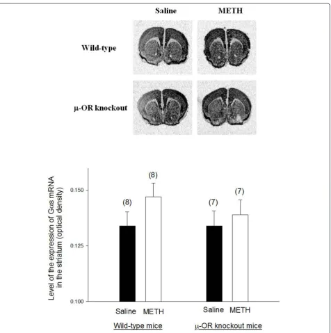
The expression of the stimulatory G proteinasubunit (Gas) mRNA in the striatum of METH-sensitized wild-type andμ-OR knockout mice
Representative autoradiograms ofin situhybridization of Gas mRNA in the brain of wild-type andμ-OR knockout
mice are presented in Figure 3. Gas mRNA is widely expressed in most brain areas including striatum and cer-ebral cortex. The expression of Gas in the striatum was similar in both wild-type andμ-OR knockout animals treated with saline. METH treatment did not alter the
Figure 2Binding of dopamine D1 ligand [3H]SCH23390 in the brains of METH-sensitized wild-type andμ-OR knockout mice. Binding of
dopamine D1 ligand [3H]SCH23390 in the brains of METH-sensitized wild-type andμ-OR knockout mice. Both strains of mice were pretreated
with daily injections saline or METH (10 mg/kg) for 7 consecutive days. Mice were killed 4 days after the final injection and brain tissues were
taken for autoradiographic analysis of [3H]SCH23390 binding. Representative autoradiograms of [3H]SCH23390 binding are shown on the top.
Mean values ± SEM are presented. Numbers in parentheses represent the number of animals/brains studied. * indicates a significant difference (P
expression of Gas mRNA in the striatum wild type mice or inμ-OR knockout mice.
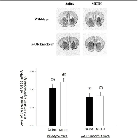
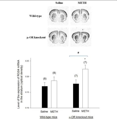
The expression of RGS2 and RGS4 mRNAs in the striatum of METH-sensitized wild-type andμ-OR knockout mice
Representative autoradiograms ofin situhybridization sig-nals for RGS2 and RGS4 mRNAs in the brain of wild-type andμ-OR knockout animals are presented in Figures 4
and 5, respectively. Both of RGS2 and RGS4 mRNAs were highly expressed in the striatum. There was no significant difference in the expression of RGS2 mRNA in the stria-tum ofμ-OR knockout or wild-type mice treated with sal-ine or METH. Basal expression of RGS4 was also similar inμ-OR knockout and wild-type mice treated with saline. However, the expression of RGS4 mRNA in the striatum increased inμ-OR knockout mice treated with METH but
Figure 3The expression of Gas mRNA in the brains of METH-sensitized wild-type andμ-OR knockout mice. The expression of Gas
mRNA in the brains of METH-sensitized wild-type andμ-OR knockout mice. Animal treatments were the same as described in Fig. 2. Gas mRNA
levels in the brain sections were analyzed byin situhybridization analysis. Representative autoradiograms of Gas mRNA expression in the brain
remained unchanged in wild-type mice sensitized with METH.
Discussion
The CNS stimulant-METH induces behavioral sensitiza-tion which is associated persistent hyperlocomotor activ-ity and stereotyped behaviors. Behavior sensitization is a widely used in rodents model for study of drug addiction
and drug seeking behaviors [5,37]. In the present study we confirmed previous finding thatμ-OR knockout mice demonstrate significantly decreased behavioral sensitiza-tion to METH as compared with wild-type mice. This was associated with a significant reduction in dopamine D1 receptor density in the striatum of approximately 30% inμ-OR knockout mice when compared to wild-type mice exposed to METH. By way of contrast, METH had
Figure 4The expression of RGS2 mRNA in the brains of METH-sensitized wild-type andμ-OR knockout mice. The expression of RGS2
mRNA in the brains of METH-sensitized wild-type andμ-OR knockout mice. Animal treatments and preparation of brain sections for analysis of
no effect on dopamine D1 receptor density in the stria-tum of wild-type mice.
We also found that the expression of Gas mRNA was unaltered by METH exposure in wild type or knockout mice, as was the expression of mRNA of the regulator of G-protein signaling, RGS2. However, the expression of RGS4 mRNA was significantly increased in the
striatum of METH treatedμ-OR knockout mice as com-pared to saline treated controls, whereas METH treat-ment had no effect on RGS4 mRNA in wild-type controls. These data suggest that in μ-OR knockout mice dopamine D1 receptor function in the striatum can be more readily down-regulated than in wild- type mice after repeated METH exposure. This may, in part,
Figure 5The expression of RGS4 mRNA in the brains of METH-sensitized wild-type andμ-OR knockout mice. The expression of RGS4
mRNA in the brains of METH-sensitized wild-type andμ-OR knockout mice. Animal treatments and preparation of brain sections for analysis of
RGS4 mRNA levels were the same as described in Fig. 3. Representative autoradiograms of RGS4 mRNA expression are shown at the top. Mean
values ± SEM are presented. Numbers in parentheses represent the number of animals/brains studied. * indicates a significant difference (P<
explain the decreased behavioral sensitization observed after METH treatment ofμ-OR knockout mice.
Dopamine is an important neurotransmitter in the CNS where it plays essential roles in numerous physiolo-gical, neuronal, and behavioral processes. One important component of the pathways in the CNS is the nigrostria-tal dopaminergic system, projecting from the substantia nigra to the striatum (putamen and caudate nucleus) that is known to be crucial for the induction of stereotyped behaviors [10]. Previously, we and others have performed dose response studies and found that 2.5 mg/kg METH is sufficient to elicit a locomotor response [28,38] but higher doses (10 mg/kg) are needed to induce behavioral sensitization to METH [28,30]. Repeated stimulation of dopamine receptors with agonists has been shown to cause down-regulation in expression of these receptors [39]. As an indirect dopamine receptor agonist, METH is known to stimulate the release and inhibit reuptake of dopamine from synaptic cleft [40], increasing extracellu-lar dopamine levels and activating postsynaptic striatal dopamine receptors. Thus, repeated METH exposure lead to down regulation of the expression of dopamine receptors in the striatum. In other studies, we found that METH (10 mg/kg) was associated with decreased tyro-sine hydroxylase (the rate limiting enzyme of dopamine synthesis) in wild-type mice but not in theμ-OR knock-out mice. These results along with our present findings indicate that the changes of dopaminergic system in mice chronically exposed to METH is related to a decrease in the expression of the enzyme involved in the synthesis of dopamine and its actions on dopamine D1 receptors rather than to the loss of dopaminergic neurons. None-theless, these data demonstrate that theμ-opioid receptor modulates the response of dopamine neurons to METH.
There are two types of dopamine receptors in the stria-tum: D1 and D2. Striatonigral neurons largely express dopamine D1 receptors whereas most striatopallidal neu-rons express dopamine D2 receptors [41]. Although con-current activation of dopamine D1 and D2 receptors is thought to be required for the full induction of stereotyped behaviors [11], activation of dopamine D1 receptors is pri-marily responsible for the induction of dopamine-mediated stereotypy [42]. Therefore, the down regulation of dopa-mine D1 receptor we found in the striatum of METH-sen-sitizedμ-OR knockout mice compared with wild-type is consistent with the view that this contributes to the less of METH-induced stereotyped behaviors in this strain of mice. Surprisingly, METH exposure in wild-type mice did not down-regulate D1 dopamine receptors. Previously our lab reported that quantitative autoradiographic analysis of striatum and nucleus accumbens showed that METH treatment leads to a decrease in dopamine D1 receptor ligand binding inμ-OR knockout mice but not in wild-type mice at low concentration (0.4 nM) of dopamine D1
receptor antagonist SCH 23390 [36]. This suggests that interactions between opiodergic receptors/neurons and neurons of the nigrostriatal pathway occur that stabilize receptor density. These interactions between these path-ways and the mechanisms involved deserve further study.
Chronic treatment (2-3 weeks) with dopamine D1 receptor antagonist SCH 23390 has been reported to increase the expression of mRNA for preproenkephalin in the rat striatum [43,44]. Recently, we found that there was an increase in expression of preproenkephalin mRNA in the nucleus accumbens and striatum in METH-sensitized wild-type mice but not inμ-OR knockout mice [36]. Also, METH induced hyperlocomotor activity at low doses and stereotyped behaviors at high doses in wild-type mice [28] but not inμ-OR knockout mice. These results indicate that a decrease in striatal and nucleus accumbens D1 receptors in METH-sensitizedμ-OR knockout mice is associated with a decrease in the behavioral response in these animals. The exact mechanism of how theμ-opioid system modulates dopaminergic neurotransmission and thus influences METH-produced behavioral responses is unclear. Based on data in the literature, however, it can be proposed that METH-induced changes in G protein sig-naling and RGS proteins might play a role in the develop-ment of behavioral sensitization to the drug.
Dopamine receptors are members of the GPCR family. Stimulation of the dopamine D1 receptors activates ade-nylyl cyclase via Gas, increasing intracellular cAMP that activates cAMP-dependent protein kinase A and its down-stream effectors [45]. There is evidence that G pro-tein signaling may be disrupted in drug addiction and neuropsychiatric disorders [46,47]. For example, post-mortem brain studies have revealed increased levels of Gas in bipolar disorder, a type of mood disorder with unknown etiology as well as being inducible by CNS sti-mulants [48]. Elevation of Gas levels is thought to enhance signaling through the dopamine D1 receptor and contribute to dopamine D1 receptor activation-mediated behavioral responses [49]. Therefore, we exam-ined the expression of Gas mRNA in the striatum of METH-sensitized mice. The results of the present study indicate that the expression of Gas mRNA in the stria-tum was not altered by repeated METH exposure in eitherμ-OR knockout or wild-type mice.
dopamine D1 and D2 receptors in the striatum of rat is coupled to RGS2 and RGS4 [55].
Amphetamine-like stimulants alters RGS mRNA expression in the brain and triggers GPCR signaling [56-58]. There are several lines of evidence that acute or repeated treatment with amphetamine modulates drug-induced behavioral and changes in gene and protein expression of RGS4 in prefrontal cortex and dorsal tum [59-61]. RGS4 mRNA was decreased in the stria-tum lasting from 1 to 6 hr after acute amphetamine [62]. RGS4 may belong to the growing family of factors regulating convergence of dopamine signaling in the striatum [63].
In the present study, METH (10 mg/kg) exposure had no influence on the expression of RGS2 mRNA in the striatum of either μ-OR knockout or wild-type mice. However, there was a higher expression of RGS4 mRNA in the striatum of METH-sensitized μ-OR knockout mice but not of wild-type mice. Increased expression of RGS4 is consistent with a reduction in signaling via dopamine D1 receptors in the striatum that may already be reduced due to the decreased density of dopamine D1 receptors in METH treatedμ-OR knockout mice. Down-regulation of dopamine D1 receptor binding in combination with increased RGS4 mRNA levels is con-sistent with diminished dopamine D1 receptor function in METH-exposed μ-OR knockout mice that would decrease the occurrence of behavioral sensitization.
Conclusions
In conclusion, the present study indicates that knockout of μ-OR in mice reduces their sensitivity to METH-induced stereotyped behaviors. Down-regulation of the expression of the dopamine D1 receptor in combination with up-regulation of the expression of RGS4 in the striatum of METH-sensitizedμ-OR knockout mice may contributes to the resistance to the behavioral responses to METH in this strain. The results suggest that theμ -opioid system and RGS proteins might be targets for the development of drugs that might reduce the reward potential and compulsive drug seeking behavior in METH abusers.
Acknowledgements
The authors gratefully thank Drs. Jerry M. Farley and Ian A. Paul at UMMC for their valuable comments on this study and Dr. Horace H. Loh at the
University of Minnesota Medical School for providing withμ-OR knockout
mice.
Author details
1Department of Pharmacology and Toxicology, University of Mississippi
Medical Center, Jackson, MS 39216, USA.2Department of Physiology, Medical
school, Soochow University, Jiangsu, P.R China.3School of Medicine, Fu Jen
Catholic University, Taipei, Taiwan.
Authors’contributions
SP carried out thein situhybridization studies, performed the statistical
analysis, and drafted the manuscript. XS participated in the animal treatment and study. LT carried out the ligand binding assay. RR participated in the data analysis and editing the manuscript. TM conceived the study and supervised the study. All authors read and approved the final manuscript.
Competing interests
The authors declare that they have no competing interests.
Received: 27 June 2011 Accepted: 10 November 2011 Published: 10 November 2011
References
1. Volkow ND:Communities across the country are trying to respond to
increased abuse of methamphetamine, a powerfully addictive stimulant.
NIDA webpage: message from the Director on Methamphetamine abuse2005 [http://www.nida.nih.gov/about/welcome/messagemeth405.html].
2. Reiner BC, Keblesh JP, Xiong H:Methamphetamine abuse, HIV infection,
and neurotoxicity.Int J Physiol Pathophysiol Pharmacol2009,25:162-179.
3. Shippenberg TS, Heidbreder C:Sensitization to the conditioned rewarding
effects of cocaine: pharmacological and temporal characteristics.J Pharmacol Exp Ther1995,273:808-815.
4. Bartlett E, Hallin A, Chapman B, Angrist B:Selective sensitization to the
psychosis-inducing effects of cocaine: a possible marker for addiction relapse vulnerability?Neuropsychopharmacology1997,16:77-82.
5. Steketee JD, Kalivas PW:Drug Wanting: Behavioral Sensitization and
Relapse to Drug-Seeking Behavior.Pharmacol Rev2011,63:A-R.
6. Robinson TE, Berridge KC:The neural basis of drug craving: an
incentive-sensitization theory of addiction.Brain Res Brain Res Rev1993,18:247-291.
7. Robinson TE, Berridge KC:The psychology and neurobiology of addiction:
an incentive-sensitization view.Addiction2000,95(Suppl 2):S91-117.
8. Itzhak Y, Martin JL, Ali SF:Methamphetamine-induced dopaminergic
neurotoxicity in mice: long-lasting sensitization to the locomotor stimulation and desensitization to the rewarding effects of methamphetamine.Prog Neuropsychopharmacol Biol Psychiatry2002,
26:1177-1183.
9. Bjorklund A, Lindvall O:In Chemical Neuroanatomy: Classical Transmitters
in the CNS, Part 1.Edited by: Bjorklund A, Lindvall. Elsevier, Amsterdam; 1984:55-122.
10. Kelley AE, Christopher GL, Gauthier AM:Induction of oral stereotypy
following amphetamine microinjection into a discrete subregion of the striatum.Psychopharmacology1988,95:556-559.
11. Marshall JF, Ruskin DN, LaHoste GJ:D1/D2 dopamine receptor interactions
in basal ganglia functions. In the dopamine receptors.Edited by: Neve KA, Neve RL. New Jersey: Humana Press; 1997:193-219.
12. Jadhav SA, Gaikwad RV, Gaonkar RK, Thorat VM, Gursale SC, Balsara JJ:
Dose-dependent response of central dopaminergic systems to buspirone in mice.Indian J Exp Biol2008,46:704-714.
13. Beaulieu J, Gainetdinov RR:The physiology, signaling, and pharmacology
of dopamine receptors.Pharmacol Rev2011,63:182-217.
14. Abramow-Newerly M, Roy AA, Nunn C, Chidiac P:There RGS proteins have
a signaling complex; interactions between RGS proteins and GPCRs, effectors, and auxiliary proteins.Cell Signal2006,18:579-591.
15. Burchett SA:Psychostimulants, madness, memory, and RGS proteins?
Neuromol Med2005,7:101-127.
16. Schwendt M, McGinty JF:Regulator of G-protein signaling 4 interacts
with metabotropic glutamate receptor subtype 5 in rat striatum: relevance to amphetamine behavioral sensitization.J Pharmacol Exper Therap2007,323:650-657.
17. Braun AR, Chase TN:Behavioral effects of chronic exposure to selective
D1 and D2 dopamine receptor agonists.Eur J Pharmacol1988,
147:441-451.
18. Rouillard C, Bedard P, Falardeau P, DiPaolo T:Repeated stimulation of D1
dopamine receptors increases the circling response to bromocriptine in rats with a 6-OHDA lesion.Eur J Pharmacol1988,157:125-133.
19. Xiao-Da Z, Guo-Zhang J:Enhanced stereotypic behavior by chronic
20. Parenti M, Flauto C, Parati E, Vescovi A, Groppetti A:Differential effect of repeated treatment with L-dopa on dopamine-D1 or D2 receptors.
Neuropharmacology1986,25:331-334.
21. Le Merrer J, Becker JA, Befort K, Kieffer BL:Reward processing by the
opioid system in the brain.Physiol Rev2009,89:1379-1412.
22. Trujillo KA, Belluzzi JD, Stein L:Naloxone blockade of amphetamine place
preference conditioning.Psychopharmacology (Berlin)1991,104:265-274.
23. Bals-Kubik R, Ableitner A, Herz A, Shippenberg TS:Neuroanatomical sites
mediating the motivational effects of opioids as mapped by conditioned place preference paradigm in rats.J Pharmacol Exp Ther1993,264:489-495.
24. Suzuki T, Mori T, Tsuji M, Misawa M, Nagase H:The role of the delta-opioid
receptor subtypes in cocaine and methamphetamine induced place preferences.Life Sci1994,55:L339-L344.
25. Knapp RJ, Malatynska E, Collins N, Fang L, Wang JY, Hruby VJ, Roeske WR,
Yamamura HI:Molecular biology and pharmacology of cloned opioid
receptors.The FASEB J1995,9:516-525.
26. Wang JQ, McGinty JF:Differential effects of D1 and D2 dopamine
receptor antagonists on acute amphetamine- or methamphetamine-induced up-regulation of zif/268 mRNA expression in rat forebrain.J Neurochem1995,65:2706-2715.
27. Hodler C, Paquet B, Gilbert F, Drolet G, Levesque D, Rouillard C:Role of
endogenous enkephalin and dynorphin in amphetamine induced behavioral sensitization [abstract].Society of Neuroscience 35th Annual Meeting, Washington, DC2005, 1029.7.
28. Shen X, Purser C, Tien LT, Chiu CT, Paul IA, Baker R, Loh HH, Ho IK, Ma T:
mu-Opioid receptor knockout mice are insensitive to methamphetamine-induced behavioral sensitization.J Neurosci Res2010,88:2294-2302.
29. Loh HH, Liu HC, Cavalli A, Yang W, Chen YF, Wei LN:μ-opioid receptor
knockout in mice: effects on ligand-induced analgesia and morphine lethality.Brain Res Mol Brain Res1998,54:321-326.
30. Tien L, Ho IK, Ma T:Methamphetamine-induced expression of zif268
mRNA is prevented by haloperidol in mice lacking mu-opioid receptor.
Neurotoxicology2010,31:326-330.
31. Costall B, Naylor RJ, Olley JE:Substantia nigra and stereotyped behavior.
Eur J Pharmacol1972,18:95-106.
32. Qian Y, Hitzemann B, Hitzemann R:D1 and D2 dopamine receptor
distribution in the neuroleptic nonresponsive and neuroleptic responsive lines of mice, a quantitative receptor autoradiographic study.
J Pharmacol Exp Ther1992,261:341-348.
33. Ongali B, Ase AR, Hebert C, Amdiss F, Reader TA:Dopamine D1 and D2
receptors in the forebrain ofdystonia musculorummutant mice: an autoradiographic survey in relation to dopamine contents.Synapse2000,
37:1-15.
34. Przewlocka B, Lason W, Przewlocki R:The effect of chronic morphine and
cocaine administration on the Gαs and Gαo protein messenger RNA levels in the rat hippocampus.Neuroscienc1994,63:1111-1116.
35. Tervonen T, Kerman K, Oostra BA, Castre’n M:Rgs4 mRNA expression is
decreased in the brain of Fmr1 knockout mouse.Molecular Brain Research
2005,133:162-165.
36. Tien LT, Ho IK, Loh HH, Ma T:Role of mu-opioid receptor in modulation
of preproenkephalin mRNA expression and opioid and dopamine receptor binding in methamphetamine-sensitized mice.J Neurosci Res
2007,85:673-680.
37. Robinson TE, Berridge KC:The psychology and neurobiology of addiction:
an incentive-sensitization view.Addiction2000,95(Suppl 2):S91-117.
38. Chiu C, Ma T, Ho IK:Methamphetamine-induced behavioral sensitization
in mice: alterations in mu-opioid receptor.J Biomedical Sci2006,
13:797-811.
39. Lin CW, Bianchi BA, Miller TR, Stashko MA, Wang SS, Curzon P, Bednarz L,
Asin KE, Britton DR:Persistent activation of the dopamine D1 receptor
contributes to prolonged receptor desensitization: studies with A-77636.
J Pharmacol Exper Therap1996,276:1022-1029.
40. Hyman SE, Malenka RC, Nestler EJ:Neural mechanisms of addiction: the
role of reward-related learning and memory.Annu Rev Neurosci2006,
29:565-598.
41. Gerfen CR, Engber TM, Mahan LC, Susel Z, Chase TN, Monsma FJ Jr,
Sibley DR:D1 and D2 dopamine receptor-regulated gene expression of
striatonigral and striatopallidal neurons.Science1990,250:1429-1432.
42. Chatoff EH, Marck BT, Matsumoto AM, Dorsa DM, Palmiter RD:Induction of
stereotypy in dopamine-deficient mice requires striatal D1 receptor activation.PNAS2001,98:10451-10456.
43. Tang F, Costa E, Schwarts JP:Increase of proenkephalin mRNA and
enkephalin content of rat striatum after daily injection of haloperidol for 2-3 weeks.Proc Natl Acad Sci USA1983,80:3841-3844.
44. Mocchetti I, Naranjo JR, Costa E:Regulation of striatal enkephalin turnover
in rats receiving antagonists of specific dopamine receptor subtypes.J Pharmacol Exp Ther1987,241:1120-1124.
45. Sunahara RK, Guan HC, O’Dowd BF, Seeman P, Laurier LG, Ng G, George SR,
Torchia J, Van Tol HH, Niznik HB:Cloning of the gene for a human
dopamine D5 receptor with higher affinity for dopamine than D1.
Nature1991,350:614-619.
46. Zhang K, Tarazi FI, Campbell A, Baldessarini RJ:GABA(B) receptors: altered
coupling to G-proteins in rats sensitized to amphetamine.Neuroscience
2000,101:5-10.
47. Mirnics K, Middleton FA, Stanwood GD, Lewis DA, Levitt P:Disease-specific
changes in regulator of G-protein signaling 4 (RGS4) expression in schizophrenia.Mol Psychiatry2001,6:293-301.
48. Young LT, Li PP, Kish SJ, Siu KP, Kamble A, Hornykiewicz O, Warsh JJ:
Cerebral cortex Gs alpha protein levels and forskolin-stimulated cyclic AMP formation are increased in bipolar affective disorder.J Neurochem
1993,61:890-898.
49. Butkerait P, Friedman E:Repeated reserpine increases striatal dopamine
receptor and guanine nucleotide binding protein RNA.J Neurochem
1993,60:566-571.
50. Hollinger S, Hepler JR:Cellular regulation of RGS proteins: Modulators
and integrators of G protein signaling.Pharmacol Rev2002,54:527-559.
51. Hooks SB, Waldo GL, Krumins AM, Harden TK:RGS6, RGS7, RGS9, and
RGS11 stimulate GTPase activity of Gifamily G-proteins with differential
selectivity and maximal activity.JBC2003,278:10087-10093.
52. Ross EM, Wilkie TM:GTPase-activating proteins (GAPs) for heterotrimeric
G proteins: regulators of G protein signaling (RGS) and RGS-like proteins.
Annu Rev Biochem2000,69:795-827.
53. Gold SJ, Ni YG, Dohlman HG, Nestler EJ:Regulators of G-protein signaling
(RGS) proteins: region-specific expression of nine subtypes in rat brain.J Neurosci1997,17:8024-8037.
54. Grafstein-Dunn E, Kathleen HY, Cockett MI, Khawaja XZ:Regional
distribution of regulators of G-protein signaling (RGS) 1, 2, 13, 14, 16, and GAIP messenger ribonucleic acids by in situ hybridization in rat brain.Molecular Brain Research2001,88:113-123.
55. Taymans JM, Leysen JE, Langlois X:Striatal gene expression of RGS2 and
RGS4 is specifically mediated by dopamine D1 and D2 receptors: Clues for RGS2 and RGS4 functions.J Neurochem2003,84:1118-1127.
56. Burchett SA, Bannon MJ, Grannenab JG:RGS expression in rat striatum:
modulation by dopamine receptors and effects of repeated amphetamine administration.J Neurochem1999,72:1529-1533.
57. Burchett SA, Volk ML, Bannon MJ, Granneman JG:Regulators of G protein
signaling: rapid changes in mRNA abundance in response to amphetamine.J Neurochem1998,70:2216-2219.
58. Bishop GB, Cullinan WE, Curran E, Gustein HB:Abused drugs modulate
RGS4 mRNA levels in rat brain: comparison between acute drug treatment and a drug challenge after chronic treatment.Neurobiol Dis
2002,10:334-343.
59. Schwendt M, Gold SJ, McGinty JF:Acute amphetamine down-regulates
RGS4 mRNA and protein expression in rat forebrain: distinct roles of D1 and D2 dopamine receptors.J Neurochem2006,96:1606-1615.
60. Schwendt M, Hearing MC, See RE, McGinty JF:Chronic cocaine reduces
RGS4 mRNA in rat prefrontal cortex and dorsal striatum.NeuroReport
2007,18:1261-1265.
61. Schwendt M, McGinty JF:Regulator of G-protein signaling 4 interacts
with mGluR5 receptors in rat striatum: Relevance to amphetamine behavioral sensitization.J Pharmacol Exper Therap2007,323:650-657.
62. Gonzalez-Nicolini V, McGinty JF:Gene expression profile from the
striatum of amphetamine-treated rats: a cDNA array and in situ hybridization histochemical study.Gene Expr Patterns2002,1:193-198.
63. Girault JA, Valjent E, Caboche J, Herve D:ERK2: a logical AND gate critical
for drug-induced plasticity?Curr Opin Pharmacol2007,7:77-85.
doi:10.1186/1423-0127-18-83
Cite this article as:Parket al.:Methamphetamine-induced changes in the striatal dopamine pathway inμ-opioid receptor knockout mice.