R E S E A R C H
Open Access
Dynamics of
DNMT3A
mutation and
prognostic relevance in patients with
primary myelodysplastic syndrome
Ming-En Lin
1,2,3, Hsin-An Hou
1*, Cheng-Hong Tsai
4, Shang-Ju Wu
1, Yuan-Yeh Kuo
5, Mei-Hsuan Tseng
1,
Ming-Chih Liu
6, Chia-Wen Liu
6, Wen-Chien Chou
1,7, Chien-Yuan Chen
1, Jih-Luh Tang
1, Ming Yao
1, Chi-Cheng Li
1,4,
Shang-Yi Huang
1, Bor-Sheng Ko
1, Szu-Chun Hsu
7, Chien-Ting Lin
1,4and Hwei-Fang Tien
1Abstract
Background:DNMT3Agene mutation has been associated with poor prognosis in acute myeloid leukemia, but its clinical implications in myelodysplastic syndrome (MDS) and dynamic changes during disease progression remain controversial.
Results:In this study,DNMT3Amutation was identified in 7.9% of 469 de novo MDS patients.DNMT3A-mutated patients had higher platelet counts at diagnosis, and patients with ring sideroblasts had the highest incidence of DNMT3Amutations, whereas those with multilineage dysplasia had the lowest incidence. Thirty-one (83.8%) of 37 DNMT3A-mutated patients had additional molecular abnormalities at diagnosis, andDNMT3Amutation was highly associated with mutations ofIDH2andSF3B1. Patients withDNMT3Amutations had a higher risk of leukemia
transformation and shorter overall survival. Further,DNMT3Amutation was an independent poor prognostic
factor irrespective of age, IPSS-R, and genetic alterations. The sequential study demonstrated that the original DNMT3Amutations were retained during follow-ups unless allogeneic hematopoietic stem cell transplantation
was performed, whileDNMT3Amutation was rarely acquired during disease progression.
Conclusions:DNMT3Amutation predicts unfavorable outcomes in MDS and was stable during disease evolutions. It may thus be a potential biomarker to predict prognosis and monitor the treatment response.
Keywords:DNMT3A, Myelodysplastic syndrome, Prognosis, Paired samples
Background
Alterations of epigenetic regulation may result in aberra-tions of gene expression and malignant transformation of cells [1,2]. DNA methylation, one of the mechanisms for epigenetic control of gene expression, regulates important physiological development, such as gene imprint and X-chromosome inactivation [3, 4]. In mammalians, three DNA methyltransferase (DNMT), namely DNMT1, 3A, and 3B have been identified [5]. Mutation of DNMT3A gene has been reported in patients with myeloid malig-nancies, including myelodysplastic syndromes (MDS) and acute myeloid leukemia (AML) [6–13].
MDS represent a clinically heterogeneous hematologic neoplasm characterized by variable degrees of cytopenias and risk of leukemia transformation [14]. The incidence (2.6 to 20.2%) of DNMT3A mutation in MDS varied widely, possibly due to different patient population and methods used [12, 13, 15, 16]. Regarding the prognostic relevance, DNMT3A mutation has been reported to pre-dict poor prognosis in AML patients [7–11]. However, the prognostic implications of DNMT3A mutation in MDS are still controversial [12, 13, 15, 17]. Walter et al. [12] and Thol et al. [13] reported thatDNMT3Amutation was associated with higher risk of leukemia transformation and shorter survival, but the other studies failed to find these associations [15, 17]. Besides, sequential studies to evaluate the dynamic changes of DNMT3A mutations during disease evolution in MDS are limited. In the * Correspondence:hsinanhou@ntu.edu.tw
1Division of Hematology, Department of Internal Medicine, National Taiwan
University Hospital, No.7, Chung Shan S. Rd., Zhongzheng Dist, Taipei 10002, Taiwan
Full list of author information is available at the end of the article
present study, we investigated theDNMT3A mutation in 469 patients with de novo MDS and analyzed its associa-tions with the clinical characteristics, outcomes, and other genetic alterations. We also performed sequential analysis of theDNMT3Agene mutation for 431 samples from 148 patients to evaluate the stability of DNMT3A mutation during the clinical course.
Methods
Subjects
This study was approved by the Institutional Review Board/Ethical Committee of the National Taiwan Univer-sity Hospital (NTUH). Diagnosis and classification of MDS were made according to the French-American-British (FAB) Cooperative Group Criteria and the WHO 2016 classification [18,19]. From May 1985 to December 2010, a total of 469 adult patients with newly diagnosed MDS at the NTUH who had enough cryopreserved cells for analysis were enrolled. Patients with secondary or therapy-related MDS were excluded. The disease of 362 patients fulfilled the criteria of MDS according to the 2016 WHO classification. Most patients (77.4%) received only palliative treatment, including transfusions, hematopoietic growth factors, immunosuppressive therapy, and low-intensity chemotherapy. Thirty (6.4%) patients received in-tensive chemotherapy, 7.2% received hypomethylating agents (HMA), and 9.0% received allogeneic hematopoietic stem cell transplantation (HSCT).
Analyses of mutations
Mutational analysis ofDNMT3A gene exons 2-23 by PCR and direct sequencing was done as described previously [9]. Analysis of the mutations in other genes involving in activated signaling pathways, such as FLT3-ITD [20], NRAS[21],KRAS[21],JAK2 [21], andPTPN11[22]; the transcription factor, such asRUNX1[23]; splicing factors, including SRSF2, U2AF1, and SF3B1[24]; and epigenetic modifications, including MLL/PTD [25], ASXL1 [26], EZH2[27],IDH1[28],IDH2[29], andTET2[30], as well as SETBP1 [21], WT1 [31], NPM1 [32], and TP53 [33], were performed as previously described. To detect DNMT3Amutation, we used DNA amplified in vitro from bone marrow (BM) cells with the Illustra GenomiPhi V2 DNA-amplification kit (GE Healthcare, UK). All muta-tions detected were verified in the original non-amplified samples [34]. Abnormal sequencing results were con-firmed by at least two repeated analyses. All nonsense or frameshift mutations were regarded as true mutations. Missense mutations were regarded as true only if they were documented in other studies or could be verified by sequencing of matched normal somatic tissues. Serial ana-lyses of DNMT3A mutations during the clinical course were also performed in 431 samples from 148 patients.
TA cloning analysis
For the patients with discrepancy of the mutation status of the DNMT3A in sequential samples, TA cloning was performed in the samples without detectable mutation followed by direct sequencing. More than 30 clones were selected for sequencing as previously described [9].
Illumina next generation sequencing (NGS) for serial studies of patients withDNMT3Amutation
Serial analyses of mutations at diagnosis, disease pro-gression, and/or remission were further performed using Illumina next generation sequencing in 32 samples from 13 patients with DNMT3A mutation at diagnosis and one during follow-up. Genomic DNA extracted from BM mononuclear cells was analyzed for mutations in 54 genes involved in myeloid malignancies by TruSight Myeloid Panel (Illumina, San Diego, CA, USA). HiSeq platform (Illumina) was used for sequencing with a me-dian reading depth of 12,000×.
Statistical analysis
The discrete variables of patients with and without gene mutations were compared using the χ2 tests, and the Fisher’s exact test was used if the expected values of contingency tables were smaller than 5. The continuous variables of patients with and without gene mutations were compared using Student’s t test. If the data were not normally distributed, Mann-Whitney U tests were used to compare continuous variables and medians of distributions. Overall survival (OS) was measured from the date of first diagnosis to the date of last follow-up or death from any cause. Time to leukemia transformation was measured from the date of MDS diagnosis to the date confirmed of acute leukemic change. Kaplan-Meier estimation was used to plot survival curves, and log-rank tests were used to calculate the difference of OS and time to leukemia transformation between groups. Multivariate Cox proportional hazard regression analysis was used to investigate independent prognostic factors for OS and time to leukemia transformation. All tests were 2-tailed, and P< 0.05 was considered statistically significant. All statistical analyses were performed with SPSS Version 17 software.
Results
DNMT3Amutations in patients with de novo MDS
Table 1Comparison of clinical features between MDS patients with and withoutDNMT3Amutation
Variables Total DNMT3Amutated DNMT3Awild Pvalue
(n= 469) (n= 37, 7.9%) (n= 432, 92.1%)
Gender† 0.201
Male 315 (67.2) 21 (6.7) 294 (93.3)
Female 154 (32.8) 16 (10.4) 138 (89.6)
Age (year)# 65.5 (16~98) 69.6 (35~89) 65.5 (16~98) 0.151
Lab data#
WBC (× 109/L) 3.84 (0.44~355.3) 5.02 (0.49~59.83) 3.73 (0.44~355.3) 0.942
Hb (g/dL) 8.3 (3~15) 8.1 (5~11) 8.3 (3~15) 0.555
Platelet (× 109
/L) 74 (2~931) 162 (14~460) 74 (2~931) 0.045
LDH (mckat/L) 8.133 (2.422~113.677) 8.208 (3.507~24.733) 8.133 (2.422~113.677) 0.739
FAB subtype†† 0.002
RA 171 (36.5) 4 (2.3) 167 (97.7) 0.001
RARS 32 (6.8) 6 (18.8) 26 (81.2) 0.031
RAEB 159 (33.9) 18 (11.3) 141 (88.7) 0.069
RAEB-T 53 (11.3) 5 (9.4) 48 (90.6) 0.593
CMML 54 (11.5) 4 (7.4) 50 (92.6) > 0.999
2016 WHO classification†† (n= 362) (n= 28, 7.7%) (n= 334, 92.3%) 0.011
MDS-SLD 60 (16.6) 1 (1.7) 59 (98.3) 0.062
MDS-MLD 106 (29.3) 3 (2.8) 103 (97.2) 0.029
MDS-RS-SLD 18 (5.0) 3 (16.7) 15 (83.3) 0.154
MDS-RS-MLD 13 (3.6) 3 (23.1) 10 (76.9) 0.070
MDS with isolated del(5q) 2 (0.6) 0 (0.0) 2 (100.0) > 0.999
MDS-EB-1 78 (21.5) 8 (10.3) 70 (89.7) 0.343
MDS-EB-2 81 (22.4) 10 (12.3) 71 (87.7) 0.097
MDS-U 4 (1.1) 0 (0.0) 4 (100.0) > 0.999
Karyotype risk††Φ (n= 437) (n= 35) (n= 402) 0.883
Good 263 (60.2) 20 (7.6) 243 (92.4) 0.721
Intermediate 88 (20.1) 7 (8.0) 81 (92.0) > 0.999
Poor 86 (19.7) 8 (9.3) 78 (90.7) 0.658
IPSS††§ (n= 437) (n= 35) (n= 402) 0.372
Low 68 (15.6) 4 (5.9) 64 (94.1) 0.630
INT-1 181 (41.4) 11 (6.0) 170 (94.0) 0.283
INT-2 107 (24.5) 11 (10.3) 96 (89.7) 0.412
High 81 (18.5) 9 (11.1) 72 (88.9) 0.259
IPSS-R††ζ (n= 437) (n= 35) (n= 402) 0.692
Very low 13 (3.0) 1 (7.7) 12 (99.3) > 0.999
Low 104 (23.8) 7 (6.7) 97 (93.3) 0.682
Intermediate 106 (24.2) 6 (5.7) 100 (94.3) 0.411
High 113 (25.9) 10 (8.8) 103 (91.2) 0.841
Very high 101 (23.1) 11 (10.9) 90 (89.1) 0.294
Abbreviations:FAB, French-American-British classification;RA, refractory anemia;RARS, refractory anemia with ring sideroblasts;RAEB, refractory anemia with excess blasts;RAEB-T, refractory anemia with excess blasts in transformation;CMML, chronic myelomonocytic leukemia;MDS-SLD, MDS with single lineage dysplasia;MDS-MLD, MDS with multilineage dysplasia;MDS-RS-SLD, MDS with ring sideroblasts with single lineage dysplasia;MDS-RS-MLD, MDS with ring sideroblasts with multilineage dysplasia;MDS-EB1, MDS with excess blasts-1;MDS-EB2, MDS with excess blasts-2;MDS-U, MDS, unclassified
†Number of patients (% among the males or females) #
Median (range)
††Number of patients (% among patients of each subgroup)
ΦGood: normal karyotype, isolated -Y, del(5q), or del(20q); Poor: complex (≧3 abnormalities) or chromosome 7 anomalies; Intermediate, other abnormalities § IPSS (international prognosis scoring system): low, 0; intermediate (INT)-1, 0.5–1; INT-2, 1.5–2; and high,≥2.5
identified in 37 (7.9%) of the 469 patients, including 7 mis-sense mutations, 2 nonmis-sense mutations, and 10 frameshift mutations (Fig.1). In addition to the 13 single-nucleotide polymorphisms without amino acid residue alterations, 9 missense mutations with uncertain biologic significance were excluded because they were not reported previously and could not be verified for lack of matched normal som-atic tissues or remission BM samples. Thirty-six of the 37 DNMT3A-mutated patients had single heterozygous mu-tation, and the remaining one patient had double muta-tions. The most common mutation was R882H (n= 11), followed by R882C (n= 8), G543C (n= 2), and Y735C (n = 2). All other mutations were detected in only one patient each (Fig.1; Additional file1: Table S1). According to the 2016 WHO classification,DNMT3Amutations were iden-tified in 28 (7.7%) of the 362 patients (Table1).
Correlation ofDNMT3Amutations with clinical features
DNMT3A-mutated patients had higher platelet counts at diagnosis than DNMT3A-wild patients (Table 1). Ac-cording to the FAB classification, DNMT3A mutation was highly associated with RARS subtype. Patients with RARS had the highest incidence (18.8%, P= 0.031) of DNMT3A mutations, whereas those with RA had the lowest incidence (2.3%, P= 0.001). By the 2016 WHO classification, patients with MDS with multilineage dys-plasia (MDS-MLD) had lower incidence of DNMT3A mutations (2.8%, P= 0.029). No association between age or gender of patients andDNMT3Amutation status was found (Table1). There was also no difference in the dis-tribution of risk groups according to the international prognostic scoring system (IPSS) or revised IPSS (IPSS-R) between patients with and without DNMT3A muta-tions (Table1).
Chromosome data were available in 437 (93.2%) pa-tients at diagnosis, and clonal chromosomal abnormal-ities were detected in 193 (44.2%) patients. There were no association of theDNMT3Amutations with common chromosomal abnormalities, including loss of Y, −20/ del(20q),−5/del(5q), + 8, and−7/del(7q) (Additional file 1: Table S2), or risks of karyotype (Table1).
Association ofDNMT3Amutation with other genetic mutations
Among the 37 patients with DNMT3A mutations, 33 (89.2%) patients had additional molecular abnormalities at diagnosis, including SF3B1 (n= 11), TET2 (n= 7), IDH2(n= 7),RUNX1(n= 7),ASXL1(n= 5),SRSF2 (n= 5), TP53 (n= 4), N-RAS (n= 2), MLL/PTD (n= 2), U2AF1 (n= 2), NPM1 (n= 2), K-RAS (n=1), and IDH1 mutations (n= 1) (Additional file 1: Table S1). Fifteen patients had 1 additional mutation, 13 had 2, and 5 had 3 (Additional file 1: Table S1). Patients with DNMT3A mutations had a significantly higher incidence of SF3B1 and IDH2 mutations than those withoutDNMT3A mu-tations (P< 0.001 andP< 0.001, respectively; Table2).
Correlation ofDNMT3Amutation with clinical outcome
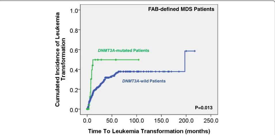
We could not find the difference in treatment regimens between the patients withDNMT3A mutations and those without. With a median follow-up of 43.9 months (range 0.1–250.7 months), patients with DNMT3A mutations had a higher risk to transform to AML (5-year AML transformation rate, 34.4 versus 22.5%, P= 0.013; Fig. 2). MDS patients, based on either the FAB or the 2016 WHO classification, had a significantly shorter OS if they har-bored DNMT3A mutation than those who did not (15.0 versus 32.5 months, P= 0.024, and 16.3 versus 41. 6 months, P= 0.011, respectively; Figs.3 and 4). Further,
Table 2Comparison of other genetic alterations between MDS patients with and without theDNMT3Amutation
Number and percentage of patients with the mutation (%)
Mutation No. examined Total patients DNMT3A-mutated patients DNMT3A-wild patients Pvalue
IDH1 468 4 (0.9) 1 (2.7) 3 (0.7) 0.281
IDH2 464 19 (4.1) 7 (18.9) 12 (2.8) < 0.001
ASXL1 459 108 (23.5) 5 (13.5) 103 (24.4) 0.160
EZH2 469 29 (6.2) 0 (0.0) 29 (6.7) 0.153
TET2 469 61 (13.0) 7 (18.9) 54 (12.5) 0.304
FLT3/ITD 465 5 (1.1) 0 (0) 5 (1.2) > 0.999
JAK2 467 4 (0.9) 0 (0.0) 4 (0.9) > 0.999
NRAS 469 25 (5.3) 2 (5.4) 23 (5.3) > 0.999
KRAS 465 8 (1.7) 1 (2.7) 7 (1.6) 0.488
PTPN11 119 1 (0.8) 0 (0) 1 (1.0) > 0.999
WT1 256 1 (0.4) 0 (0) 1 (0.4) > 0.999
MLL/PTD 447 5 (1.1) 2 (5.4) 3 (0.7) 0.057
RUNX1 462 61 (13.2) 7 (18.9) 54 (12.7) 0.308
U2AF1 469 35 (7.5) 2 (5.4) 33 (7.6) > 0.999
SRSF2 469 60 (12.8) 5 (13.5) 55 (12.7) 0.801
SF3B1 469# 48 (10.2) 11 (29.7) 37 (8.6) < 0.001
Lower-risk IPSS 249 33 (13.3) 7 (46.7) 26 (11.1) 0.001
Higher-risk IPSS 188 11 (5.9) 3 (15) 8 (4.8) 0.098
SETBP1 466 15 (3.2) 0 (0) 15 (3.5) 0.621
TP53 465 42 (9.0) 4 (10.8) 38 (8.9) 0.763
Abbreviations: No.number,ITDinternal tandem duplication,PTDpartial tandem duplication #
Four hundred and thirty-seven of them had cytogenetic data and could be assigned to the IPSS-R risk groups
we could not find the survival difference between the pa-tients with frameshift and non-frameshift mutations. Interestingly, patients withDNMT3Amutations had a bet-ter OS if they received allogenic HSCT than those who did not (P= 0.038, Additional file1: Figure S1).
BecauseDNMT3Amutation was closely associated with SF3B1mutation, a good prognostic factor in MDS patients [35, 36], we divided the whole cohort to two subgroups, SF3B1-mutated and SF3B1-wild type, to evaluate the
prognostic significance ofDNMT3Amutation independent ofSF3B1mutation. In theSF3B1-wild patients,DNMT3A mutation predicted worse prognosis (OS, 14.6 ± 4.7 months versus 30.9 ± 3.2 months,P= 0.005). On the other hand, in the 48 SF3B1-mutated patients, DNMT3A mutation had no prognostic implication (OS, 17.7 ± 11.0 months versus 39.7 ± 4.2 months,P= 0.858) (Additional file1: Figure S2).
Intriguingly, the impact of DNMT3A mutation on OS and time to leukemia transformation remained significant Fig. 3Kaplan-Meier curves stratified by the status ofDNMT3Amutations for overall survival among the whole cohort of 469 MDS patients according to the FAB classification
after adjusting the effects of age, gender, IPSS-R [37, 38], and mutations with prognostic significance in multivariate Cox regression analysis (FAB defined patients: OS: hazard ratio, HR 1.733, 95% CI 1.118–2.688, P= 0.014; time to leukemia transformation: HR 3.088, 95% CI 1.574–6.056, P= 0.001; 2016 WHO classification defined patients: OS: HR 1.800, 95% CI 1.080–3.000, P= 0.024; time to leukemia transformation: HR 2.360, 95% CI 1.129–4.933, P= 0.022; Table3).
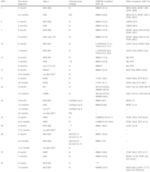
Sequential studies ofDNMT3Amutations
To investigate the role ofDNMT3Amutation in clinical evolution, DNMT3A gene mutation status was sequen-tially tested during the clinical course in 431 samples from 148 patients, including 13 patients with DNMT3A mutations at diagnosis and 135 patients without the mu-tation. In the 13 DNMT3A-mutated patients, 8 had dis-ease progression, including 6 [unique patient numbers (UPNs) 1, 5, 7, 13, 24, and 37] with AML transform-ation. Four patients (UPNs 17, 24, 30, and 36) lost the originalDNMT3Aand other concurrent mutations/cyto-genetic abnormalities when complete remission (CR)
was achieved following curative-intent chemotherapy and/or allogeneic HSCT (Table 4). On the other hand, the other 9 patients withDNMT3Amutations at diagno-sis retained their mutations during follow-ups. Among the eight with disease progression, one (UPN 37) ac-quired a novelRUNX1mutation when the disease trans-formed to AML.
Among the 135 patients without DNMT3A mutation at diagnosis, 1 (0.7%) patient (UPN 47) acquired a novel DNMT3A mutation during sequential follow-up. This patient had MDS with excess blasts-1 (MDS-EB1) at diagnosis when no DNMT3A mutation was detectable even using more sensitive cloning method and next gen-eration sequencing. He acquired GNAS, ASXL1, and ZRSR2 mutations in addition to DNMT3A mutation in the 19th month and died of progressive cytopenia in the 29th month.
We further analyzed the variant allele frequencies of the mutations in the 48DNMT3A-mutated patients by NGS (Table 4). The mutant burden of DNMT3A mutations at diagnosis ranged from 8.4 to 45.24% with a median of 31. 1%. Among the 13 patients with serial studies during the
Table 3Multivariate analysis (Cox regression) for the overall survival and time to leukemia transformation in MDS patients
Overall survival Time to leukemia transformation
Variable HR (95% CI) Pvalue* HR (95% CI) Pvalue*
FAB-defined MDS patients
Age > 65 1.512 (1.134–2.016) 0.005 0.656 (0.417–1.031) 0.067
Male vs female 1.104 (0.818–1.491) 0.517 0.967 (0.615–1.521) 0.886
IPSS-R higher risk# 3.239 (2.199–4.772) < 0.001 5.258 (2.639–10.477) < 0.001
DNMT3Amutation 1.733 (1.118–2.688) 0.014 3.088 (1.574–6.056) 0.001
ASXL1mutation 2.010 (1.434–2.818) < 0.001 3.396 (2.075–5.556) < 0.001
EZH2mutation 1.019 (0.597–1.741) 0.945 0.885 (0.416–1.880) 0.750
TET2mutation 1.416 (0.974–2.059) 0.068 1.398 (0.739–2.644) 0.303
RUNX1mutation 1.134 (0.773–1.663) 0.519 1.751 (1.026–2.987) 0.040
SF3B1mutation 1.071 (0.679–1.691) 0.767 1.392 (0.699–2.771) 0.347
TP53mutation 8.254 (5.338–12.762) < 0.001 6.653 (3.183–13.909) < 0.001
2016 WHO-defined MDS patients
Age > 65 1.649 (1.173–2.319) 0.004 0.731 (0.444–1.203) 0.217
Male vs female 1.184 (0.822–1.705) 0.364 0.988 (0.596–1.637) 0.963
IPSS-R higher risk# 3.840 (2.432–6.063) < 0.001 5.078 (2.390–10.787) < 0.001
DNMT3Amutation 1.800 (1.080–3.000) 0.024 2.360 (1.129–4.933) 0.022
ASXL1mutation 1.830 (1.208–2.774) 0.004 3.596 (2.092–6.182) < 0.001
EZH2mutation 1.196 (0.536–2.668) 0.662 0.927 (0.299–2.875) 0.896
TET2mutation 1.367 (0.841–2.223) 0.208 1.420 (0.643–3.139) 0.386
RUNX1mutation 1.165 (0.723–1.876) 0.531 1.426 (0.753–2.702) 0.276
SF3B1mutation 1.251 (0.743–2.153) 0.387 1.423 (0.613–3.305) 0.412
TP53mutation 8.517 (5.005–14.492) < 0.001 8.885 (4.077–19.365) < 0.001
Abbreviation: HR, hazard ratio;CI, confidence interval;IPSS-R, Revised international prognostic scoring system *Pvalue < 0.05 was considered significant
#
Table 4Sequential studies in MDS patients withDNMT3Amutations at diagnosis and/or at follow-ups
UPN Time from
diagnosis
Status Chromosome
change
DNMT3Amutation (VAF, %)
Other mutations (VAF, %)
1 0 month MDS-EB2 N R882H (41.3) NRAS(38.4),RUNX1(39),
SF3B1(38.8)
6.5 months AML ND R882H (28.5) NRAS(29.1),RUNX1(26.7),
SF3B1(28.3)
5 0 month MDS-EB2 N R882H (25.2) CEBPA(23.5)
5 months AML N R882H (41.8) CEBPA(38.3)
7 0 month MDS-EB2 N R882H (34.2) RUNX1(30.3), IDH2(32.8),
SF3B1(32.5)
9 months AML (s/p C/T) N R882H (27.4) RUNX1(28.6), IDH2(27.6),
SF3B1(28.3)
10 0 month MDS-EB1 N L723TfsX56 (17.1),
1554+1G>T (17)
U2AF1(16.5), RUNX1(9.8)
7 months MDS-EB1 N L723TfsX56 (6.3),
1554+1G>T (6.3)
U2AF1(6.4),RUNX1(3.6)
13 0 month MDS-EB1 N R882H (12.4) MLL-PTD
7 months AML + 8 R882H (20.9) MLL-PTD
9 months s/p C/T in CR N R882H* MLL-PTD
17 0 month RAEB-T N R882H (35.3) IDH2(7.6),NPM1(39.5)
19.5 months s/p allo-HSCT N – –
21 0 month RARS N Y735C (34.5) SF3B1(44.4),TET2(42.3)
34 months RARS N Y735C (31.1) SF3B1(44),TET2(44.3)
23 0 month RA N W313X (45.24),
W860R (45.55)
ASXL1(35.12), IDH2(48.19)
3.5 months CMML N W313X (47.74),
W860R (44.6)
ASXL1(33.31), IDH2(45.29)
24 0 month MDS-EB1 inv(9)(p11q12) R882H (8.4) NPM1 (7)
7.5 months AML inv(9)(p11q12) R882H(33.8) NPM1 (31.3)
12 months s/p C/T inv(9)(p11q12) – –
20 months s/p allo-HSCT ND – –
27 0 month RARS N L508SfsX143 (41.1) SF3B1 (39.9), TET2(42.6)
43.5 months RARS N L508SfsX143 (39.9) SF3B1 (39.2), TET2(41.2)
30 0 month MDS-EB2 −7 R882C (30.5) U2AF1 (29.2)
13.5 months s/p allo-HSCT N – –
36 0 month MDS-EB1 der(7)t(1;7)
(q12;q11),+ 21
R882C (25.4) –
9.5 months MDS-EB2 der(7)t(1;7)
(q12;q11),+ 21
R882C (19) –
13.5 months s/p allo-HSCT N – –
37 0 month RARS N R882H (38.2) SF3B1 (38.2), TET2 (41.7)
7 months AML N R882H (43.4) RUNX1 (21.6), SF3B1 (43),
TET2 (45.9)
47 0 month MDS-EB1 N –** –
19 months MDS-EB1 N N838D (39.7) GNAS (36.5), ASXL1 (17.3),
ASXL1 (9), ZRSR2 (6)
The data of patients who were sequentially studied but had noDNMT3Amutation at both diagnosis and follow-ups are not shown
Abbreviations:UPNunique patient number;−, negative; +, positive;RA, refractory anemia;RARS, refractory anemia with ring sideroblasts;RAEB, refractory anemia with excess blasts;RAEB-T, refractory anemia with excess blasts in transformation;CMML, chronic myelomonocytic leukemia;MDS-EB1, MDS with excess blasts-1;MDS-EB2, MDS with excess blasts-2, s/p, status post;allo-HSCT, allogeneic-hematopoietic stem cell transplantation;AML, acute myeloid leukemia;C/T, chemotherapy;N, normal karyotype;ND, no data;PTD, partial tandem duplication;VAF, variant allele frequency
*In this sample,DNMT3Amutation was not detected by direct sequencing, but 1 of 23 clones showedDNMT3Amutation by TA cloning technique. The disease of this patient relapsed at the 10th month from diagnosis (BM blast 31.6%), and he died of AML at 14th month from diagnosis
clinical courses, the mutation burden at subsequent follow-ups, compared to that at diagnosis, was increased in 3 patient (UPNs 5, 13, and 24), decreased in 6 patients (UPNs 1, 7, 10, 17, 30, and 36, Table4) and stationary in 4 patients (UPNs 21, 23, 27, and 37). All of the three pa-tients with increased DNMT3A mutation burden had leukemia transformation. Their variant allele frequencies of DNMT3A and other co-occurring mutations were in-creased at least 10% (10.0–347.1%) at leukemia transform-ation compared with those at baseline. The patient (UPN 37) who had least increase in variant allele frequency of DNMT3A mutation during disease progression acquired RUNX1mutation at leukemia transformation. In contrast, the variant allele frequencies ofDNMT3Aand other con-current mutations were relative stationary or even de-creased during follow-up in the patients without leukemia transformation.
Discussion
In the present study, we identified 19 differentDNMT3A mutations in 37 (7.9%) of the 469 FAB-defined and 7.7% of the 2016 WHO-defined MDS patients. Similar to previ-ous studies on AML or MDS cohorts [7–10,12,13, 17], most mutations are located in the MTase domain, espe-cially at amino acid R882 locus. Of these 19 mutations, 10 are frameshift and 2 are nonsense mutations. They gener-ate truncgener-ated peptides with complete or partial deletion of the MTase and are expected to abolish the normal func-tion ofDNMT3Agene. The R882 mutations result in im-paired gene function [7, 39], but the influence of the remaining missense mutations on the enzyme activity are unclear. In this study, the prevalence ofDNMT3A muta-tion is 7.9 and 7.7% in MDS according to the FAB and 2016 WHO classification, respectively (Table1), similar to most of the previous reports (7.8 to 10%) [12,40–42] but higher than that of Thol et al. (2.6%) [13].
The reports with detailed demographics of MDS pa-tients with DNMT3A mutation in literature are limited. In the report of Walter et al., but not in the current study and other studies [40, 42], DNMT3A mutations were associated with older age; in contrast, DNMT3A mutations were associated with higher platelet count in our study but not in other studies [12,40,42]. The asso-ciation of DNMT3A mutations with higher platelet count was also shown in AML in previous studies [8,9]. No comparison of age and hemogram between patients with and without DNMT3A was done in the study of Thol et al. [13] in which only five patients were found to have DNMT3A mutation. The causes of differences in the incidence of DNMT3A mutation and the clinical characteristics of DNMT3A-mutated patients might re-sult from the differences in patient population recruited, detection platform used, sample size, and DNMT3A re-gions screened. In the study of Thol et al. [13], exons
15-23 instead of exons 2-23 ofDNMT3Agene were ana-lyzed in most patients (173 of 193 patients). Therefore, some patients harboringDNMT3Amutations might not be detected, and this might partially explain the lower incidence ofDNMT3Amutation in their cohort (2.6%).
In this study, DNMT3A mutations were positively as-sociated with IDH2and SF3B1mutations (Table2). The close association of DNMT3Aand IDH2mutations was also shown in AML [9]. Mutations of DNMT3A and SF3B1, a component of spliceosome complex frequently mutated in RARS, have been reported to occur concur-rently more often than expected by chance in lower-risk MDS patients [17]. In our cohort, the positive associ-ation of these two genetic alterassoci-ations could also be found in lower-risk MDS patients (P< 0.001; Table2). In addition, we could find a trend of positive correlation between these two mutations in higher-risk MDS pa-tients (P= 0.098; Table 2). The close associations
be-tween DNMT3A mutation and RARS and between
DNMT3A and SF3B1 mutations in this study (Table 1) might be related with each other. To investigate the as-sociations among the RARS subtype, DNMT3A muta-tion, and SF3B1mutation, we divided the whole cohort to RARS and non-RARS patients. The close association ofDNMT3AandSF3B1mutations retained in both sub-groups. In contrast, no association between DNMT3A mutation and RARS subtype was found when we divided the whole population to SF3B1-mutated and SF3B1 wild-type patients. In the studies of more than 100 genes by high-throughput DNA sequencing, Haferlach et al. [43] and Papaemmanuil et al. [44] also found a positive correlation betweenDNMT3AandSF3B1mutations, in-dicating that interaction between these two gene muta-tions may play a role in the pathogenesis of MDS, but further investigations are needed to elucidate its mech-anism, especially in RARS subtype. No data regarding the association between DNMT3A mutation and RARS were shown in these two studies.
not find the prognostic significance of DNMT3A muta-tion. The same was also true in another study, in which 24% of patients had SF3B1 mutation [44]. Both cohorts had significantly higher incidence ofSF3B1mutation than ours (10.2%). It may be possible thatDNMT3A mutation would have prognostic effect only in MDS cohorts with low prevalence of SF3B1mutation. Nevertheless, we dis-tinctly showed thatDNMT3Amutation was an independ-ent poor prognostic factor for OS irrespective of the status ofSF3B1mutation and other prognostic factors.
Based on the finding of higher risk of AML transform-ation and shorter survival inDNMT3A-mutated patients, as shown in current study, it would be interesting to in-vestigate the effect of allogenic HSCT in these patients. We found that patients with DNMT3A mutations had a better OS if they received allogenic HSCT than those who did not. It implied that HSCT might ameliorate the poor survival impact of the adverse-risk genotype. Further pro-spective studies with more patients recruited are needed to verify this point. In a study of 46 decitabine-treated AML patients, Metzeler proposed thatDNMT3A-mutated patients might have better treatment response and longer OS [45]. Subsequently, Traina et al. reported DNMT3A mutation as an independent predictor of better response and improved progression-free survival in MDS patients treated with DNMT inhibitors [41]. In our study, only 2 of 36 patients treated with HMA had DNMT3A mutation. These two patients had treatment response and OS simi-lar to others. The influence ofDNMT3Amutation on the treatment response to DNMT inhibitors was not evalu-ated because of the small number ofDNMT3A-mutated patients.
DNMT3Amutation was found quite stable during dis-ease evolution in AML patients [9, 46], but to the best of our knowledge, the dynamic change of this mutation in MDS patients has not been reported yet in literature. Here we showed thatDNMT3Amutation was also quite stable in the clinical course of MDS patients; all DNMT3A-mutated patients retained the original muta-tions during sequential follow-ups unless CR was achieved after allogeneic HSCT or intensive chemother-apy. On the other hand, DNMT3A mutation was rarely acquired during disease evolution; only one (0.7%) of the 145 DNMT3A-wild patients acquired the mutation sub-sequently (Table4).
It is well known that age-related clonal hematopoiesis is associated with increase in the risk of hematologic cancer and the majority of the variants occurred in three genes: DNMT3A,TET2, and ASXL1 [47–49]. Hematologic can-cers were more common in persons with a variant allele fraction of 0.10 or greater. Therefore, it was proposed that DNMT3Amutation is relevant for initiating hematopoietic stem cell clonal expansion and an early initiation event for hematological malignancies. Our finding that DNMT3A
mutation was retained unless CR was achieved was consist-ent with this hypothesis. In paticonsist-ents who failed to achieve remission, the clone harboring DNMT3A mutation sur-vived and may contribute to subsequent relapse. Persist-ence ofDNMT3Amutation in some AML patients in CR was described by us and other researchers [9,50–54]. In a recent study of Gaidzik et al.,DNMT3Amutant transcript levels in CR did not predict outcome in AML patients [54]. In contrast, Thol et al. showed that patients with DNMT3A-mutated lympho-myeloid clonal hematopoiesis (LM-CH) in CR had a higher cumulative incidence of re-lapse at 10 years compared with those withoutDNMT3A -mutated LM-CH (75 versus 27%) [55]. In the present study, we aimed to delineate the dynamic pattern of DNMT3Amutation in MDS development and progression. By NGS, the only patient (UPN 13) who retained his ori-ginal DNMT3Amutation after high intensity chemother-apy finally relapsed. On the other hand, none of the patients in CR who lost their original DNMT3Amutation after allogeneic HSCT experienced disease relapse. Our data suggested that DNMT3Amutation might be used to assess the treatment response and the risk of relapse after curative-intent treatments in MDS patients. Together, whether retaining of DNMT3A mutations after curative-intent treatment is informative for the assessment of the relapse risk in MDS patients remains unclear. It should be cautious to interpret in clinical decision-making and more large-scale studies in MDS patients are warranted to clarify this point.
Conclusions
We identified associations of DNMT3A mutations with distinct clinical features and mutations of SF3B1 and IDH2genes. In addition, we demonstrated thatDNMT3A mutations independently predicted poor outcomes and were stable in the clinical course. It may be used as a bio-marker to monitor the response after curative-intent treat-ment. Additional file 1, is available at Clinical Epigenetics’ website.
Additional file
Additional file 1:Table S1.The mutation patterns in 37 MDS patients withDNMT3Amutations at diagnosis.Table S2.Cytogenetics between
MDS patients with and withoutDNMT3Amutation.Figure S1. Kaplan–Meier
survival curves for overall survival among patients withDNMT3Amutations
stratified by whether receiving allogeneic HSCT or not.Figure S2.
Kaplan-Meier curves stratified by the status ofDNMT3Amutations for overall survival
among the 421SF3B1-wild type MDS patients (A) and among the 48SF3B1
-mutated MDS patients (B). (DOC 242 kb)
Abbreviations
AML:Acute myeloid leukemia; BM: Bone marrow; CMML: Chronic
EB1: Myelodysplastic syndrome with excess blasts-1; MDS-MLD: Myelodysplastic syndrome with multilineage dysplasia; NGS: Next generation sequencing; NTUH: National Taiwan University Hospital; OS: Overall survival; RA: Refractory anemia; RAEB: Refractory anemia with excess blasts; RAEB-T: Refractory anemia with excess blasts in transformation; RARS: Refractory anemia with ring sideroblasts; UPN: Unique patient number
Acknowledgements
We would like to acknowledge the service provided by the DNA Sequencing Core of the First Core Laboratory, National Taiwan University College of Medicine.
Funding
This work was partially sponsored by grants MOST 103-2628-B-002-008-MY3, 103-2923-B-002-001, MOST 103-2314-B-002- 130-MY3, 103-2314-B-002-131 MY3, 104-2314-B-002-128-MY4, and 106- 2314-B-002-226-MY3 from the Ministry of
Science and Technology (Taiwan), National Taiwan University Hospital−National
Taiwan University joint research grant (UN103-051), and MOHW 105-TDU-B-211-134004 from the Ministry of Health and Welfare (Taiwan), NTUH 102P06, from the Department of Medical Research, National Taiwan University Hospital, and Taiwan Health Foundation.
Availability of data and materials
The datasets generated and/or analyzed during the current study are not publicly available due to individual privacy but are available from the corresponding author on reasonable request.
Authors’contributions
M-EL was responsible for the data management and interpretation, mutation analysis, statistical analysis, and manuscript writing; H-AH was responsible for the study design, study plan and coordination, data management and interpretation, mutation analysis, statistical analysis, and manuscript writing; S-JW contributed patient samples and clinical data and was responsible for the data interpretation; C-HT and Y-YK were responsible for the mutation analysis and interpretation; J-LT, MY, C-CL, W-CC, S-YH, B-SK, S-CH, C-TL, and C-YC contributed patient samples and clinical data; M-HT, C-WL, and M-CL performed the gene mutation and chromosomal studies; H-FT designed and coordinated the study over the entire period and wrote the manuscript. All authors read and approved the final manuscript.
Ethics approval and consent to participate
This study was approved by the Institutional Review Board/Ethical Committee of the National Taiwan University Hospital (NTUH20150709RINA).
Consent for publication Not applicable
Competing interests
The authors declare that they have no competing interests.
Publisher’s Note
Springer Nature remains neutral with regard to jurisdictional claims in published maps and institutional affiliations.
Author details
1Division of Hematology, Department of Internal Medicine, National Taiwan
University Hospital, No.7, Chung Shan S. Rd., Zhongzheng Dist, Taipei 10002, Taiwan.2Division of Hematology, Department of Internal Medicine, National
Taiwan University Hospital, Hsin-Chu Branch, Hsinchu City, Taiwan.3Graduate
Institute of Clinical Medicine, College of Medicine, National Taiwan University, Taipei, Taiwan.4Tai-Cheng Stem Cell Therapy Center, National Taiwan University, Taipei, Taiwan.5Graduate Institute of Oncology, College of
Medicine, National Taiwan University, Taipei, Taiwan.6Departments of
Pathology, National Taiwan University Hospital, Taipei, Taiwan.7Department
of Laboratory Medicine, National Taiwan University Hospital, Taipei, Taiwan.
Received: 16 January 2018 Accepted: 21 March 2018
References
1. Jones PA, Laird PW. Cancer epigenetics comes of age. Nat Genet. 1999;21:163–7.
2. Seligson DB, Horvath S, Shi T, et al. Global histone modification patterns
predict risk of prostate cancer recurrence. Nature. 2005;435:1262–6.
3. Razin A, Cedar H. DNA methylation and genomic imprinting. Cell. 1994;77:473–6.
4. Lee JT, Jaenisch R. The (epi)genetic control of mammalian X-chromosome
inactivation. Curr Opin Genet Dev. 1997;7:274–80.
5. Okano M, Xie S, Li E. Cloning and characterization of a family of novel
mammalian DNA (cytosine-5) methyltransferases. Nat Genet. 1998;19:219–20.
6. Yamashita Y, Yuan J, Suetake I, et al. Array-based genomic resequencing of
human leukemia. Oncogene. 2010;29:3723–31.
7. Ley TJ, Ding L, Walter MJ, et al. DNMT3A mutations in acute myeloid
leukemia. N Engl J Med. 2010;363:2424–33.
8. Thol F, Damm F, Ludeking A, et al. Incidence and prognostic influence of
DNMT3A mutations in acute myeloid leukemia. Journal of clinical oncology :
official journal of the American Society of Clinical Oncology. 2011;29:2889–96.
9. Hou HA, Kuo YY, Liu CY, et al. DNMT3A mutations in acute myeloid
leukemia: stability during disease evolution and clinical implications. Blood.
2012;119:559–68.
10. Ribeiro AF, Pratcorona M, Erpelinck-Verschueren C, et al. Mutant DNMT3A: a
marker of poor prognosis in acute myeloid leukemia. Blood. 2012;119:5824–31.
11. Shivarov V, Gueorguieva R, Stoimenov A, et al. DNMT3A mutation is a poor
prognosis biomarker in AML: results of a meta-analysis of 4500 AML
patients. Leuk Res. 2013;37:1445–50.
12. Walter MJ, Ding L, Shen D, et al. Recurrent DNMT3A mutations in patients
with myelodysplastic syndromes. Leukemia. 2011;25:1153–8.
13. Thol F, Winschel C, Ludeking A, et al. Rare occurrence of DNMT3A
mutations in myelodysplastic syndromes. Haematologica. 2011;96:1870–3.
14. Tefferi A, Vardiman JW. Myelodysplastic syndromes. N Engl J Med. 2009;
361:1872–85.
15. Roller A, Grossmann V, Bacher U, et al. Landmark analysis of DNMT3A
mutations in hematological malignancies. Leukemia. 2013;27:1573–8.
16. Chesnais V, Renneville A, Toma A, et al. Effect of lenalidomide treatment on
clonal architecture of myelodysplastic syndromes without 5q deletion.
Blood. 2016;127:749–60.
17. Bejar R, Stevenson KE, Caughey BA, et al. Validation of a prognostic model
and the impact of mutations in patients with lower-risk myelodysplastic syndromes. Journal of clinical oncology: official journal of the American
Society of Clinical Oncology. 2012;30:3376–82.
18. Bennett JM, Catovsky D, Daniel MT, et al. Proposals for the classification of
the myelodysplastic syndromes. Br J Haematol. 1982;51:189–99.
19. Arber DA, Orazi A, Hasserjian R, et al. The 2016 revision to the World Health
Organization classification of myeloid neoplasms and acute leukemia. Blood.
2016;127:2391–405.
20. Hou HA, Lin CC, Chou WC, et al. Integration of cytogenetic and molecular
alterations in risk stratification of 318 patients with de novo non-M3 acute
myeloid leukemia. Leukemia. 2014;28:50–8.
21. Hou HA, Kuo YY, Tang JL, et al. Clinical implications of the SETBP1 mutation
in patients with primary myelodysplastic syndrome and its stability during
disease progression. Am J Hematol. 2014;89:181–6.
22. Hou HA, Chou WC, Lin LI, et al. Characterization of acute myeloid leukemia
with PTPN11 mutation: the mutation is closely associated with NPM1
mutation but inversely related to FLT3/ITD. Leukemia. 2008;22:1075–8.
23. Tang JL, Hou HA, Chen CY, et al. AML1/RUNX1 mutations in 470 adult
patients with de novo acute myeloid leukemia: prognostic implication and
interaction with other gene alterations. Blood. 2009;114:5352–61.
24. Hou HA, Liu CY, Kuo YY, et al. Splicing factor mutations predict poor prognosis
in patients with de novo acute myeloid leukemia. Oncotarget. 2016;7:9084–101.
25. Shiah HS, Kuo YY, Tang JL, et al. Clinical and biological implications of
partial tandem duplication of the MLL gene in acute myeloid leukemia
without chromosomal abnormalities at 11q23. Leukemia. 2002;16:196–202.
26. Chen TC, Hou HA, Chou WC, et al. Dynamics of ASXL1 mutation and other
associated genetic alterations during disease progression in patients with primary myelodysplastic syndrome. Blood cancer journal. 2014;e177:4.
27. Ernst T, Chase AJ, Score J, et al. Inactivating mutations of the histone
methyltransferase gene EZH2 in myeloid disorders. Nat Genet. 2010;42:722–6.
28. Lin CC, Hou HA, Chou WC, et al. IDH mutations are closely associated
with mutations of DNMT3A, ASXL1 and SRSF2 in patients with
myelodysplastic syndromes and are stable during disease evolution. Am J
Hematol. 2014;89:137–44.
29. Chou WC, Lei WC, Ko BS, et al. The prognostic impact and stability of
Isocitrate dehydrogenase 2 mutation in adult patients with acute myeloid
30. Chou WC, Chou SC, Liu CY, et al. TET2 mutation is an unfavorable prognostic factor in acute myeloid leukemia patients with intermediate-risk
cytogenetics. Blood. 2011;118:3803–10.
31. Hou HA, Huang TC, Lin LI, et al. WT1 mutation in 470 adult patients
with acute myeloid leukemia: stability during disease evolution and implication of its incorporation into a survival scoring system. Blood.
2010;115:5222–31.
32. Falini B, Mecucci C, Tiacci E, et al. Cytoplasmic nucleophosmin in acute
myelogenous leukemia with a normal karyotype. N Engl J Med. 2005;352: 254–66.
33. Hou HA, Chou WC, Kuo YY, et al. TP53 mutations in de novo acute myeloid
leukemia patients: longitudinal follow-ups show the mutation is stable during disease evolution. Blood cancer journal. 2015;e331:5.
34. Hou HA, Tsai CH, Lin CC, Chou WC, Kuo YY, Liu CY, Tseng MH, Peng YL, Liu
MC, Liu CW, Liao XW, Lin LI, Yao M, Tang JL, Tien HF. Incorporation of mutations in five genes in the revised international prognostic scoring system can improve risk stratification in the patients with myelodysplastic syndrome. Blood Cancer J. 2018.
35. Papaemmanuil E, Cazzola M, Boultwood J, et al. Somatic SF3B1 mutation in
myelodysplasia with ring sideroblasts. N Engl J Med. 2011;365:1384–95.
36. Malcovati L, Karimi M, Papaemmanuil E, et al. SF3B1 mutation identifies a
distinct subset of myelodysplastic syndrome with ring sideroblasts. Blood.
2015;126:233–41.
37. Greenberg PL, Tuechler H, Schanz J, et al. Revised international prognostic
scoring system for myelodysplastic syndromes. Blood. 2012;120:2454–65.
38. Yang YT, Hou HA, Liu CY, et al. IPSS-R in 555 Taiwanese patients with
primary MDS: integration of monosomal karyotype can better risk-stratify
the patients. Am J Hematol. 2014;89:E142–9.
39. Kim SJ, Zhao H, Hardikar S, et al. A DNMT3A mutation common in AML
exhibits dominant-negative effects in murine ES cells. Blood. 2013;122:4086–9.
40. Lin J, Yao DM, Qian J, et al. Recurrent DNMT3A R882 mutations in Chinese
patients with acute myeloid leukemia and myelodysplastic syndrome. PLoS One. 2011;6:e26906.
41. Traina F, Visconte V, Elson P, et al. Impact of molecular mutations on
treatment response to DNMT inhibitors in myelodysplasia and related
neoplasms. Leukemia. 2014;28:78–87.
42. Tefferi A, Lasho TL, Patnaik MM, et al. Targeted next-generation sequencing
in myelodysplastic syndromes and prognostic interaction between
mutations and IPSS-R. Am J Hematol. 2017;92(12):1311–7.
43. Haferlach T, Nagata Y, Grossmann V, Okuno Y, Bacher U, Nagae G,
Schnittger S, Sanada M, Kon A, Alpermann T, Yoshida K, Roller A, Nadarajah N, Shiraishi Y, Shiozawa Y, Chiba K, Tanaka H, Koeffler HP, Klein H-U, Dugas M, Aburatani H, Kohlmann A, Miyano S, Haferlach C, Kern W, Ogawa S. Landscape of genetic lesions in 944 patients with myelodysplastic
syndromes. Leukemia. 2014;28(2):241–247.
44. Papaemmanuil E, Gerstung M, Malcovati L, et al. Clinical and biological
implications of driver mutations in myelodysplastic syndromes. Blood. 2013;
122:3616–27. quiz 3699
45. Metzeler KH, Walker A, Geyer S, et al. DNMT3A mutations and response to
the hypomethylating agent decitabine in acute myeloid leukemia.
Leukemia. 2012;26:1106–7.
46. Gaidzik VI, Weber D, Paschka P, et al. DNMT3A mutant transcript levels
persist in remission and do not predict outcome in patients with acute
myeloid leukemia. Leukemia. 2017;32:30–7.
47. Genovese G, Kahler AK, Handsaker RE, et al. Clonal hematopoiesis and
blood-cancer risk inferred from blood DNA sequence. N Engl J Med. 2014; 371:2477–87.
48. Xie M, Lu C, Wang J, et al. Age-related mutations associated with clonal
hematopoietic expansion and malignancies. Nat Med. 2014;20:1472–8.
49. Jaiswal S, Fontanillas P, Flannick J, et al. Age-related clonal
hematopoiesis associated with adverse outcomes. N Engl J Med. 2014; 371:2488–98.
50. Ploen GG, Nederby L, Guldberg P, et al. Persistence of DNMT3A mutations
at long-term remission in adult patients with AML. Br J Haematol. 2014; 167:478–86.
51. Ivey A, Hills RK, Simpson MA, et al. Assessment of minimal residual disease
in standard-risk AML. N Engl J Med. 2016;374:422–33.
52. Jeziskova I, Musilova M, Culen M, et al. Distribution of mutations in DNMT3A
gene and the suitability of mutations in R882 codon for MRD monitoring in
patients with AML. Int J Hematol. 2015;102:553–7.
53. Bhatnagar B, Eisfeld AK, Nicolet D, et al. Persistence of DNMT3A R882
mutations during remission does not adversely affect outcomes of patients
with acute myeloid leukaemia. Br J Haematol. 2016;175:226–36.
54. Gaidzik VI, Weber D, Paschka P, et al. DNMT3A mutant transcript levels
persist in remission and do not predict outcome in patients with acute
myeloid leukemia. Leukemia. 2018;32:30–7.
55. Thol F, Klesse S, Kohler L, et al. Acute myeloid leukemia derived from
lympho-myeloid clonal hematopoiesis. Leukemia. 2017;31:1286–95.
• We accept pre-submission inquiries
• Our selector tool helps you to find the most relevant journal
• We provide round the clock customer support
• Convenient online submission
• Thorough peer review
• Inclusion in PubMed and all major indexing services
• Maximum visibility for your research
Submit your manuscript at www.biomedcentral.com/submit