R E S E A R C H
Open Access
Growth differentiation factor 15 contributes
to marrow adipocyte remodeling in
response to the growth of leukemic cells
Wei Lu
1†, Yun Wan
1†, Zhiqiang Li
2†, Bin Zhu
3, Chunrong Yin
4, Haiyan Liu
1, Shaoxin Yang
1, Yuanmei Zhai
3,
Yehua Yu
5, Yanyu Wei
1and Jun Shi
1*Abstract
Background:The adipocyte remodeling, including of the morphological change, might indicate special pathological function. Our previous study found that the morphological remodeling of larger marrow adipocytes into small marrow adipocytes correlates with a poor prognosis for acute myeloid leukemia (AML) patients. However, the mechanisms contributed to the marrow adipocyte remodeling are still poorly understood.
Methods: GDF15 expression was analyzed by RT-qPCR and western blotting assays in the leukemic cells. The
enhancing and antibody neutralization tests in vitro were employed to evaluate the effect of GDF15 on the morphology of mature adipocytes. CCK8 test was used to detect the proliferation of leukemic cells after co-cultivation with small marrow adipocytes. Flow cytometry was used to analysis the proportion of cell cycle of leukemic cells. Immunofluorescence staining and linear analysis were applied to verify the GDF15 expression and the relationship between GDF15 and small marrow adipocytes in AML patients.
Results:In this study, we found that leukemic cell lines not only expressed significantly higher growth differentiation factor 15 (GDF15) than the other three cytokines associated with adipocyte differentiation in RNA level but also secreted GDF15 factor. Furthermore, the in vitro experiments demonstrated that GDF15 was involved in the conversion of small marrow adipocytes from larger marrow adipocytes. Correspondingly, the leukemic cells proliferated more rapidly through regulating the cell cycle when co-cultured with GDF15-induced small marrow adipocytes. The immunofluorescence staining on the bone marrow sections of AML patients further exhibited that GDF15 was partly produced by leukemic cells. The positive correlation between the concentration of GDF15 in the marrow aspirates and the number and the volume of small marrow adipocytes might suggest the contribution of GDF15 in AML patients (r= 0.72,r= 0.67).
Conclusions:GDF15 secreted by leukemic cells was involved in the morphological remodeling of marrow adipocytes, which can in turn promote leukemic cell growth, indicating that GDF15 may be a promising treatment target for AML patients.
Keywords: Acute myeloid leukemia, GDF15, Marrow adipocyte, Adipocyte remodeling
* Correspondence:[email protected]
†Equal contributors
1Department of Hematology, Shanghai Jiao Tong University Affiliated Sixth
People’s Hospital, Shanghai, China
Full list of author information is available at the end of the article
Background
Acute myeloid leukemia (AML) is a malignant hematological disease that occurs primarily in the bone marrow (BM). Marrow adipocytes, as an important component of the BM niche, have been suggested to contribute to the proliferation and anti-chemotherapy of AML cells by providing energy or secreting adipokines [1, 2]. Our previous study found that only small marrow adipo-cytes, not the total marrow adipoadipo-cytes, were correlated with a poor prognosis for AML patients [3], suggesting that understanding the generating mechanism of small marrow adipocytes may be useful for improving the prog-nosis of AML patients.
In prostate cancer and ovarian cancer, it has been re-ported that adipocytes surrounding the tumor cells be-come small and are involved in the metastasis and growth of prostate cancers or ovarian cancers [4, 5]. In leukemia, Shafat et al. reported that marrow adipocytes transfer fatty acids to AML blasts by activating lipolysis in adipocytes [2]. These reports indicate that the reduc-tion of adipocyte size is due to transfer of their lipid droplets to tumor cells. Additionally, in breast cancer, tumor cells release inflammatory factors, such as TNFα and Wnt3a, which are involved in the regulation of mor-phological remodeling of the mature adipocytes, includ-ing the reduction of adipocyte size and acquisition of fibroblast-like morphology [6]. This implies that the morphological remodeling of adipocytes are not only due to the transfer of their lipid droplets to the sur-rounding tumor cells but also highly dependent on ex-trinsic signals from tumor cells.
Growth differentiation factor-15 (GDF15), a TGF-β/ bone morphogenetic protein (BMP) superfamily mem-ber, is a 40-kDa secretory propeptide that is cleaved in the endoplasmic reticulum to release a 25-kDa circulat-ing protein [7]. Under physiological conditions, GDF15 is abundantly expressed only in placenta and macro-phage cells [8]. However, recent studies have reported that GDF15 is highly expressed in many types of cancer tissues, including colorectal, gastric, esophageal, oral, pancreatic and so on [9–14]. We previously found the expression of GDF15 was high in the residual acute lymphoblastic leukemic cells in mice [15]. However, its expression in AML cells has not been studied in depth. Studies have shown that similar to other TGF-βfamily members, GDF15 is involved in the inhibition of cell growth, induction of apoptosis, and enhancement of cancer invasiveness in different cancer cell lines [16–19]. Until now, little attention was paid to the effect of GDF15 on the adipocytes. Increased serum concentra-tions of GDF15 have been reported in patients with an-orexia nervosa and obesity [20], inspiring us to explore the function of GDF15 on the adipocytes. Here, we dem-onstrate that leukemic cells highly express GDF15, and
in turn, GDF15-induced small adipocytes may promote the growth of leukemic cells.
Methods
Cell culture and regents
The leukemic cell lines THP-1, K562, HEL, HL-60 and Kasumi (Chinese Academy of Sciences Cell Bank, Shanghai, China) were cultured in 1640 supplemented with 10% fetal bovine serum (FBS) (Gibco, Grand Island, NY, USA) and penicillin-streptomycin at 37 °C in 5% CO2. Primary AML blasts and mesenchymal stem cells in bone marrow (BMSCs) were isolated by Ficoll-Hypaque (Axis-Shield Diagnostics, Dundee, Scotland, UK) density-gradient centrifugation. BMSCs were differentiated into adipocytes as previously described [21]. The differentiated adipocytes were stained with Oil Red O (ORO). Condi-tioned medium (CM) from the leukemic cell lines was ob-tained from cells cultured with high glucose Dulbecco’s modified Eagle’s medium (DMEM) supplemented with 1% FBS. Mature adipocytes were cultured with high glucose DMEM supplemented with 10% FBS alone or mixed with the leukemic cell lines CM at the ratio of 4:1. Mature adi-pocytes were treated with recombinant human GDF15 (rhGDF15 200 ng/ml, Peprotech, Cat#120–28, USA) or a neutralizing anti-GDF15 antibody (8μg/ml, R&D Systems, Cat#MAB957, USA) for 5 days to observe the effect of GDF15 factor on the marrow adipocytes.
Cell cycle analysis
The measurements were made using a flow cytometry (Beckman, Urbana, IL, USA). In brief, THP-1 and K562 cells were cultured with the CM from mature adipocytes and small adipocytes for 48 h respectively, washed with PBS, then fixed with 70% ethanol for 24 h. Cells were in-cubated with propyl iodide (Sigma, St Louis, MO, USA) organism dye for 30 min at 37 °C, followed by flow cyto-metric analysis.
Free fat acid detection
Adipocytes were treated with or without rhGDF15 (200 ng/ml) for 5 days, then the cultured medium was replaced by high glucose DMEM with 1% FBS for 48 h. The supernatant of adipocytes was collected and stored in −80 °C.The concentration of FFA was detected by using a colorimetric method via a commercial kit (Sigma Aldrich, St. Louis, Missouri). The assay was carried out according to manufacturer’s specifications.
Cell proliferation assay
cells (3 × 10³) were seeded into 96-well plates, and the leukemic cell proliferation was evaluated by a CCK8 kit (Dojindo, Japan). CCK8 reagent was added to each well and incubated for 2 h at 37 °C. The measurement of ab-sorption at 450 nm was performed using a microplate reader (Multiscan FC, Thermo Fisher, USA).
Transfection assays
Transfection assays were set up for 4 groups, scrambled siRNA (negative control/NC), GDF15-homo-161 (siRNA-161); 290 (siRNA-290) and GDF15-homo-860 (siRNA-GDF15-homo-860) groups. GDF15-siRNA and scrambled siRNA were obtained from Shanghai GenePharma Co., Ltd (Shanghai, China) and blended into 20μM with DEPC water and stored in - 20 °C. The THP-1 cells were seeded in 24-well plates at a density of 2 × 10⁵cells/well for 24 h prior to transfection and then the medium was replaced with 0.5 mL of Opti-MEM medium. Subsequently, 1μL of Lipofectamine 2000, 2 μL of GDF15-siRNA was added into the corresponding well. After 48 h and 72 h of transfection, the cells were collected for further gene expression assays.
PCR and real-time quantitative PCR (RT-qPCR)
Total RNA was extracted using Trizol (Invitrogen, Paisley, UK), and the RNA was converted into cDNA using the PrimeScript™RT reagent Kit (Takara Bio Inc, Shiga, Japan) for RT-qPCR and using the PrimeScript™ II 1st Strand cDNA Synthesis Kit (Takara Bio Inc, Shiga, Japan) for PCR. All RT-qPCR reactions were performed using an ABI 7500 system (Biosystems, Foster City, CA, USA) and the SYBR Premix Ex Taq reagent kit (Takara Bio Inc, Shiga, Japan). Premix Taq™(Takara Bio Inc, Shiga, Japan) for electrophoresis. All the primers used in this study are presented in the Table1.
Western blotting analysis and ELISA
Mononuclear cells from the controls and primary AML patients were washed twice with phosphate buffer saline and resuspended in 200 μl of lysis buffer containing a mix of protease inhibitors (Beyotime, Haimen, Jiangsu, China). Immunoblots were prepared as previously
described [22]. Rabbit anti-GDF15 monoclonal antibody (Abcam, Cambridge, MA, USA), rabbit anti-β-tubulin antibody (Abcam, Cambridge, MA, USA), rabbit anti-Cdk2 antibody (Abcam, Cambridge, MA, USA), rabbit anti-Cyclin D1 antibody (Abcam, Cambridge, MA, USA), rabbit anti-P21 antibody (Abcam, Cam-bridge, MA, USA) and rabbit anti-β-actin monoclo-nal antibody (Cell Sigmonoclo-naling Technology, Danvers, MA, USA). The THP-1 cell CM was obtained from cells cul-tured in regular medium with 1% FBS at different cell densities (2 × 10⁵/ml, 5 × 10⁵/ml, 1 × 10⁶/ml, 2 × 10⁶/ ml). The ELISA analysis for GDF15 was performed according to the manufacturer’s instructions (R&D systems).
Immunofluorescence
Adipocytes differentiated on coverslips grown alone or co-cultivated with leukemic cells were fixed with 4% parafor-maldehyde and blocked with goat serum. The process was carried out as previously described [23]. Cells were stained with neutral lipid specific BODIPY®493/503 (4, 4-difluoro-1,3,5,7,8-pentamethyl-4-bora-3a,4a-diaza-s-indacene) dye, tubulin and DAPI. The immunofluorescence of BM paraf-fin sections from AML patients was performed as previ-ously described [24]. The sections were stained with GDF15 (goat anti-GDF15 multiclonal antibody, Abcam, Cambridge, MA, USA), CD34 (rabbit anti-CD34 mono-clonal antibody, Abcam, Cambridge, MA, USA), CD117 (rabbit anti-CD117 polyclonal antibody, Proteintech, Wuhan, China) and DAPI (Solarbio, Beijing, China). Fluorescent images were captured using a confocal laser microscopy system (Leica SP2).
Adipocyte measurements
BM trephine biopsies of 20 AML patients were obtained from the posterior iliac crest, and BM tissues were fixed, decalcified and embedded with paraffin or plastic slices according to the conventional methods. This study was approved by the Medical Ethical Committee of our insti-tute. The adipocyte number and adipocyte volume were measured as previously reported. The number and area of per adipocyte cultured in vitro were measured by
Table 1Sequences of the primers used to detect genes expression by RT-qPCR
Name Forward Reverse GeneBank Accession
GDF15 GACCCTCAGAGTTGCACTCC GCCTGGTTAGCAGGTCCTC NM_004864
FABP4 AACCTTAGATGGGGGTGTCCTG CTCTCTCATAAACTCTCGTG NM_001442
PPARγ GGGATCAGCTCCGTGGATCT TGCACTTTGGTACTCTTGAAGTT NM_138711
C/EBPα GAACAGCAACGAGTACCGGGTA GCCATGGCCTTGACCAAGGAG NM_004364
HSL GACCCCTGCACAACATGATG TGAGCAGCACCCTTTGGATG NM_005357
ATGL GGCTTCCTCGGCGTCTACTA TTTACCAGGTTGAAGGAGGGG NM_020376
using Image-Pro Plus 5.1. Ten fields were analyzed at × 400 magnification.
Statistical analysis
Values were calculated as the mean ± SEM. The Spearman’s correlation test was used to analyze the correlation between small adipocyte volume or small adipocyte number and GDF15 level in the BM. For all analyses, P< 0.05 was considered to be significant. All statistical ana-lyses were performed using the SPSS 20.0 software program (Statistical Package for Social Science, SPSS Inc. Chicago, IL., USA).
Results
The soluble cytokines secreted by leukemic cells contribute to adipocyte remodeling
Our previous study found that leukemic cells could in-duce the formation of small adipocytes in vitro [3]. To investigate the effect of soluble cytokines secreted by leukemic cells on BMSC-derived mature adipocytes, we cultured the mature adipocytes with the CM of different leukemic cells, including THP-1, K562, HL-60, Kasumi, primary AML blasts (LC) and healthy mononuclear cells (MNC) for 5 days. The ORO staining and the quantitative analysis of adipocyte area showed that except Kasumi cells, the average area of adipocytes cultured with the CM of different leukemic cell lines was significantly de-creased (reduced by 48%~ 64%, P< 0.05, Fig. 1a-b)
when compared with the control. Similar results were also observed when mature adipocytes cultured with LC CM, but not with MNC CM. Meanwhile, the dra-matic reduction of the content of lipid-droplets in mature adipocytes was also observed by the detection of OD values and RT-qPCR analysis of adipogenic genes, including fatty acid binding protein (FABP4), peroxisome proliferator-activated receptor gamma (PPARγ) and CCAAT/enhancer binding protein alpha (C/EBPα) (Fig. 1c-d), suggesting that the soluble cyto-kines secreted by leukemic cells promote the remod-eling of small adipocytes from larger adipocytes.
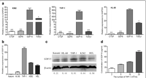
GDF15 is highly expressed in leukemic cell lines
To explore the critical cytokines secreted by leukemic cells, the mRNA levels of four cytokines that inhibited adipogenesis or promoted the transition to fibroblasts, such as TGF-β1, GDF15, growth differentiation factor 6 (GDF6) and connective tissue growth factor (CTGF) were detected by PCR and RT-qPCR assay. As shown in Fig. 2a, the expression of GDF15 was obviously higher than the other three factors in leukemic cells (P< 0.05 or
P< 0.001). Furthermore, RT-qPCR and Western blotting analysis were performed to detect the GDF15 expression in different AML cell lines, including THP-1, K562, HEL, HL-60 and Kasumi. Our results demonstrated that THP-1 cells expressed the highest GDF15 among these cells (Fig.2b-c); therefore, they were used in the following
Fig. 1The soluble cytokines secreted by leukemic cells contribute to the adipocyte remodeling.aBMSC-derived adipocytes cultured with HG-DMEM
studies. ELISA assay showed that GDF15 factor was con-tinuously released into the supernatant with the density of THP-1 cells increasing (Fig.2d), indicating that AML cells can highly express and secrete GDF15.
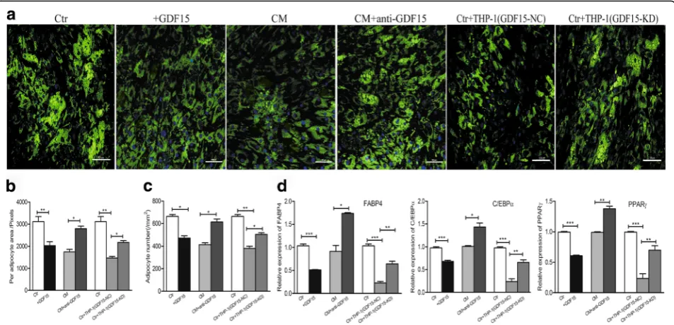
GDF15 is involved in the remodeling of larger adipocytes to small adipocytes
The biological effects of GDF15 on the adipocytes was further examined. After adding rhGDF15 into the cul-ture medium of macul-ture adipocytes for 5 days, adipocytes exhibited a decrease in the number and size of lipid droplets, as seen by the BODIPY staining (Fig. 3a), co-culturing mature adipocytes with the supernatant of THP-1 cells caused a reduction in the number and size of lipid droplets, while the addition of neutralizing anti-GDF15 antibody into the medium resulted in an in-crease in the number and size of lipid droplets (Fig.3a). To further verify the function of GDF15 secreted by leukemic cells on mature adipocytes, we knocked down (KD) the GDF15 expression in THP-1 cells (Additional file1: Figure S1) and assessed its effects on adipocytes. Adipo-cytes co-cultured with GDF15-KD THP-1 cells exhibited an increase in the number and size of lipid droplets when compared with adipocytes co-cultured with the negative control (NC) by the BODIPY staining (Fig. 3a). Subse-quently, quantitative analysis showed a reduction in the
area of each adipocyte and adipocyte number when adipo-cytes treated with rhGDF15 (Fig. 3b-c). However, an obvi-ous increase in the area of each adipocyte and adipocyte number was observed when adipocytes were treated with GDF15 neutralizing antibody or co-cultured with GDF15-KD THP-1 cells (Fig.3b-c). Additionally, we also observed that mature adipocytes exhibited a dramatic reduction in the adipogenic markers, including FABP4, PPARγ and C/ EBPα by RT-qPCR analysis when they were treated with rhGDF15 (Fig.3d). The expression of adipogenic markers increased when adipocytes treated with GDF15 neu-tralizing antibody or co-cultured with GDF15-KD THP-1 cells (Fig. 3d). These results suggested that it was GDF15 secreted by leukemic cells that promoted the remodeling of small adipocytes.
GDF15-induced small adipocytes promote the growth of AML cells
Generally, the change of adipocyte morphology might indicate its abnormity in function [25–27]. We found that small adipocytes induced by GDF15 produced more free fatty acids (FFAs), resulting from the increase of lipolytic gene expression (hormone-sensitive triglycer-ide lipase, HSL and adipose triglycertriglycer-ide lipase, ATGL) (Fig. 4a-b). To investigate the effects of GDF15-induced small adipocytes on the proliferation of AML cells, we
Fig. 2AML cell lines highly express GDF15.aRT-qPCR analysis of different cytokines associated with the regulation of adipogenesis in AML cell
collected the CM from GDF15-induced small adipocytes and mature adipocytes. As shown in Fig.4c, leukemic cell proliferation significantly increased when cultured with the CM from GDF15-induced small adipocytes compared with that from mature adipocytes, suggesting small adipo-cytes can promote the growth of leukemic cells. To study the mechanism of small adipocytes to enhance the leukemic cell proliferation, we analyzed the change of cell cycle of leukemic cells. The proportion of THP-1 cells and K562 cells in S phase was increased after treatment with the CM from small adipocytes when compared with that from mature adipocytes (40.03 ± 2.72% vs 33.63 ± 2.51%; 44.67 ± 2.31% vs 35.77 ± 2.07%, P< 0.05, respectively) (Fig. 4d). Furthermore, the protein ex-pression of the cycle proteins CDK2 and Cyclin D1 were observed to increase while cyclin-dependent kin-ase inhibitor P21 decrekin-ased (Fig. 4e).
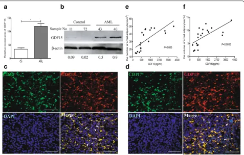
GDF15 is associated with small adipocytes in AML patients To further investigate the contribution of GDF15 to small marrow adipocytes in AML patients, the expres-sion of GDF15 was measured in the BM of AML pa-tients. We collected the mononuclear cells from BM of the AML patients (n= 15) and controls (n= 12). Com-pared to the controls, primary AML blasts exhibited higher expression of GDF15 by RT-qPCR and Western blotting analyses (Fig. 5a-b). On the BM sections of
AML patients, CD34+ or CD117+ cells, which were regarded as leukemic cells, were positive for GDF15 (Fig. 5c-d). However, part of GDF15+ cells exhibited no expression of CD34 or CD117, indicating that GDF15 can also be expressed from other cells besides primary AML blasts. Furthermore, we detected the level of GDF15 in BM aspirates and quantified both the number and the volume of small marrow adipocytes on the BM sections from the same AML patients. Linear analysis showed that the levels of GDF15 in BM aspirates were positively correlated with the number and the volume of small marrow adipocytes on the BM sections (Fig. 5e-f), suggesting that increased GDF15 was associated with small marrow adipocytes in AML.
Discussion
In the present study, we investigated the possible mechanism on the generation of small adipocytes in AML patients by focusing on extracellular regulatory factors. We found that small marrow adipocytes were remodeled from larger marrow adipocytes in response to the release of GDF15 from leukemic cells. Accord-ingly, GDF15-induced small adipocytes could promote the proliferation of leukemic cells, indicating that GDF15 plays a critical role in the crosstalk between leukemic cells and adipocytes.
Fig. 3Contribution of GDF15 secreted by leukemic cells to the adipocyte remodeling.aAdipocytes treated with rhGDF15 (+GDF15) or neutralizing
anti-GDF15 antibody (CM + anti-GDF15) or co-cultured with GDF15 knock down THP-1 cells (Ctr + THP-1 GDF15-KD) were stained with Alexa Fluor 493/503-conjugated BODIPY. Adipocytes cultured alone (Ctr) or cultured with the conditioned medium of THP-1 cells (CM) or co-cultured with negative control (Ctr + THP-1 GDF15-NC) were used as the controls respectively. Scale bar represents 100μm.bThe area of each adipocyte in each group was analyzed by using Image-Pro-Plus 5.1. *P< 0.05, **P< 0.01.cAdipocyte number in each group was analyzed by using Image-Pro-Plus 5.1. *P< 0.05, **P< 0.01.dThe adipogenic gene (FABP4, PPARγ, C/EBPα) expression was detected by RT-qPCR analysis in each group. *P< 0.05, **P< 0.01,
In this study, both on BM sections and in AML cell lines, our experiments showed that AML cells highly expressed GDF15. As a secretory protein, GDF15 might be released into the BM cavity by leukemic cells and have an effect on other cells in BM, including adipo-cytes. Certainly, non-leukemic cells also expressed GDF15 on BM sections (Fig.5c-d). Our previous studies have demonstrated that GDF15 was highly expressed in leukemia-activated fibroblasts, suggesting that GDF15 was from many kinds of cells in BM of AML patients. Additionally, it has been reported that mature adipocytes can undergo morphologic changes, from mature adipo-cytes to small adipoadipo-cytes (cancer-associated adipoadipo-cytes) to fibroblast-like cells (adipocytes-derived fibroblasts) when they were activated by soluble factors derived from solid tumor cells [6]. Therefore, we guessed that GDF15 secreted by leukemic cells might play an important role in the conversion from adipocytes to leukemia-activated fibroblasts. Functionally, GDF15 was involved in the fibrosis of many organs [28–30]. Although both the fi-broblasts and adipocytes can be differentiated from BMSCs, it is less known about the function of GDF15 in adipogenesis. However, it is well known that TGF-β1 and GDF15 are belonged to the TGF-β superfamily,
which can prevent preadipocyte differentiation through cooperation with the Wnt signaling pathway [27]. Simi-larly, CTGF, a downstream mediator of TGF-β1 signal-ing in many cell types, has anti-adipogenic effects in primary adipocytes [31,32]. It leads us to infer the possi-bility that GDF15 may play an important role in the marrow adipocyte differentiation of AML patients. In-deed, our results, acquired by exogenous addition rhGDF15 or neutralizing anti-GDF15 antibody, indicated that GDF15 can induce the transition of larger adipo-cytes into small adipoadipo-cytes. This was further confirmed by the positive correlation between the levels of GDF15 and the number and volume of small adipocytes in AML patients. Meanwhile, our results were consistent with the findings reported by Chrysovergis K et al. In that study, they found that the transgenic mice treated with GDF15 expressing xenografts had smaller adipocytes compared to wild-type (WT) littermates [33].
Our results indicated that GDF15 could induce the adipocyte remodeling, that is the morphological transi-tion of larger adipocytes into small adipocytes. It has been showed that the small adipocytes in mice highly expressed lipolytic genes [33]. Our results also showed that the expression of lipolytic genes including HSL and
Fig. 4GDF15-induced small adipocytes promote the AML cells growth by increasing the lipolysis.aRT-qPCR analysis of lipolytic genes (HSLand
ATGL increased in the GDF15-induced small adipocytes, indicating that the marrow adipocytes remodeling might be dependent on lipolysis. However, much remains to be uncovered on the mechanism of lipolysis regulation by GDF15. It has been reported that GDF15 induced a sig-nificant ERK activation in the malignant progression of human cancer cell [34–36]. What’s more, ERK activation could cause HSL phosphorylation which leads to in-creased activity of the enzyme [37]. Thus, whether the increased HSL in GDF15 induced-small adipocytes is also dependent on ERK signal pathway requires further study. Adipose tissue is a plastic organ with the ability of a continuous remodeling, including of extension and re-gression depending on nutrient intake [38, 39]. The morphological, transcriptional and functional remodel-ing of adipocytes may be concurrent [39]. Indeed, we found that when the adipocytes were becoming small in-duced by GDF15, they had a stronger ability to promote the proliferation of leukemic cells (Fig.4). Smaller adipo-cytes are frequently linked to higher metabolic activity
since smaller adipocytes suggest greater utilization of fat storage for metabolism [40,41]. Until now, the function of FFAs on the tumor cells was still controversial. On one hand, some reported that FFAs could inhibit the growth and progression of breast cancer cells [42, 43]. On the other hand, FFAs could promote the prolifera-tion of leukemic cells and the metastasis of ovarian can-cer cells [2, 44]. In our study, we found that with the high expression of lipolytic genes, the FFA levels in-creased during the process of small adipocyte formation. Since it has been reported that FFAs can be transported by FABP4 to AML cells [2], we considered that the leukemic cell rapid proliferation might rely on FFAs pro-viding energy.
Conclusions
In conclusion, leukemic cells mediated the marrow pocyte remodeling from larger adipocytes to small adi-pocytes by secreting GDF15. In turn, small adiadi-pocytes enhanced the leukemic cell proliferation by providing
Fig. 5The relationship of GDF15 expression and small marrow adipocytes in AML patients.aRT-qPCR analysis ofGDF15mRNA expression in BM
FFA. From the point of bone marrow microenviron-ment, GDF15 might be a new target to treat the leukemia. Since our study was focused only on clinical samples, animal studies in the future would provide a better insight into the mechanism of GDF15 regulation on marrow adipocytes remodeling.
Additional file
Additional file 1:Figure S1.The GDF15 expression in THP-1 cells with different treatment. (DOCX 172 kb)
Abbreviations
AML:Acute myeloid leukemia; ATGL: Adipose triglyceride lipase; BM: Bone marrow; BMP: Bone morphogenetic protein; C/EBPα: CCAAT/ enhancer binding protein alpha; CM: Conditioned medium; CTGF: Connective tissue growth factor; DMEM: Dulbecco’s modified Eagle’s medium; FABP4: Fatty acid-binding protein 4; FBS: Fetal bovine serum; FFA: Free fat acid; GDF15: Growth differentiation factor-15; GDF6: Growth differentiation factor-6; HSL: Hormone-sensitive triglyceride lipase; MSC: Mesenchymal stem cells; ORO: Oil Red O; PPARγ: Peroxisome proliferator activated receptor gamma; RT-qPCR: Real-time quantitative PCR
Acknowledgements
Not applicable.
Funding
The authors would like to thank for the support by the National Natural Science Foundation of China (Grant No. 81570135), National Science and Technology Major Equipment Projects of China (Grant No. 2013YQ03065109) and Shanghai Public Health Project (Grant No. 15GWZK0501).
Availability of data and materials
All data generated or analyzed during this study are included in this published article.
Authors’contributions
WL performed the laboratory tests and wrote the manuscript. YW performed the immunofluorescence. ZL analyzed the results and revised the paper. BZ and CY collected patient samples and analyzed clinical information. HL and SY isolated the BMSCs. YZ performed the ELISA assay. YY and YW performed the experiments. JS designed the experiments, analyzed the results and revised the paper. All authors read and approved the final version of the submitted manuscript.
Ethics approval and consent to participate
Not applicable.
Consent for publication
Not applicable.
Competing interests
The authors declare that they have no competing interests.
Publisher’s Note
Springer Nature remains neutral with regard to jurisdictional claims in published maps and institutional affiliations.
Author details
1Department of Hematology, Shanghai Jiao Tong University Affiliated Sixth
People’s Hospital, Shanghai, China.2Department of Blood Tranfusion, Shanghai Jiao Tong University Affiliated Sixth People’s Hospital, Shanghai, China.3Department of Hematology, Shanghai Jiao Tong University Affiliated
Sixth People’s Hospital South Campus, Shanghai, China.4Department of
Hematology, Tongren Hospital Shanghai Jiao Tong University School of Medicine, Shanghai, China.5Department of Hematology, Shanghai Jiao Tong
University School of Medicine Affiliated Ninth People’s Hospital, Shanghai, China.
Received: 5 November 2017 Accepted: 15 March 2018
References
1. Tabe Y, Konopleva M, Munsell MF, Marini FC, Zompetta C, McQueen T, Tsao T, Zhao S, Pierce S, Igari J, et al. PML-RARalpha is associated with leptin-receptor induction: the role of mesenchymal stem cell-derived adipocytes in APL cell survival. Blood. 2004;103:1815–22.
2. Shafat MS, Oellerich T, Mohr S, Robinson SD, Edwards DR, Marlein CR, Piddock RE, Fenech M, Zaitseva L, Abdul-Aziz A, et al. Leukemic blasts program bone marrow adipocytes to generate a pro-tumoral microenvironment. Blood. 2017; 129:1320–32.
3. Lu W, Weng W, Zhu Q, Zhai Y, Wan Y, Liu H, Yang S, Yu Y, Wei Y, Shi J. Small bone marrow adipocytes predict poor prognosis in acute myeloid leukaemia. Haematologica. 2018;103:e21–4.
4. Herroon MK, Rajagurubandara E, Hardaway AL, Powell K, Turchick A, Feldmann D, Podgorski I. Bone marrow adipocytes promote tumor growth in bone via FABP4-dependent mechanisms. Oncotarget. 2013;4:2108–23. 5. Gazi E, Gardner P, Lockyer NP, Hart CA, Brown MD, Clarke NW. Direct evidence
of lipid translocation between adipocytes and prostate cancer cells with imaging FTIR microspectroscopy. J Lipid Res. 2007;48:1846–56. 6. Bochet L, Lehuédé C, Dauvillier S, Wang YY, Dirat B, Laurent V, Dray C, Guiet
R, Maridonneau-Parini I, Le Gonidec S, et al. Adipocyte-derived fibroblasts promote tumor progression and contribute to the desmoplastic reaction in breast Cancer. Cancer Res. 2013;73:5657–68.
7. Eling TE, Baek SJ, Shim M, Lee CH. NSAID activated gene (NAG-1), a modulator of tumorigenesis. J Biochem Mol Biol. 2006;39:649–55.
8. Breit SN, Johnen H, Cook AD, Tsai VW, Mohammad MG, Kuffner T, Zhang HP, Marquis CP, Jiang L, Lockwood G, et al. The TGF-βsuperfamily cytokine, MIC-1/GDF15: a pleotrophic cytokine with roles in inflammation, cancer and metabolism. Growth Factors. 2011;29:187–95.
9. Li C, Wang X, Casal I, Wang J, Li P, Zhang W, Xu E, Lai M, Zhang H. Growth differentiation factor 15 is a promising diagnostic and prognostic biomarker in colorectal cancer. J Cell Mol Med. 2016;20:1420–6.
10. Mohamed AA, Soliman H, Ismail M, Ziada D, Farid TM, Aref AM, Al Daly ME, Abd Elmageed ZY. Evaluation of circulating ADH and MIC-1 as diagnostic markers in Egyptian patients with pancreatic cancer. Pancreatology. 2015;15: 34–9.
11. Fisher OM, Levert-Mignon AJ, Lord SJ, Lee-Ng KK, Botelho NK, Falkenback D, Thomas ML, Bobryshev YV, Whiteman DC, Brown DA, et al. MIC-1/GDF15 in Barrett’s oesophagus and oesophageal adenocarcinoma. Br J Cancer. 2015; 112:1384–91.
12. Yang CZ, Ma J, Luo QQ, Neskey DM, Zhu DW, Liu Y, Myers JN, Zhang CP, Zhang ZY, Zhong LP. Elevated level of serum growth differentiation factor 15 is associated with oral leukoplakia and oral squamous cell carcinoma. J Oral Pathol Med. 2014;43:28–34.
13. Blanco-Calvo M, Tarrío N, Reboredo M, Haz-Conde M, García J, Quindós M, Figueroa A, Antón-Aparicio L, Calvo L, Valladares-Ayerbes M. Circulating levels of GDF15, MMP7 and miR-200c as a poor prognostic signature in gastric cancer. Future Oncol. 2014;10:1187–202.
14. Qian Y, Jung YS, Chen X. Differentiated embryochondrocyte expressed gene 1 regulates p53-dependent cell survival versus cell death through macrophage inhibitory cytokine-1. Proc Natl Acad Sci U S A. 2012;109:11300–5.
15. Duan CW, Shi J, Chen J, Wang B, Yu YH, Qin X, Zhou XC, Cai YJ, Li ZQ, Zhang F, et al. Leukemia propagating cells rebuild an evolving niche in response to therapy. Cancer Cell. 2014;25:778–93.
16. Baek KE, Yoon SR, Kim JT, Kim KS, Kang SH, Yang Y, Lim JS, Choi I, Nam MS, Yoon M, et al. Upregulation and secretion of macrophage inhibitory cytokine-1 (MIC-1) in gastric cancer. Clin Chim Acta. 2009;401:128–33.
17. Golkar L, Ding XZ, Ujiki MB, Salabat MR, Kelly DL, Scholtens D, Fought AJ, Bentrem DJ, Talamonti MS, Bell RH, et al. Resveratrol inhibits pancreatic cancer cell proliferation through transcriptional induction of macrophage inhibitory cytokine-1. J Surg Res. 2007;138:163–9.
18. Kadara H, Schroeder CP, Lotan D, Pisano C, Lotan R. Induction of GDF-15/ NAG-1/MIC-1 in human lung carcinoma cells by retinoid-related molecules and assessment of its role in apoptosis. Cancer Biol Ther. 2006;5:518–22. 19. Lambert JR, Kelly JA, Shim M, Huffer WE, Nordeen SK, Baek SJ, Eling TE,
20. Karczewska-Kupczewska M, Kowalska I, Nikolajuk A, Adamska A, Otziomek E, Gorska M, Straczkowski M. Hyperinsulinemia acutely increases serum macrophage inhibitory cytokine-1 concentration in anorexia nervosa and obesity. Clin Endocrinol. 2012;76:46–50.
21. Meulle A, Salles B, Daviaud D, Valet P, Muller C. Positive regulation of DNA double strand break repair activity during differentiation of long life span cells: the example of adipogenesis. PLoS One. 2008;3:e3345.
22. Muller C, Monferran S, Gamp AC, Calsou P, Salles B. Inhibition of Ku heterodimer DNA end binding activity during granulocytic differentiation of human promyelocytic cell lines. Oncogene. 2001;20:4373–82.
23. Dirat B, Bochet L, Dabek M, Daviaud D, Dauvillier S, Majed B, Wang YY, Meulle A, Salles B, Le Gonidec S, et al. Cancer-associated adipocytes exhibit an activated phenotype and contribute to breast Cancer invasion. Cancer Res. 2011;71:2455–65.
24. Saiz-Lopez P, Chinnaiya K, Campa VM, Delgado I, Ros MA, Towers M. An intrinsic timer specifies distal structures of the vertebrate limb. Nat Commun. 2015;6:8108.
25. Lonn M, Mehlig K, Bengtsson C, Lissner L. Adipocyte size predicts incidence of type 2 diabetes in women. FASEB J. 2010;24:326–31.
26. Naveiras O, Nardi V, Wenzel PL, Hauschka PV, Fahey F, Daley GQ. Bone-marrow adipocytes as negative regulators of the haematopoietic microenvironment. Nature. 2009;460:259–63.
27. Zhou S, Eid K, Glowacki J. Cooperation between TGF-βand Wnt pathways during chondrocyte and adipocyte differentiation of human marrow stromal cells. J Bone Miner Res. 2004;19:463–70.
28. Kumar A, Ruan M, Clifton K, Syed F, Khosla S, Oursler MJ. TGF-βmediates suppression of Adipogenesis by estradiol through connective tissue growth factor induction. Endocrinology. 2012;153:254–63.
29. Wang Z, Wang C, Liu S, He W, Wang L, Gan J, Huang Z, Wang Z, Wei H, Zhang J, et al. Specifically formed corona on silica nanoparticles enhances transforming growth factorβ1 activity in triggering lung fibrosis. ACS Nano. 2017;11:1659–72.
30. Wu N, Meng F, Invernizzi P, Bernuzzi F, Venter J, Standeford H, Onori P, Marzioni M, Alvaro D. Franchitto a,et al. the secretin/secretin receptor axis modulates liver fibrosis through changes in transforming growth factor-β1 biliary secretion in mice. Hepatology. 2016;64:865–79.
31. Tan JT, McLennan SV, Song WW, Lo LW, Bonner JG, Williams PF, Twigg SM. Connective tissue growth factor inhibits adipocyte differentiation. Am J Physiol Cell Physiol. 2008;295:C740–51.
32. Song WW, McLennan SV, Tam C, Williams PF, Baxter RC, Twigg SM. CCN2 requires TGF-βsignalling to regulate CCAAT/enhancer binding proteins and inhibit fat cell differentiation. J Cell Commun Signal. 2015;9:27–36. 33. Chrysovergis K, Wang X, Kosak J, Lee SH, Kim JS, Foley JF, Travlos G, Singh S,
Baek SJ, Eling TE. NAG-1/GDF-15 prevents obesity by increasing thermogenesis, lipolysis and oxidative metabolism. Int J Obes. 2014;38:1555–64.
34. Lee DH, Yang Y, Lee SJ, Kim KY, Koo TH, Shin SM, Song KS, Lee YH, Kim YJ, Lee JJ, et al. Macrophage inhibitory cytokine-1 induces the invasiveness of gastric cancer cells by up- regulating the urokinase-type plasminogen activator system. Cancer Res. 2003;63:4648–55.
35. Jin YJ, Lee JH, Kim YM, Oh GT, Lee H. Macrophage inhibitory cytokine-1 stimulates proliferation of human umbilical vein endothelial cells by up-regulating cyclins D1 and E through the PI3K/Akt-, ERK-, and JNK-dependent AP-1 and E2F activation signaling pathways. Cell Signal. 2012;24:1485–95. 36. Kim KK, Lee JJ, Yang Y, You KH, Lee JH. Macrophage inhibitory cytokine-1
activates AKT and ERK-1/2 via the transactivation of ErbB2 in human breast and gastric cancer cells. Carcinogenesis. 2008;29:704–12.
37. Greenberg AS, Shen WJ, Muliro K, Patel S, Souza SC, Roth RA, Kraemer FB. Stimulation of lipolysis and hormone-sensitive lipase via the extracellular signal-regulated kinase pathway. J Biol Chem. 2001;276:45456–61. 38. Patil YN, Dille KN, Burk DH, Cortez CC, Gettys TW. Cellular and molecular
remodeling of inguinal adipose tissue mitochondria by dietary methionine restriction. J Nutr Biochem. 2015;26:1235–47.
39. Lee MJ, Wu Y, Fried SK. Adipose tissue remodeling in pathophysiology of obesity. Curr Opin Clin Nutr Metab Care. 2010;13:371–6.
40. Feldman BJ, Streeper RS, Farese RV Jr, Yamamoto KR. Myostatin modulates adiogenesis to generate adipocytes with favorable metabolic effects. Proc Natl Acad Sci U S A. 2006;103:15675–80.
41. Stienstra R, Joosten LA, Koenen T, van Tits B, van Diepen JA, van den Berg SA, Rensen PC, Voshol PJ, Fantuzzi G, Hijmans A, et al. The inflammasome-mediated caspase-1 activation controls adipocyte differentiation and insulin sensitivity. Cell Metab. 2010;12:593–605.
42. Wu Y, Yu X, Yi X, Wu K, Dwabe S, Atefi M, Elshimali Y, Kemp KT 2nd, Bhat K, Haro J, et al. Aberrant phosphorylation of SMAD4 Thr277-mediated USP9x-SMAD4 interaction by free fatty acids promotes breast Cancer metastasis. Cancer Res. 2017;77:1383–94.
43. Gluschnaider U, Hertz R, Ohayon S, Smeir E, Smets M, Pikarsky E, Bar-Tana J. Long-chain fatty acid analogues suppress breast tumorigenesis and progression. Cancer Res. 2014;74:6991–7002.
44. Nieman KM, Kenny HA, Penicka CV, Ladanyi A, Buell-Gutbrod R, Zillhardt MR, Romero IL, Carey MS, Mills GB, Hotamisligil GS, et al. Adipocytes promote ovarian cancer metastasis and provide energy for rapid tumor growth. Nat Med. 2011;17:1498–503.
• We accept pre-submission inquiries
• Our selector tool helps you to find the most relevant journal
• We provide round the clock customer support
• Convenient online submission
• Thorough peer review
• Inclusion in PubMed and all major indexing services
• Maximum visibility for your research
Submit your manuscript at www.biomedcentral.com/submit


![Bis[μ bis(2,6 di 1 naphthylphenyl)phosphanido]bis[(tetrahydrofuran)sodium(I)]](data:image/gif;base64,R0lGODlhAQABAIAAAP///wAAACH5BAEAAAAALAAAAAABAAEAAAICRAEAOw==)