R E V I E W
Open Access
Light-sensitive brain pathways and aging
V. Daneault
1,2,3*, M. Dumont
2, É. Massé
1,2, G. Vandewalle
3,4and J. Carrier
1,2,3Abstract
Notwithstanding its effects on the classical visual system allowing image formation, light acts upon several
non-image-forming (NIF) functions including body temperature, hormonal secretions, sleep-wake cycle,
alertness, and cognitive performance. Studies have shown that NIF functions are maximally sensitive to blue
wavelengths (460
–
480 nm), in comparison to longer light wavelengths. Higher blue light sensitivity has been
reported for melatonin suppression, pupillary constriction, vigilance, and performance improvement but also
for modulation of cognitive brain functions. Studies investigating acute stimulating effects of light on brain
activity during the execution of cognitive tasks have suggested that brain activations progress from subcortical
regions involved in alertness, such as the thalamus, the hypothalamus, and the brainstem, before reaching
cortical regions associated with the ongoing task. In the course of aging, lower blue light sensitivity of some
NIF functions has been reported. Here, we first describe neural pathways underlying effects of light on NIF
functions and we discuss eye and cerebral mechanisms associated with aging which may affect NIF light
sensitivity. Thereafter, we report results of investigations on pupillary constriction and cognitive brain sensitivity
to light in the course of aging. Whereas the impact of light on cognitive brain responses appears to decrease
substantially, pupillary constriction seems to remain more intact over the lifespan. Altogether, these results
demonstrate that aging research should take into account the diversity of the pathways underlying the effects
of light on specific NIF functions which may explain their differences in light sensitivity.
Keywords:
Light, Aging, Brain, Non-image-forming (NIF) functions
Background
Two functional systems detecting light: photoreceptor
contribution and neural pathways
From a functional point of view, there are two
sys-tems detecting light in mammals and humans. The
first one is the classical visual system responsible for
image formation, and the second one is the
non-image-forming (NIF) system which detects
environ-mental irradiance and contributes to modulation of
many fundamental functions in living organisms. The
physiological,
behavioral, and cognitive functions
which are modulated by light but not associated with
conscious image perception are called NIF functions.
These responses include circadian entrainment and
shift the timing of circadian rhythms such as
hor-mone secretion (melatonin, cortisol), heart rate, body
temperature, and the sleep-wake cycle. These NIF
effects are detected hours or days following light
exposure. NIF responses also include acute
physio-logical effects of light detected more rapidly,
includ-ing melatonin suppression, pupillary constriction,
alertness, and performance improvement as well as
cognitive brain responses [1
–
5].
Melanopsin retinal ganglion cells
In the course of the year 2000s, the discovery of
mela-nopsin (OPN4)-photosensitive pigment expressed by
intrinsically photosensitive retinal ganglion cells (ipRGC)
contributed to a better understanding of the neural bases
of the NIF system [6]. The crucial importance of OPN4
in NIF responses has been corroborated by animal and
human studies [7
–
10]. In humans, melanopsin is
expressed in a small subset of cells representing only
1
–
2 % of all retinal ganglion cells (RGC) [1, 10
–
14].
These photoreceptors measure the intensity of light
(ir-radiance detection) with a maximum sensitivity toward
short light wavelength (blue ~ 460
–
480 nm) [6, 7, 11].
* Correspondence:[email protected] 1
Functional Neuroimaging Unit, University of Montreal Geriatric Institute, Montreal, QC, Canada
2Center for Advanced Research in Sleep Medicine, Hôpital du Sacré-Cœur de Montréal, Montreal, QC, Canada
Full list of author information is available at the end of the article
Melanopsin ipRGC have a low spatial resolution and
long latencies as compared to cone and rod responses,
and they show the ability to integrate photic energy
over long periods of time [6, 7, 13, 14]. To date, five
ipRGC subtypes (M1
–
M5) have been identified
accord-ing to morphological, molecular, and functional
charac-teristics [8, 11, 15]. M1 have more melanopsin pigment
than all other subtypes, and they can be subdivided
according to the transcription factor Brn3b (Brn3b
positive-M1 versus Brn3b-negative M1) [16
–
18]. M2
have extended dendrites and soma. M2 also shows
more complex connections than M1 including afferents
from the rods and cones suggesting that their intrinsic
photic response might be more modulated by inputs
from classical photoreceptors [18]. M3 has similar
characteristics to M2, with intermediate levels of
mela-nopsin [15, 19] and M4
–
M5 possess long dendrites,
abundant arborization, and very low levels of
melanop-sin (i.e., low intrinsic light response) [15, 18
–
23]. M1 to
M5 project to specific subcortical brain areas and play
different functional roles in the NIF and in the classical
visual systems [16, 22].
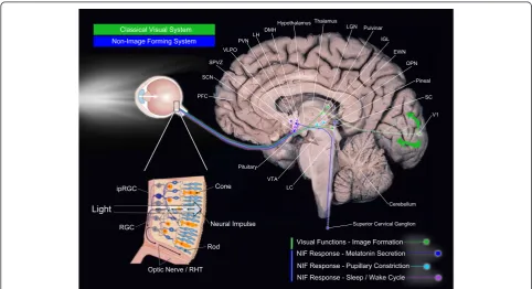
Visual and non-visual neural pathways
Classical visual system: image forming system
Specific neural pathways are described for visual and
non-visual systems (Fig. 1). Beginning with the eye, the
classical visual system uses mainly rods and cones for
image formation but also ipRGC for rudimentary visual
functions [20, 22]. Cones are responsible for photopic
vision (higher light intensity) with high spatial acuity
and color discrimination. The classical photopic system
in humans includes three types of cones showing mean
peak sensitivity (
λmax
) at 555 nanometers (nm), i.e., the
green part of the light visible spectrum. S-cones express
the short-wavelength-sensitive opsin cyanolabe (
λmax
420 nm), M-cones express chlorolabe opsin (
λmax
535 nm), and L-cones express a red-shifted opsin, the
erythrolabe (
λmax
565 nm) [24]. Scotopic vision (i.e.,
contrast detection, dim light vision) is sustained by rods
[25] using rhodopsin photopigment (
λmax
507 nm in
humans) [24]. Using the optic tract, the brain pathways
of the classical visual system project to subcortical
nu-cleus, such as the thalamic lateral geniculate nucleus
(LGN), the superior colliculus (SC), and the lateral
posterior pulvinar complex (Pul-LP), before reaching the
primary visual occipital area (V1) and then at other
neo-cortical regions engaged in dorsal and ventral visual
at-tentional brain pathways [26
–
29] (Fig. 1). Animal studies
show that ipRGC (possibly non-M1 subtypes [22, 23])
also send projections to dorsal LGN (dLGN) and SC [16,
17, 22, 23, 30, 31]. These ipRGC projections play a role
in conscious perception of spatial brightness and speed
motion [16, 31
–
33]. Recent animal evidences also
sup-port the functional role of melanopsin-expressing ipRGC
projections to dLGN in visual responses optimization
with irradiance detection [33]. Overall, complex
interac-tions between classical (cones, rods) and non-classical
(melanopsin-expressing
ipRGC)
photoreceptors
and
their projections contribute to the classical visual system
[16, 17, 20, 32].
Non-visual system/non-image-forming system
The second system, namely, the NIF system, uses ipRGC
in addition to rods and cones and shows a peak
sensitiv-ity in the blue part of the light spectrum (~460
–
480 nm)
[6, 7, 11, 13, 14, 31, 34]. A monosynaptic pathway, the
retinohypothalamic tract (RHT), conveys light
informa-tion from ipRGC axons [35, 36]. As illustrated in Fig. 1,
the NIF system directly projects via the RHT to
sub-cortical regions engaged in melatonin secretion,
pupillary constriction, and the regulation of the
sleep-wake cycle [2, 37, 38].
RHT directly connects the ipRGC from the retina to
the suprachiasmatic nuclei (SCN) of the anterior
hypo-thalamus, the master circadian oscillator (biological
clock) [1, 11, 39]. SCN is the endogenous master
biological clock that allows temporal organization of
liv-ing organisms, synchronizliv-ing circadian rhythms among
themselves as well as with the external environment.
SCN sends efferent projections to the hypothalamic and
non-hypothalamic structures [30], including the
para-ventricular nucleus of the hypothalamus (PVN), the
dor-somedial nucleus of the hypothalamus (DMH), and
finally, the intergeniculate leaflet (IGL) of the thalamus
which also sends projections to SCN [40]. Interactions
between the SCN, the PVN, the superior cervical
gan-glion (SCG), and the pineal gland support the neural
network of melatonin suppression [41] (see Fig. 1
mela-tonin suppression). Without being exhaustive here,
many brain areas other than the SCN also receive direct
projections from the ipRGC. Thus, olivary pretectal
nucleus (OPN), the crucial node of the pupillary
con-striction pathway, receives direct projections from the
ipRGC. OPN sends projections to the Edinger-Westphal
nucleus (EWN) which in turn, innervate the sphincter
muscle of the pupil allowing pupillary constriction [42].
The ipRGC also sends direct connections to regions
en-gaged in the regulation of the sleep-wake cycle [2, 37, 38],
such as the ventrolateral preoptic nucleus (VLPO;
sleep-wake regulation core-region), the subparaventricular
nucleus/zone (SPVZ) of the hypothalamus, which is
involved in sleep regulation but also in motor activity,
as well as the lateral hypothalamus (LH), which
con-tains orexin (hypocretin) neurons regulating
wakeful-ness [20, 22, 30, 40]. Furthermore, light may also
affect the sleep-wake cycle via the connections
between the SCN and the DMH since the DMH also
sends projections to the VLPO, the LH, and the locus
coeruleus (LC) [40, 43, 44]. The amygdala, a structure
involved in emotional processes, also receives direct
projections from the ipRGC [30, 31] and might
repre-sent a key target of the NIF system by potentiating
ef-fects of light on alertness and mood. This limbic area
is part of the neural network named the
“
Salience
Net-work
”
associated with responsiveness to stimuli [45].
Photoreceptor contribution to NIF responses
Light stimulus characteristics influence the
photore-ceptor
’
s contribution to specific NIF responses. For
instance, light intensity, wavelength, and temporal
characteristics define the specific photoreceptor
’
s
con-tribution to pupil light reflex (PLR) [46
–
49]. At low
light intensities, rods and cones contribute to PLR but
cones
’
contribution decreases as the duration of light
ex-posure increases and is minimal beyond 30 s [47, 48]. At
high light intensities (>12 log units per ph/cm
2/s), ipRGC
mainly contributes to the sustained PLR [8, 47, 50], i.e., in
response to light exposure extending beyond 30 s.
Recently, complex photoreceptor
interventions/com-munications have also been reported for circadian
entrainment. Blue-yellow cone
’
s color discrimination/
opponency seem to modulate the ipRGC signal
trans-mission to SCN neurons making them sensitive to color
[51]. Thus, SCN cells would be sensitive to both
bright-ness and color. This could correspond to an evolutionary
strategy using color as a time-of-day indicator based on
spectral composition of the solar cycle and twilight
tran-sition [51].
independence, seems more likely based on the observed
data. Further research will help identify retinal and
neural networks involved in the effects of light for each
NIF functions.
Overall, as for the classical visual pathway, the
under-lying neural pathways of the NIF system are complex
and several brain areas are involved in the mechanisms
by which changes in the quality of the light environment
affect various NIF functions [22, 30, 31].
Effects of light on alertness and cognitive functions: short
versus longer wavelengths
In agreement with the peak sensitivity of each
light-detecting system, many studies have confirmed greater
sensitivity of non-visual responses under blue
mono-chromatic light exposure (~460
–
480 nm), in comparison
to longer wavelengths such as green monochromatic
light [4, 5, 53
–
59]. Hence, the impact of light on
sleepi-ness, alertsleepi-ness, performance, as well as the modulation of
cognitive brain functions are greater under blue
mono-chromatic light and blue-enriched light exposure, as
com-pared to longer light wavelengths [4, 5, 53
–
57, 60]. Lower
levels of subjective sleepiness [53, 61, 62], but also of
ob-jective alertness as measured with electroencephalogram
(EEG), are reported under blue light exposure, as
com-pared to longer light wavelength or darkness [53].
Higher performance speed to the psychomotor
vigi-lance task (PVT) is also observed when exposed to
blue-enriched light exposure as compared to
longer-enriched lights [4, 63, 64]. Likewise, blue
monochro-matic light exposure, as compared to green and red
monochromatic lights, induces higher amplitude levels
on the P300, an event-related potential associated with
attentional demands [65].
Since 2004, a number of studies investigated the
brain mechanisms underlying the stimulating effects
of light on alertness and cognitive functions in
humans [5, 56, 57, 66
–
72]. These investigations
showed that light exposure, particularly blue light,
during the execution of cognitive tasks potentiate
brain activations of subcortical structures associated
with vigilance including the hypothalamus, brainstem
(LC), thalamus, and limbic areas (the amygdala and
hippocampus) likely before spreading to cortical
re-gions engaged in the ongoing task [5]. Recently and
according to a theory of melanopsin bistable
proper-ties [59, 73, 74], long wavelength light exposure
(589 nm) administered an hour before a given test
light exposure increases the impact of that test light
on some brain responses (i.e., pulvinar, cerebellum,
frontal areas) associated with the execution of a
cogni-tive task [75]. Overall, these studies confirmed that in
young subjects, light exposure, particularly blue light,
has greater modulating effects on cognitive brain
functions than other light wavelengths most likely
through melanopsin photoreception and triggers brain
activation increases in regions related to alertness and
to executive functions (for a review, see [5, 76]).
Aging and non-image-forming system modifications
Age-related differences in the impact of light have been
reported for some acute non-visual responses, with a
decreased effect of monochromatic blue light (456 nm) on
clock gene expression, subjective alertness, sleepiness, and
mood in older, as compared to young individuals [77
–
79].
However, some investigations did not find age-related
re-duction in the impact of light when using polychromatic
white light [80
–
82]. A potential decrease in the impact of
light remains therefore debated, and it could be that
age-related changes occur for specific wavelengths of light
or for specific NIF responses but not for others.
Age-related modifications from the eye to the brain
may affect the NIF system and contribute to lower
sensi-tivity to light in aging [83
–
89]. Circadian oscillations are
driven by rhythmic expression of clock genes and
auto-regulatory transcriptional-translational feedback loops
over approximately a 24-h period. Aging appears to be
associated with changes in clock gene expression with a
reduced amplitude in
Bmal1
and
Clock
expression in
SCN [90
–
92], lower
Per2 expression
in the pituitary
gland [93], and lower
Per 1,2,3
expression at the
periph-eral level (liver, heart) [94]. Age-related differences under
light exposure were also revealed including reduction in
Per1
expression after light pulses [90
–
92] and reduction
of
Per2
expression following blue morning light
expos-ure [79]. Since
Per 1
–
2
expression is rapidly induced by
light and is required for entrainment, age-related
tem-poral disorganization may partly result from lower SCN
sensitivity to photic stimulation (for a review, see [95]).
uptake
—
the main neurotransmitter of the RHT) [109]
have been reported. Again, all these modifications might
induce functional deficits among various systems
includ-ing the non-image-forminclud-ing one.
Last but not least, many important age-related changes
also occur at the eye level: there is a decrease of
photo-receptor sensitivity, a reduction in pupil size, known as
senile miosis, and an increase of ocular crystalline lens
absorption known as
“
lens yellowing
”
[85, 86, 110
–
115].
The combination of all these changes is very likely to
re-duce the amount of light reaching the retina and may
modulate the impact of light on NIF functions.
Pupillary constriction and brain sensitivity to light in the
course of aging
In order to improve the understanding of the impact of
light on non-visual older subjects, we completed two
re-search protocols. We aimed at measuring pupillary
con-striction [116] and non-visual cognitive brain activity while
exposed to light [117]. We recruited two groups of subjects,
16 young and 14 older individuals (see [116] and [117] for
complete sample description). All were healthy, right
handed, non-smokers, slept between 7 and 9 h per night,
were non-medicated, and MRI compatible. They also
underwent an optometric exam to make sure they were free
of ocular disease. The main hypothesis of our investigations
was that in older, compared to young subjects, we would
detect a reduction in pupillary and brain responses to light.
Pupillary constriction in relation with healthy aging
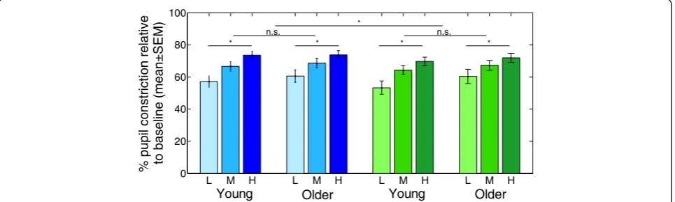
In the pupillometry protocol, subjects were first
main-tained in darkness for 15 min before we captured baseline
pupil size. Subjects were then exposed for 45 s to three
ir-radiances levels of blue (480 nm) and green (550 nm)
monochromatic light (low 7 × 10
12ph/cm
2/s, medium 3 ×
10
13ph/cm
2/s, high 1 × 10
14ph/cm
2/s). Resting period in
darkness lasted 2 min between each light exposure.
As expected, at the baseline (before any light
expos-ure), analysis of the raw pupil size area showed that
older subjects have a smaller pupil as compared to
young subjects [116]. As PLR was the NIF response of
interest,
we
subsequently
estimated
the
sustained
pupillary constriction for each age group under each
light condition. Normalized pupillary constriction was
calculated for each subject using the value under light
exposure in relation with the baseline pupil size. As
illustrated in Fig. 2, results showed that pupillary
con-striction was greater with blue than green light and
greater at higher irradiance. However, analysis did not
reveal significant age-related differences for sustained
pupillary constriction. Our results concur with senile
miosis, as absolute pupil size was smaller with age.
According to the peak sensitivity of the NIF system, we
also observed greater effects of blue rather than green
lights and higher rather than lower irradiances. However,
similar sustained pupillary constriction was observed in
both age groups suggesting that despite a reduction of
the amount of light reaching the retina, this non-visual
response to light is maintained in healthy aging.
Our first study confirms the reduction in pupil size
with aging and the greater impact of blue versus. green
light on PLR [116] but does not reveal significant
age-related differences in pupil dynamic under light
expos-ure. This original result indicates that PLR might
dif-fer from other acute non-visual responses showing a
decrease in sensitivity to blue light with age (i.e.,
sup-pression of melatonin secretion, modulation subjective
alertness, mood and the expression of certain clock
genes) [77
–
79].
As previously exposed, different NIF responses are
regulated by partially independent neural networks
L M H L M H L M H L M H
0 20 40 60 80 100
e
vit
al
er
n
oit
cir
t
s
n
o
c
li
p
u
p
%
to bas
el
in
e (mean±
S
E
M)
Young
Older
Young
Older
* * *
*
*
n.s. n.s.
[23, 30, 118
–
120]. These anatomical differences
sup-port the possibility of variations in the age-related
changes in effects of light on various NIF functions,
sustained for instance by the OPN (PLR) or the SCN
(entrainment). Specific light sensitivities for different
NIF responses [121, 122] might also contribute to the
diversity in the changes in the impact of light in aging.
Animal evidences revealed indeed higher sensitivity
thresholds (i.e., requiring higher light level) for the
circa-dian entrainment phase response and masking (i.e., motor
activity suppression in nocturnal animals under light
ex-posure) than for pupillary constriction [121, 122]. It is
plausible that the sensitivity threshold of the pupillary
re-flex is low enough to trigger a pupillary response similar
to that of young people despite the reduction of photic
in-put reaching the retina.
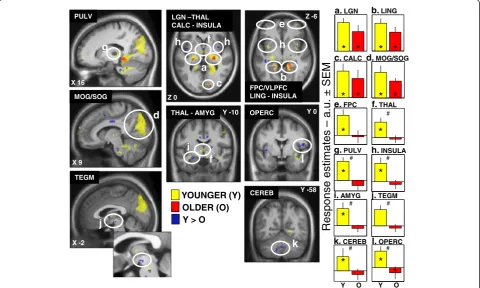
Brain sensitivity to light, cognition, and healthy aging
For the neuroimaging study, the same two groups of
subjects completed an fMRI recording at night, 1 h after
their habitual sleep time. They had to follow a regular
sleep schedule 7 days prior to the experiment and were
maintained in darkness 2 h before the experimental light
exposure. In the scanner, subjects completed 28 blocks
of 45 s of the auditory working memory two-back task
while maintained in a darkness condition or under blue
monochromatic light of three irradiance levels (low 7 ×
10
12ph/cm
2/s, medium 3 × 10
13ph/cm
2/s, high 1 ×
10
14ph/cm
2/s). The two-back task required the subjects
to answer, with a response box, whether each letter
pre-sented was the same as the two prior letters. This task
engaged auditory processing, attention, storing,
compar-ing, and updating information in working memory [123].
Subjects were well trained to the task prior to the fMRI
recordings. Consequently, behavioral analyses revealed
no significant differences between the two groups and
between the four light conditions for accuracy and
re-sponse time values [117]. This was intended and
consist-ent with a ceiling effect in both groups, so that the
limited amount of light we administered could not
sig-nificantly impact performance. This situation was ideal
for the purpose of our study which was to investigate
R
e
s
p
ons
e
es
ti
m
a
tes
– a.u. ±
SEM
MOG/SOG
LGN –THAL CALC - INSULA
b
OPERC THAL - AMYG
PULV
TEGM
CEREB
YOUNGER (Y)
OLDER (O)
Y > O
a.
LGNb.
LINGc.
CALCd.
MOG/SOGf.
THALk.
CEREBl.
OPERCg.
PULVh.
INSULA#
a
c
d
f
g
h
f
l
j
k
X 9 X 16
X -2
Y -10
Y -58 Y 0
e.
FPC
e
FPC/VLPFC LING - INSULA Z 0
Z -6
* *
* *
* *
* *
*
*
*
# #
# #
#
#
Y O Y O
h
h
i.
AMYGj.
TEGMi
*
*
*
*
*
the brain mechanisms involved in the impact of light as
we are sure that behavior did not significantly bias our
fMRI results.
In accordance with literature, and supporting that the
subjects performed the task correctly, we first showed
brain activations in areas known to be involved in the
task including the frontal gyrus, the superior parietal
and temporal gyrus, the intraparietal sulcus (IPS), the
motor and sensorimotor cortices as well as the thalamus,
and the cerebellum [117]. We also investigated which
brain areas responded to the presence of light during the
execution of the task, independently of the irradiance
levels, in young and older subjects. Results indicated
common brain activations in young and older
individ-uals in the LGN, the lingual gyrus, the calcarine
sul-cus, and in the occipital gyrus. These common brain
activations in relation with the effects of light are
pre-sented in Fig. 3.
Analysis also revealed significant age-related
differ-ences as young subjects presented a higher impact of
light than older subjects (represented in blue in Fig. 3)
in the thalamus and a region compatible with the ventral
tegmental area (VTA), important areas for arousal
regu-lation [124], in the amygdala and the insular cortex,
re-gions involved in emotional regulation [125], as well as
in the frontal operculum and in the cerebellum. Some of
these regions have been previously reported in
non-visual effects of light in young subjects and are part of
the salience brain network engaged in the selection of
most relevant information to guide behavior [45, 126].
Less brain sensitivity to light among regions of this
net-work might have important impacts on brain sensitivity
to light in aging on alertness and attention.
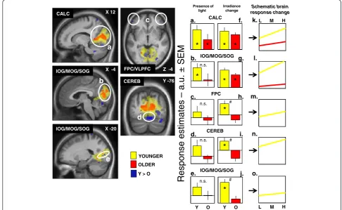
We also investigated which brain areas responded
dif-ferently with age to increasing blue light irradiance levels
during the ongoing cognitive task. Again, results showed
R
e
s
pons
e es
ti
m
a
te
s
– a.
u. ±
SEM
YOUNGER
OLDER
Y > O
IOG/MOG/SOG CALC
IOG/MOG/SOG CEREB
CALC
a
X 12
IOG/MOG/SOG
b
FPC/VLPFC
c
Z -4
CEREB
d
Y -76
* *
*
*
#
Schematic brain response change
L M H
L M H
k.
Y O Y O
X -4
X -20
e
IOG/MOG/SOG
Presence of light
Irradiance change
*
*
*
*
*
# n.s.
*
n.s. n.s.
n.s.
#
o.
FPC
l.
n.
m.
a.
f.
g.
c.
h.
d.
i.
e.
j.
b.
common brain activations in young and older subjects
in the calcarine sulcus, as well as in the inferior, median,
and superior occipital gyrus. As represented in Fig. 4,
these regions seem to increase their activation with
in-creased light intensity in both groups. More importantly,
our results also pointed toward age-related differences in
the prefrontal cortex, an important region for higher
cognitive functions [127], in the occipital cortex, a
re-gion related to the visual system, and finally, in the
cere-bellum. Our results suggested an increase in frontal,
occipital, and cerebellum brain activations in young
sub-jects following light increase intensity, while in older
subjects, this phenomenon was absent.
Overall, these results indicated that light is still able to
modify ongoing brain activity in older individuals in the
context of our protocol. Age-related modifications are
also evident at the irradiance levels we used. Based on
our results, one could argue that light impact is better
conserved in aging in brain areas that are typically
asso-ciated with vision (LGN, calcarine sulcus, and occipital
areas), while areas involved in alertness and cognition
regulation seem to undergo a more pronounced
dimin-ution in their response to light.
Reduced age-related effects of blue monochromatic
light on the thalamus and VTA activity might be related
to various molecular and neural changes in the arousal
system. Hypocretin/orexin neurons, the expression of
which decreases with age [128], innervate many cell
groups including
“
wake-active
”
monoaminergic
popula-tions of the VTA [129
–
132]. A reduced impact of blue
monochromatic light in the VTA-compatible area
sug-gests that the dopaminergic system could be involved in
age-related changes of the stimulating effect of light on
brain responses. The VTA is an important source of
dopamine in the brain and is crucial both for the
regula-tion of sleep and alertness and for cogniregula-tion and mood
[124]. It is notable that the VTA sends projections to the
SCN [133]. Since dopamine dysfunction is thought to
play an important role in the cognitive decline found
in healthy aging [134], the reduced effect of light
upon brain-related dopamine regions might contribute
to reduce the stimulating effect of blue light on
cog-nitive functions.
Conclusions
Lighting-up the aging brain
Light is a simple mean that could easily be used to
im-prove cognition, sleepiness, mood, and sleep in normal
and pathological aging. Daytime sleepiness is a
signifi-cant characteristic of specific neurodegenerative
disor-ders and is associated with not only current cognitive
impairments but also increased risks for developing
cog-nitive decline [135
–
141]. In Alzheimer
’
s and Parkinson
’
s
disease patients, excessive sleepiness and fatigue have
been associated with increased functional impairment
[142] and cognitive dysfunction [143]. While Parkinson
’
s
disease is directly related to dopamine dysfunction [144],
a slow degeneration of hypocretin neurons has been
re-ported over the course of Alzheimer
’
s disease [130].
Im-portantly, light exposure has a positive effect on sleep
and mood in Parkinson
’
s disease patients and
improve-ment of cognitive functions have been reported using
2 h of bright light therapy (polychromatic light
—
3000 lx
and over) in Alzheimer
’
s disease patients [145, 146].
Qualitative positive effects of light exposure on sleep,
mood, and cognition have also been reported in
Alzhei-mer
’
s disease patients with greater effect of blue-green
bright light exposure in the morning as compared to
dim red light [147].
The spectral quality of light may be a crucial factor to
consider when dealing with light in aging. Besides
monochromatic light, one could use polychromatic light,
enriched in blue wavelength for instance. These would
be more applicable to real life and have been reported to
improve some aspects of cognitive performance relative
to classical incandescent light [64, 148]. Each non-visual
response to light will require special attention as they
may be differently affected by age since they rely in part
on different photoreceptor contributions and partly
in-dependent brain pathways.
Furthermore, investigations need to identify light
characteristics (quality, quantity, duration) that can
effectively modulate alertness and cognitive
perform-ance in aging. In order to reach a better
understand-ing of the eye factors upon brain sensitivity to light,
future investigations need to measure pupil size at
the time of the experience or to include older
sub-jects who underwent lens replacement following a
cataract surgery. As it is now recognized that
mela-nopsin gene polymorphism (OPN4) influences pupil
size under light exposure [149, 150] and that clock
gene polymorphism (PER3) influences non-visual
sen-sitivity to light according to sleep pressure and circadian
phase [57, 63, 75], it is also crucial to consider
genetic-s
—
age interactions. Aside from pharmacology, we may
then be in a position to provide light tools to improve life
quality in aging.
Ethics approval and consent to participate
Our
experiments
received
Institutional
ethics
ap-proval from the Research ethics board of the
Comité mixte
d'éthique de la recherche du Regroupement Neuroimagerie
Québec (CMER-RNQ)
and written informed consent was
obtained from each participant.
Competing interests
Authors’contributions
JC, GV, and MD had the original idea for the studies. EM and VD created Fig. 1. VD drafted the manuscript, which was revised by all authors. All authors read and approved the final manuscript.
Acknowledgements
Work was performed at University of Montreal Geriatric Institute, Montreal, Quebec, Canada. The authors thank André Cyr and Carollyn Hurst for their help with data collection. We also thank Jean Paquet from the Center for Advanced Research in Sleep Medicine, Hôpital du Sacré-Coeur de Montréal, for his support on the statistical analyses.
Funding
This was not an industry-supported study. Our work was funded by the Can-adian Institutes of Health Research (CIHR), Natural Sciences and Engineering Research Council of Canada (NSERC), Fonds de la recherche du Québec-Santé (FRQS), and by the International Office of the University of Montreal.
Author details
1Functional Neuroimaging Unit, University of Montreal Geriatric Institute, Montreal, QC, Canada.2Center for Advanced Research in Sleep Medicine, Hôpital du Sacré-Cœur de Montréal, Montreal, QC, Canada.3Department of Psychology, University of Montreal, Montreal, QC, Canada.4Cyclotron Research Centre, University of Liège, Liège, Belgium.
Received: 8 January 2016 Accepted: 1 March 2016
References
1. Hankins MW, Peirson SN, Foster RG. Melanopsin: an exciting photopigment. Trends Neurosci. 2008;31(1):27–36.
2. Altimus CM, Guler AD, Villa KL, McNeill DS, Legates TA, Hattar S. Rods-cones and melanopsin detect light and dark to modulate sleep independent of image formation. Proc Natl Acad Sci U S A. 2008;105(50):19998–20003. 3. Leary SM, Gilpin P, Lockley L, Rodriguez L, Jarrett L, Stevenson VL. Intrathecal
baclofen therapy improves functional intelligibility of speech in cerebral palsy. Clin Rehabil. 2006;20(3):228–31.
4. Chellappa SL, Steiner R, Blattner P, Oelhafen P, Gotz T, Cajochen C. Non-visual effects of light on melatonin, alertness and cognitive performance: can blue-enriched light keep us alert? PLoS One. 2011;6(1):e16429. 5. Vandewalle G, Maquet P, Dijk DJ. Light as a modulator of cognitive brain
function. Trends Cogn Sci. 2009;13(10):429–38.
6. Provencio I, Rodriguez IR, Jiang G, Hayes WP, Moreira EF, Rollag MD. A novel human opsin in the inner retina. J Neurosci. 2000;20(2):600–5.
7. Panda S, Sato TK, Castrucci AM, Rollag MD, DeGrip WJ, Hogenesch JB, Provencio I, Kay SA. Melanopsin (Opn4) requirement for normal light-induced circadian phase shifting. Science. 2002;298(5601):2213–6. 8. Lucas RJ, Hattar S, Takao M, Berson DM, Foster RG, Yau KW. Diminished
pupillary light reflex at high irradiances in melanopsin-knockout mice. Science. 2003;299(5604):245–7.
9. Ruby NF, Brennan TJ, Xie X, Cao V, Franken P, Heller HC, O’Hara BF. Role of melanopsin in circadian responses to light. Science. 2002;298(5601):2211–3. 10. Guler AD, Ecker JL, Lall GS, Haq S, Altimus CM, Liao HW, Barnard AR, Cahill
H, Badea TC, Zhao H, Hankins MW, Berson DM Lucas RJ, et al. Melanopsin cells are the principal conduits for rod-cone input to non-image-forming vision. Nature. 2008;453(7191):102–5.
11. Berson DM. Strange vision: ganglion cells as circadian photoreceptors. Trends Neurosci. 2003;26(6):314–20.
12. Hankins MW, Lucas RJ. The primary visual pathway in humans is regulated according to long-term light exposure through the action of a nonclassical photopigment. Curr Biol. 2002;12(3):191–8.
13. Hattar S, Liao HW, Takao M, Berson DM, Yau KW. Melanopsin-containing retinal ganglion cells: architecture, projections, and intrinsic photosensitivity. Science. 2002;295(5557):1065–70.
14. Berson DM, Dunn FA, Takao M. Phototransduction by retinal ganglion cells that set the circadian clock. Science. 2002;295(5557):1070–3.
15. Sand A, Schmidt TM, Kofuji P. Diverse types of ganglion cell photoreceptors in the mammalian retina. Prog Retin Eye Res. 2012;31(4):287–302. 16. Zhao X, Stafford BK, Godin AL, King WM, Wong KY. Photoresponse diversity
among the five types of intrinsically photosensitive retinal ganglion cells. J Physiol. 2014;592(Pt 7):1619–36.
17. Brown TM, Gias C, Hatori M, Keding SR, Semo M, Coffey PJ, Gigg J, Piggins HD, Panda S, Lucas RJ. Melanopsin contributions to irradiance coding in the thalamo-cortical visual system. PLoS Biol. 2010;8(12):e1000558.
18. Sexton T, Buhr E, Van Gelder RN. Melanopsin and mechanisms of non-visual ocular photoreception. J Biol Chem. 2012;287(3):1649–56.
19. Munch M, Kawasaki A. Intrinsically photosensitive retinal ganglion cells: classification, function and clinical implications. Curr Opin Neurol. 2013;26(1):45–51.
20. Schmidt TM, Chen SK, Hattar S. Intrinsically photosensitive retinal ganglion cells: many subtypes, diverse functions. Trends Neurosci. 2011;34(11):572–80. 21. Baver SB, Pickard GE, Sollars PJ. Two types of melanopsin retinal ganglion
cell differentially innervate the hypothalamic suprachiasmatic nucleus and the olivary pretectal nucleus. Eur J Neurosci. 2008;27(7):1763–70. 22. Ecker JL, Dumitrescu ON, Wong KY, Alam NM, Chen SK, LeGates T, Renna
JM, Prusky GT, Berson DM, Hattar S. Melanopsin-expressing retinal ganglion-cell photoreceptors: ganglion-cellular diversity and role in pattern vision. Neuron. 2010;67(1):49–60.
23. Schmidt TM, Do MT, Dacey D, Lucas R, Hattar S, Matynia A. Melanopsin-positive intrinsically photosensitive retinal ganglion cells: from form to function. J Neurosci. 2011;31(45):16094–101.
24. Lucas RJ, Peirson SN, Berson DM, Brown TM, Cooper HM, Czeisler CA, Figueiro MG, Gamlin DP, Lockley SW, O’Hagan JB, Price LL, Provencio I, Skene DJ, et al. Measuring and using light in the melanopsin age. Trends Neurosci. 2014;37(1):1–9.
25. Kefalov VJ. Rod and cone visual pigments and phototransduction through pharmacological, genetic, and physiological approaches. J Biol Chem. 2012; 287(3):1635–41.
26. Panetsos F, Sanchez-Jimenez A, Cerio ED, Diaz-Guemes I, Sanchez FM. Consistent phosphenes generated by electrical microstimulation of the visual thalamus. An experimental approach for thalamic visual neuroprostheses. Front Neurosci. 2011;5:84.
27. Urbanski M, Coubard OA, Bourlon C. Visualizing the blind brain: brain imaging of visual field defects from early recovery to rehabilitation techniques. Front Integr Neurosci. 2014;8:74.
28. DeSimone K, Viviano JD, Schneider KA. Population receptive field estimation reveals new retinotopic maps in human subcortex. J Neurosci. 2015;35(27):9836–47.
29. Bear MF, Connors BW, Paradiso MA. Neurosciences à la découverte du cerveau: Éditions Pradel. 2007.
30. Hattar S, Kumar M, Park A, Tong P, Tung J, Yau KW, Berson DM, et al. Central projections of melanopsin-expressing retinal ganglion cells in the mouse. J Comp Neurol. 2006;497(3):326–49.
31. Dacey DM, Liao HW, Peterson BB, Robinson FR, Smith VC, Pokorny J, Yau KW, Gamlin PD. et al. Melanopsin-expressing ganglion cells in primate retina signal colour and irradiance and project to the LGN. Nature. 2005;433(7027): 749–54.
32. Estevez ME, Fogerson PM, Ilardi MC, Borghuis BG, Chan E, Weng S, Auferkorte ON, Demb JB, Berson DM, et al. Form and function of the M4 cell, an intrinsically photosensitive retinal ganglion cell type contributing to geniculocortical vision. J Neurosci. 2012;32(39):13608–20.
33. Storchi R, Milosavljevic N, Eleftheriou CG, Martial FP, Orlowska-Feuer P, Bedford RA, Brown TM, Montemurro MA, Petersen RS, Lucas RJ, et al. Melanopsin-driven increases in maintained activity enhance thalamic visual response reliability across a simulated dawn. Proc Natl Acad Sci U S A. 2015; 112(42):E5734–5743.
34. Provencio I, Warther DM. Melanopsin, the photopigment of intrinsically photosensitive retinal ganglion cells. WIREs Membr Transp Signal. 2012;1: 228–237.
35. Hatori M, Le H, Vollmers C, Keding SR, Tanaka N, Buch T, Waisman A, Schmedt C, Jegla T, Panda S, et al. Inducible ablation of melanopsin-expressing retinal ganglion cells reveals their central role in non-image forming visual responses. PLoS One. 2008;3(6):e2451.
36. McNeill DS, Sheely CJ, Ecker JL, Badea TC, Morhardt D, Guido W, Hattar S, et al. Development of melanopsin-based irradiance detecting circuitry. Neural Dev. 2011;6:8.
37. Lupi D, Oster H, Thompson S, Foster RG. The acute light-induction of sleep is mediated by OPN4-based photoreception. Nat Neurosci. 2008;11(9):1068–73. 38. Tsai JW, Hannibal J, Hagiwara G, Colas D, Ruppert E, Ruby NF, Heller HC,
39. Ralph MR, Foster RG, Davis FC, Menaker M. Transplanted suprachiasmatic nucleus determines circadian period. Science. 1990;247(4945):975–8. 40. Morin LP. Neuroanatomy of the extended circadian rhythm system. Exp
Neurol. 2013;243:4–20.
41. Teclemariam-Mesbah R, Ter Horst GJ, Postema F, Wortel J, Buijs RM. Anatomical demonstration of the suprachiasmatic nucleus-pineal pathway. J Comp Neurol. 1999;406(2):171–82.
42. Markwell EL, Feigl B, Zele AJ. Intrinsically photosensitive melanopsin retinal ganglion cell contributions to the pupillary light reflex and circadian rhythm. Clin Exp Optom. 2010;93(3):137–49.
43. Chou TC, Scammell TE, Gooley JJ, Gaus SE, Saper CB, Lu J. Critical role of dorsomedial hypothalamic nucleus in a wide range of behavioral circadian rhythms. J Neurosci. 2003;23(33):10691–702.
44. Sakurai T. The neural circuit of orexin (hypocretin): maintaining sleep and wakefulness. Nat Rev Neurosci. 2007;8(3):171–81.
45. Seeley WW, Menon V, Schatzberg AF, Keller J, Glover GH, Kenna H, Reiss AL, Greicius MD, et al. Dissociable intrinsic connectivity networks for salience processing and executive control. J Neurosci. 2007;27(9):2349–56. 46. Allen AE, Brown TM, Lucas RJ. A distinct contribution of
short-wavelength-sensitive cones to light-evoked activity in the mouse pretectal olivary nucleus. J Neurosci. 2011;31(46):16833–43.
47. Gooley JJ, Ho Mien I, St Hilaire MA, Yeo SC, Chua EC, van Reen E, Hanley CJ, Hull JT, Czeisler CA, Lockley SW, et al. Melanopsin and rod-cone
photoreceptors play different roles in mediating pupillary light responses during exposure to continuous light in humans. J Neurosci. 2012;32(41): 14242–53.
48. McDougal DH, Gamlin PD. The influence of intrinsically-photosensitive retinal ganglion cells on the spectral sensitivity and response dynamics of the human pupillary light reflex. Vis Res. 2010;50(1):72–87.
49. Szkudlarek HJ, Orlowska P, Lewandowski MH. Light-induced responses of slow oscillatory neurons of the rat olivary pretectal nucleus. PLoS One. 2012; 7(3):e33083.
50. Tsujimura S, Ukai K, Ohama D, Nuruki A, Yunokuchi K. Contribution of human melanopsin retinal ganglion cells to steady-state pupil responses. Proc Biol Sci. 2010;277(1693):2485–92.
51. Walmsley L, Hanna L, Mouland J, Martial F, West A, Smedley AR, Bechtold DA, Webb AR, Lucas RJ, Brown TM, et al. Colour as a signal for entraining the mammalian circadian clock. PLoS Biol. 2015;13(4):e1002127.
52. Chen SK, Badea TC and Hattar S. Photoentrainment and pupillary light reflex are mediated by distinct populations of ipRGCs. Nature. 2011;476:92–95. 53. Lockley SW, Evans EE, Scheer FA, Brainard GC, Czeisler CA, Aeschbach
D. Short-wavelength sensitivity for the direct effects of light on alertness, vigilance, and the waking electroencephalogram in humans. Sleep. 2006;29(2):161–8.
54. Lockley SW, Brainard GC, Czeisler CA. High sensitivity of the human circadian melatonin rhythm to resetting by short wavelength light. J Clin Endocrinol Metab. 2003;88(9):4502–5.
55. Cajochen C, Munch M, Kobialka S, Krauchi K, Steiner R, Oelhafen P, Orgul S, Wirz-Justice A, et al. High sensitivity of human melatonin, alertness, thermoregulation, and heart rate to short wavelength light. J Clin Endocrinol Metab. 2005;90(3):1311–6.
56. Vandewalle G, Schwartz S, Grandjean D, Wuillaume C, Balteau E, Degueldre C, Schabus M, Phillips C, Luxe A, Dijk DJ, Maquet P, et al. Spectral quality of light modulates emotional brain responses in humans. Proc Natl Acad Sci U S A. 2010;107(45):19549–54.
57. Vandewalle G, Archer SN, Wuillaume C, Balteau E, Degueldre C, Luxen A, Dijk DJ, Maquet P, et al. Effects of light on cognitive brain responses depend on circadian phase and sleep homeostasis. J Biol Rhythm. 2011; 26(3):249–59.
58. Brainard GC, Hanifin JP, Greeson JM, Byrne B, Glickman G, Gerner E, Rollag MD, et al. Action spectrum for melatonin regulation in humans: evidence for a novel circadian photoreceptor. J Neurosci. 2001;21(16):6405–12. 59. Mure LS, Cornut PL, Rieux C, Drouyer E, Denis P, Gronfier C, Cooperet HM,
et al. Melanopsin bistability: a fly’s eye technology in the human retina. PLoS One. 2009;4(6):e5991.
60. Chellappa SL, Steiner R, Oelhafen P, Lang D, Gotz T, Krebs J, Cajochen C, et al. Acute exposure to evening blue-enriched light impacts on human sleep. J Sleep Res. 2013;22(5):573–80.
61. Viola AU, James LM, Schlangen LJ, Dijk DJ. Blue-enriched white light in the workplace improves self-reported alertness, performance and sleep quality. Scand J Work Environ Health. 2008;34(4):297–306.
62. Revell VL, Arendt J, Fogg LF, Skene DJ. Alerting effects of light are sensitive to very short wavelengths. Neurosci Lett. 2006;399(1–2):96–100.
63. Chellappa SL, Viola AU, Schmidt C, Bachmann V, Gabel V, Maire M, Reichert CF, Valomon A, Gotz T, Landolt HP, Cajochen C, et al. Human melatonin and alerting response to blue-enriched light depend on a polymorphism in the clock gene PER3. J Clin Endocrinol Metab. 2012;97(3):E433–437. 64. Chellappa SL, Gordijn MC, Cajochen C. Can light make us bright? Effects of
light on cognition and sleep. Prog Brain Res. 2011;190:119–33. 65. Okamoto Y, Nakagawa S. Effects of daytime light exposure on
cognitive brain activity as measured by the ERP P300. Physiol Behav. 2015;138:313–8.
66. Vandewalle G, Schmidt C, Albouy G, Sterpenich V, Darsaud A, Rauchs G, Berken PY, Balteau E, Degueldre C, Luxen A, Maquet P, Dijk DJ, et al. Brain responses to violet, blue, and green monochromatic light exposures in humans: prominent role of blue light and the brainstem. PLoS One. 2007;2(11):e1247. 67. Vandewalle G, Gais S, Schabus M, Balteau E, Carrier J, Darsaud A,
Sterpenich V, Albouy G, Dijk DJ, Maquet P, et al. Wavelength-dependent modulation of brain responses to a working memory task by daytime light exposure. Cereb Cortex. 2007;17(12):2788–95. 68. Vandewalle G. The stimulating impact of light on brain cognition function.
Med Sci. 2014;30(10):902–9.
69. Vandewalle G, Collignon O, Hull JT, Daneault V, Albouy G, Lepore F, Phillips C, Doyon J, Czeisler CA, Dumont M, Lockley SW, Carrier J, et al. Blue light stimulates cognitive brain activity in visually blind individuals. J Cogn Neurosci. 2013;25(12):2072–85.
70. Vandewalle G, Hebert M, Beaulieu C, Richard L, Daneault V, Garon ML, Leblanc J, Grandjean D, Maquet P, Schwartz S, Dumont M, Doyon J, Carrier J, et al. Abnormal hypothalamic response to light in seasonal affective disorder. Biol Psychiatry. 2011;70(10):954–61.
71. Vandewalle G, Balteau E, Phillips C, Degueldre C, Moreau V, Sterpenich V, Albouy G, Darsaud A, Desseilles M, Dang-Vu TT, Peigneux P, Luxen A, Dijk DJ, et al. Daytime light exposure dynamically enhances brain responses. Curr Biol. 2006;16(16):1616–21.
72. Perrin F, Peigneux P, Fuchs S, Verhaeghe S, Laureys S, Middleton B, Degueldre C, Del Fiore G, Vandewalle G, Balteau E, Poirrier R, Moreau V, Luxen A, et al. Nonvisual responses to light exposure in the human brain during the circadian night. Curr Biol. 2004;14(20):1842–6.
73. Matsuyama T, Yamashita T, Imamoto Y, Shichida Y. Photochemical properties of mammalian melanopsin. Biochemistry. 2012;51(27):5454–62. 74. Mure LS, Rieux C, Hattar S, Cooper HM. Melanopsin-dependent nonvisual responses: evidence for photopigment bistability in vivo. J Biol Rhythm. 2007;22(5):411–24.
75. Chellappa SL, Ly JQ, Meyer C, Balteau E, Degueldre C, Luxen A, Phillips C, Cooper HM, Vandewalle G. Photic memory for executive brain responses. PNAS. 2014; 111(16):6087-91.
76. Gaggioni G, Maquet P, Schmidt C, Dijk DJ, Vandewalle G. Neuroimaging, cognition, light and circadian rhythms. Front Syst Neurosci. 2014;8:126. 77. Herljevic M, Middleton B, Thapan K, Skene DJ. Light-induced melatonin suppression: age-related reduction in response to short wavelength light. Exp Gerontol. 2005;40(3):237–42.
78. Sletten TL, Revell VL, Middleton B, Lederle KA, Skene DJ. Age-related changes in acute and phase-advancing responses to monochromatic light. J Biol Rhythm. 2009;24(1):73–84.
79. Jud C, Chappuis S, Revell VL, Sletten TL, Saaltink DJ, Cajochen C, Skene DJ, Albrecht U. Age-dependent alterations in human PER2 levels after early morning blue light exposure. Chronobiol Int. 2009;26(7):1462–9. 80. Benloucif S, Green K, L’Hermite-Baleriaux M, Weintraub S, Wolfe LF, Zee PC.
Responsiveness of the aging circadian clock to light. Neurobiol Aging. 2006; 27(12):1870–9.
81. Nathan PJ, Burrows GD, Norman TR. The effect of age and pre-light melatonin concentration on the melatonin sensitivity to dim light. Int Clin Psychopharmacol. 1999;14(3):189–92.
82. Klerman EB, Duffy JF, Dijk DJ, Czeisler CA. Circadian phase resetting in older people by ocular bright light exposure. J Investig Med. 2001;49(1):30–40. 83. Samuels ER, Szabadi E. Functional neuroanatomy of the noradrenergic locus
coeruleus: its roles in the regulation of arousal and autonomic function part II: physiological and pharmacological manipulations and pathological alterations of locus coeruleus activity in humans. Curr Neuropharmacol. 2008;6(3):254–85.
85. Hammond Jr BR, Nanez JE, Fair C, Snodderly DM. Iris color and age-related changes in lens optical density. Ophthalmic Physiol Opt. 2000;20(5):381–6. 86. Kessel L, Lundeman JH, Herbst K, Andersen TV, Larsen M. Age-related changes
in the transmission properties of the human lens and their relevance to circadian entrainment. J Cataract Refract Surg. 2010;36(2):308–12. 87. Turner PL, Mainster MA. Circadian photoreception: ageing and the eye’s
important role in systemic health. Br J Ophthalmol. 2008;92(11):1439–44. 88. Farajnia S, Michel S, Deboer T, van der Leest HT, Houben T, Rohling JH,
Ramkisoensing JH, Yasenkov R, Meijer JH. Evidence for neuronal desynchrony in the aged suprachiasmatic nucleus clock. J Neurosci. 2012; 32(17):5891–9.
89. Kondratova AA, Kondratov RV. The circadian clock and pathology of the ageing brain. Nat Rev Neurosci. 2012;13(5):325–35.
90. Asai M, Yoshinobu Y, Kaneko S, Mori A, Nikaido T, Moriya T, Akiyama M, Shibata S. Circadian profile of Per gene mRNA expression in the suprachiasmatic nucleus, paraventricular nucleus, and pineal body of aged rats. J Neurosci Res.
2001;66(6):1133–9.
91. Davidson AJ, Yamazaki S, Arble DM, Menaker M, Block GD. Resetting of central and peripheral circadian oscillators in aged rats. Neurobiol Aging. 2008;29(3):471–7.
92. Kolker DE, Fukuyama H, Huang DS, Takahashi JS, Horton TH, Turek FW. Aging alters circadian and light-induced expression of clock genes in golden hamsters. J Biol Rhythm. 2003;18(2):159–69.
93. Sitzmann BD, Lemos DR, Ottinger MA, Urbanski HF. Effects of age on clock gene expression in the rhesus macaque pituitary gland. Neurobiol Aging. 2010;31(4):696–705.
94. Claustrat F, Fournier I, Geelen G, Brun J, Corman B, Claustrat B. Aging and circadian clock gene expression in peripheral tissues in rats. Pathol Biol. 2005;53(5):257–60.
95. Gibson EM, Williams 3rd WP, Kriegsfeld LJ. Aging in the circadian system: considerations for health, disease prevention and longevity. Exp Gerontol. 2009;44(1–2):51–6.
96. Kawakami F, Okamura H, Tamada Y, Maebayashi Y, Fukui K, Ibata Y. Loss of day-night differences in VIP mRNA levels in the suprachiasmatic nucleus of aged rats. Neurosci Lett. 1997;222(2):99–102.
97. Kawaguchi C, Isojima Y, Shintani N, Hatanaka M, Guo X, Okumura N, Nagai K, Hashimoto H, Baba, A. PACAP-deficient mice exhibit light parameter-dependent abnormalities on nonvisual photoreception and early activity onset. PLoS One. 2010;5(2):e9286.
98. Roozendaal B, van Gool WA, Swaab DF, Hoogendijk JE, Mirmiran M. Changes in vasopressin cells of the rat suprachiasmatic nucleus with aging. Brain Res. 1987;409(2):259–64.
99. Hofman MA, Swaab DF. Alterations in circadian rhythmicity of the vasopressin-producing neurons of the human suprachiasmatic nucleus (SCN) with aging. Brain Res. 1994;651(1–2):134–42.
100. Hofman MA. The human circadian clock and aging. Chronobiol Int. 2000; 17(3):245–59.
101. Aujard F, Cayetanot F, Bentivoglio M, Perret M. Age-related effects on the biological clock and its behavioral output in a primate. Chronobiol Int. 2006; 23(1–2):451–60.
102. Cayetanot F, Bentivoglio M, Aujard F. Arginine-vasopressin and vasointestinal polypeptide rhythms in the suprachiasmatic nucleus of the mouse lemur reveal aging-related alterations of circadian pacemaker neurons in a non-human primate. Eur J Neurosci. 2005;22(4):902–10. 103. Hofman MA. Circadian oscillations of neuropeptide expression in the
human biological clock. J Comp Physiol A Neuroethol Sens Neural Behav Physiol. 2003;189(11):823–31.
104. Long X, Liao W, Jiang C, Liang D, Qiu B, Zhang L. Healthy aging: an automatic analysis of global and regional morphological alterations of human brain. Acad Radiol. 2012;19(7):785–93.
105. Peters A. The effects of normal aging on myelin and nerve fibers: a review. J Neurocytol. 2002;31(8–9):581–93.
106. Guttmann CR, Jolesz FA, Kikinis R, Killiany RJ, Moss MB, Sandor T, Albert MS. White matter changes with normal aging. Neurology. 1998;50(4):972–8. 107. Walhovd KB, Fjell AM, Reinvang I, Lundervold A, Dale AM, Eilertsen DE,
Quinn BT, Salat D, Makris N, Fischl B. Effects of age on volumes of cortex, white matter and subcortical structures. Neurobiol Aging. 2005;26(9):1261–70. discussion 1275–1268.
108. Samuels ER, Szabadi E. Functional neuroanatomy of the noradrenergic locus coeruleus: its roles in the regulation of arousal and autonomic
function part I: principles of functional organisation. Curr Neuropharmacol. 2008;6(3):235–53.
109. Deng XH, Bertini G, Palomba M, Xu YZ, Bonaconsa M, Nygard M, Bentivoglio M. Glial transcripts and immune-challenged glia in the suprachiasmatic nucleus of young and aged mice. Chronobiol Int. 2010;27(4):742–67. 110. Fotiou DF, Brozou CG, Tsiptsios DJ, Fotiou A, Kabitsi A, Nakou M, Giantselidis
C, Goula A. Effect of age on pupillary light reflex: evaluation of pupil mobility for clinical practice and research. Electromyogr Clin Neurophysiol. 2007;47(1):11–22.
111. Freund PR, Watson J, Gilmour GS, Gaillard F, Sauve Y. Differential changes in retina function with normal aging in humans. Doc Ophthalmol. 2011; 122(3):177–90.
112. Hebert M, Beattie CW, Tam EM, Yatham LN, Lam RW. Electroretinography in patients with winter seasonal affective disorder. Psychiatry Res. 2004; 127(1–2):27–34.
113. Sturr JF, Zhang L, Taub HA, Hannon DJ, Jackowski MM. Psychophysical evidence for losses in rod sensitivity in the aging visual system. Vis Res. 1997;37(4):475–81.
114. Bitsios P, Prettyman R, Szabadi E. Changes in autonomic function with age: a study of pupillary kinetics in healthy young and old people. Age Ageing. 1996;25(6):432–8.
115. Winn B, Whitaker D, Elliott DB, Phillips NJ. Factors affecting light-adapted pupil size in normal human subjects. Invest Ophthalmol Vis Sci. 1994; 35(3):1132–7.
116. Daneault V, Vandewalle G, Hebert M, Teikari P, Mure LS, Doyon J, Gronfier C Cooper HM, Dumont M, Carrier J. Does pupil constriction under blue and green monochromatic light exposure change with age? J Biol Rhythm. 2012;27(3):257–64.
117. Daneault V, Hebert M, Albouy G, Doyon J, Dumont M, Carrier J, Vandewalle G. Aging reduces the stimulating effect of blue light on cognitive brain functions. Sleep. 2014;37(1):85–96.
118. Pickard GE, Sollars PJ. Intrinsically photosensitive retinal ganglion cells. Sci China Life Sci. 2010;53(1):58–67.
119. Pickard GE, Baver SB, Ogilvie MD, Sollars PJ. Light-induced fos expression in intrinsically photosensitive retinal ganglion cells in melanopsin knockout (opn4) mice. PLoS One. 2009;4(3):e4984.
120. LeGates TA, Fernandez DC, Hattar S. Light as a central modulator of circadian rhythms, sleep and affect. Nat Rev Neurosci. 2014;15(7):443–54. 121. Butler MP, Silver R. Divergent photic thresholds in the non-image-forming
visual system: entrainment, masking and pupillary light reflex. Proc Biol Sci. 2011;278(1706):745–50.
122. Hut RA, Oklejewicz M, Rieux C, Cooper HM. Photic sensitivity ranges of hamster pupillary and circadian phase responses do not overlap. J Biol Rhythm. 2008;23(1):37–48.
123. Owen AM, McMillan KM, Laird AR, Bullmore E. N-back working memory paradigm: a meta-analysis of normative functional neuroimaging studies. Hum Brain Mapp. 2005;25(1):46–59.
124. Luo AH, Aston-Jones G. Circuit projection from suprachiasmatic nucleus to ventral tegmental area: a novel circadian output pathway. Eur J Neurosci. 2009;29(4):748–60.
125. Stein JL, Wiedholz LM, Bassett DS, Weinberger DR, Zink CF, Mattay VS, Meyer-Lindenberg A. A validated network of effective amygdala connectivity. NeuroImage. 2007;36(3):736–45.
126. Menon V, Uddin LQ. Saliency, switching, attention and control: a network model of insula function. Brain Struct Funct. 2010;214(5–6):655–67. 127. Koechlin E, Hyafil A. Anterior prefrontal function and the limits of human
decision-making. Science. 2007;318(5850):594–8.
128. Kessler BA, Stanley EM, Frederick-Duus D, Fadel J. Age-related loss of orexin/ hypocretin neurons. Neuroscience. 2011;178:82–8.
129. Kilduff TS, Peyron C. The hypocretin/orexin ligand-receptor system: implications for sleep and sleep disorders. Trends Neurosci. 2000;23(8): 359–65.
130. Shan L, Dauvilliers Y, Siegel JM. Interactions of the histamine and hypocretin systems in CNS disorders. Nat Rev Neurol. 2015;11(7):401–13.
131. Sutcliffe JG, de Lecea L. The hypocretins: excitatory neuromodulatory peptides for multiple homeostatic systems, including sleep and feeding. J Neurosci Res. 2000;62(2):161–8.
132. Ebrahim IO, Howard RS, Kopelman MD, Sharief MK, Williams AJ. The hypocretin/orexin system. J R Soc Med. 2002;95(5):227–30.
134. Eppinger B, Hammerer D, Li SC. Neuromodulation of reward-based learning and decision making in human aging. Ann N Y Acad Sci. 2011;1235:1–17. 135. Rao V, Spiro JR, Samus QM, Rosenblatt A, Steele C, Baker A, Harper M,
Brandt J, Mayer L, Rabins PV, Lyketsos CG. Sleep disturbances in the elderly residing in assisted living: findings from the Maryland Assisted Living Study. Int J Geriatr Psychiatry. 2005;20(10):956–66.
136. Foley D, Monjan A, Masaki K, Ross W, Havlik R, White L, Launer L. Daytime sleepiness is associated with 3-year incident dementia and cognitive decline in older Japanese-American men. J Am Geriatr Soc. 2001;49(12):1628–32.
137. Merlino G, Piani A, Gigli GL, Cancelli I, Rinaldi A, Baroselli A, Serafini A, Zanchettin B, Valente M. Daytime sleepiness is associated with dementia and cognitive decline in older Italian adults: a population-based study. Sleep Med. 2010;11(4):372–7.
138. Keage HA, Banks S, Yang KL, Morgan K, Brayne C, Matthews FE. What sleep characteristics predict cognitive decline in the elderly? Sleep Med. 2012; 13(7):886–92.
139. Elwood PC, Bayer AJ, Fish M, Pickering J, Mitchell C, Gallacher JE. Sleep disturbance and daytime sleepiness predict vascular dementia. J Epidemiol Community Health. 2011;65(9):820–4.
140. Abbott RD, Ross GW, White LR, Tanner CM, Masaki KH, Nelson JS, Curb JD, Petrovitch H. Excessive daytime sleepiness and subsequent development of Parkinson disease. Neurology. 2005;65(9):1442–6.
141. Ohayon MM, Vecchierini MF. Normative sleep data, cognitive function and daily living activities in older adults in the community. Sleep. 2005;28(8):981–9. 142. Lee JH, Bliwise DL, Ansari FP, Goldstein FC, Cellar JS, Lah JJ, Levey AI.
Daytime sleepiness and functional impairment in Alzheimer disease. Am J Geriatr Psychiatr. 2007;15(7):620–6.
143. Shin HY, Han HJ, Shin DJ, Park HM, Lee YB, Park KH. Sleep problems associated with behavioral and psychological symptoms as well as cognitive functions in Alzheimer’s disease. J Clin Neurol. 2014;10(3):203–9. 144. Willis GL, Moore C, Armstrong SM. A historical justification for and
retrospective analysis of the systematic application of light therapy in Parkinson’s disease. Rev Neurosci. 2012;23(2):199–226.
145. Yamadera H, Ito T, Suzuki H, Asayama K, Ito R, Endo S. Effects of bright light on cognitive and sleep-wake (circadian) rhythm disturbances in Alzheimer-type dementia. Psychiatry Clin Neurosci. 2000;54(3):352–3.
146. Ito T, Yamadera H, Ito R, Endo S. Effects of bright light on cognitive disturbances in Alzheimer-type dementia. Nihon Ika Daigaku Zasshi. 1999;66(4):229–38.
147. Nowak L, Davis J. Qualitative analysis of therapeutic light effects on global function in Alzheimer’s disease. West J Nurs Res. 2011;33(7):933–52. 148. Cajochen C, Chellappa S, Schmidt C. What keeps us awake? The role of clocks
and hourglasses, light, and melatonin. Int Rev Neurobiol. 2010;93:57–90. 149. Higuchi S, Hida A, Tsujimura S, Mishima K, Yasukouchi A, Lee SI, Kinjyo Y,
Miyahira M. Melanopsin gene polymorphism I394T is associated with pupillary light responses in a dose-dependent manner. PLoS One. 2013;8(3): e60310.
150. Lee SI, Hida A, Tsujimura S, Morita T, Mishima K, Higuchi S. Association between melanopsin gene polymorphism (I394T) and pupillary light reflex is dependent on light wavelength. J Physiol Anthropol. 2013;32:16.
• We accept pre-submission inquiries
• Our selector tool helps you to find the most relevant journal • We provide round the clock customer support
• Convenient online submission • Thorough peer review
• Inclusion in PubMed and all major indexing services • Maximum visibility for your research
Submit your manuscript at www.biomedcentral.com/submit