R E V I E W
Open Access
Metformin in colorectal cancer: molecular
mechanism, preclinical and clinical aspects
Muhamad Noor Alfarizal Kamarudin
1*, Md. Moklesur Rahman Sarker
2,3*, Jin-Rong Zhou
4and Ishwar Parhar
1Abstract
Growing evidence showed the increased prevalence of cancer incidents, particularly colorectal cancer, among type 2 diabetic mellitus patients. Antidiabetic medications such as, insulin, sulfonylureas, dipeptyl peptidase (DPP) 4 inhibitors and glucose-dependent insulinotropic peptide (GLP-1) analogues increased the additional risk of different cancers to diabetic patients. Conversely, metformin has drawn attention among physicians and researchers since its use as antidiabetic drug exhibited beneficial effect in the prevention and treatment of cancer in diabetic patients as well as an independent anticancer drug. This review aims to provide the comprehensive information on the use of metformin at preclinical and clinical stages among colorectal cancer patients. We highlight the efficacy of
metformin as an anti-proliferative, chemopreventive, apoptosis inducing agent, adjuvant, and radio-chemosensitizer in various colorectal cancer models. This multifarious effects of metformin is largely attributed to its capability in modulating upstream and downstream molecular targets involved in apoptosis, autophagy, cell cycle, oxidative stress, inflammation, metabolic homeostasis, and epigenetic regulation. Moreover, the review highlights metformin intake and colorectal cancer risk based on different clinical and epidemiologic results from different gender and specific population background among diabetic and non-diabetic patients. The improved understanding of metformin as a potential chemotherapeutic drug or as neo-adjuvant will provide better information for it to be used globally as an affordable, well-tolerated, and effective anticancer agent for colorectal cancer.
Keywords:Metformin, Colorectal cancer, Cancer, Type 2 diabetes mellitus, Chemopreventive, Anticancer
Background
Cancer remains one of the leading cause of death with high global prevalence despite numerous advancements made in the last decade. A recent cancer statistics by American Cancer Society projected a total of 1,762,450 new cancer cases with 606,880 mortality to occur in the United States alone [1]. The report estimated prostate (20%), lung and bronchus (13%), and colorectal (9%) to be the most prevalent new cancer cases in males whereas breast (30%), lung and bronchus (13%), and colorectal (8%) in females in 2019. Among this, respira-tory and digestive system cancers are projected to con-tribute the highest mortality rate among other cancers.
Colorectal or colon cancer (CRC) is projected to record the highest mortality cases (51,020) among other digest-ive system cancer (total of 165,460 cases) [1]. Factors such as bad dietary habits, smoking status, alcohol con-sumption, genetic predisposition, obesity, diabetes melli-tus, and sedentary lifestyle significantly increase the risk of developing CRC [2–4]. To date, surgery, such as right colectomy, sigmoid colectomy, and total abdominal col-ectomy with ileorectal anastomosis as well as chemo-therapy are the available treatment options. Additionally, patients with advanced stage of CRC are normally treated with chemotherapeutic drug, 5-fluorouracil (5-FU) alone or in combination of adjuvant such as oxali-platin and avastin [3, 5–7]. Although these treatment regimens are effective at improving disease and overall survival (OS), severe side-effects such as severe nausea, vomiting, weight loss, and risk of infectious complica-tions due to immunosuppression often burden the patients.
© The Author(s). 2019Open AccessThis article is distributed under the terms of the Creative Commons Attribution 4.0
International License (http://creativecommons.org/licenses/by/4.0/), which permits unrestricted use, distribution, and
reproduction in any medium, provided you give appropriate credit to the original author(s) and the source, provide a link to the Creative Commons license, and indicate if changes were made. The Creative Commons Public Domain Dedication waiver (http://creativecommons.org/publicdomain/zero/1.0/) applies to the data made available in this article, unless otherwise stated.
* Correspondence:MuhamadNoor.Alfarizal@monash.edu; moklesur2002@yahoo.com;dr.moklesur2014@gmail.com
1Brain Research Institute Monash Sunway (BRIMS), Jeffrey Cheah School of
Medicine and Health Sciences, Monash University Malaysia, 47500 Bandar Sunway, Selangor, Malaysia
2Department of Pharmacy, State University of Bangladesh, 77 Satmasjid Road,
Dhanmondi, Dhaka 1205, Bangladesh
Even though both diseases are complex and multifari-ous by nature, both CRC and diabetes mellitus share various similar clinical risk factors which include age, diet, obesity, and gender [8, 9]. Furthermore, in the last decade, the pathogenesis and pathophysiological mecha-nisms of both CRC and type 2 diabetes mellitus (T2DM) related conditions such as hyperglycaemia, hyperinsuli-nemia, and insulin resistance are found to be closely related since they both involve the regulation of the in-sulin/insulin-like growth factor (IGF) signalling pathway
[10, 11]. For instance, hyperinsulinemia and insulin
resistance are found to promote the progression of tumorigenesis via either the insulin receptor in the epi-thelial tissues or by modulating the levels of other mod-ulators, such as insulin-like growth factors (IGFs), sex hormones, inflammatory processes, and adipokines. This is due to the relative insulin sensitivity of the epithelial cells that enhances insulmediated signalling that in-duces cancer cell proliferation and metastasis [10,11].
Metformin (1,1-dimethylbiguanide), a product of French lilac (Galega officinalis), is an oral biguanide and hypoglycemic agent that is prescribed to over 120 million patients with gestational diabetes [12, 13], T2DM [14, 15], non-alcoholic fatty liver disease [16, 17], prema-ture puberty, [18] and polycystic ovarian syndrome (PCOS) [19,20] worldwide. Unlike other biguanides such as sulfo-nylurea, and thiazolidinediones, the oral consumption of metformin is beneficial since it reduces the risk of cardio-vascular disease by reducing cholesterol levels as well as inflammatory and blood clotting markers while controlling the blood glucose level [21, 22]. The presence of two methyl substitutes in metformin reduces the lipophilicity of metformin that aides the hepatic lactate clearance and excretion of metformin unchanged in the urine as com-pared to other diabetic drugs. Furthermore, metformin has several advantages in treating T2DM and associated cancer risks as compared to exogenous insulin and insulin secreta-logues such as sulfonylurea drugs, which are reported to increase cancer risk and recurrence [23,24]. Since metfor-min primary actions significantly reduce the circulating glucose and plasma insulin, hence, it improves insulin resistance in peripheral tissue. Therefore, the repurposed use of metformin may be beneficial in reducing the risk of diabetes related cancer incident [25]. Moreover, numerous lines of empirical evidence have supported the use of metformin as an anticancer agent that inhibits the transformative and hyperproliferative processes with anti-angiogenesis, radio-chemosensitizer, and antimeta-bolic effects that suppress carcinogenesis [25–27]. For example, in glioma models, the use of metformin in combination with five other repurposed drugs (itraco-nazole, naproxen, pirfenidone, rifampin, and quetiapine; known as EMT inhibiting sextet (EIS)) is shown to inhibit glioblastoma cells proliferation, invasion, chemoresistance,
and metastatic activities which further blocked the epithelial to mesenchymal transition (EMT) [28]. The anticancer property of metformin is largely attributed to its capability in modulating signaling pathways involved in cellular prolif-eration, apoptosis, and metabolism. For instance, metformin modulates the synergistic regulation between AMPK, GSK-3β, and PPAR-γ that confer its angiogenic, anti-invasive, and anti-proliferative as observed in pancreatic cancer and glioblastoma multiforme (GBM) [29].
In the last decade, mounting evidence support the use of metformin in the prevention and treatment of CRC (reviewed in later sections). Furthermore, extensive in vitro and in vivo research activities have successfully elucidated the molecular mechanisms of metformin in CRC models (discussed in later section). Moreover, the use of metformin as monotherapy or as an adjuvant in CRC intervention has led to further dose reduction and increased radio-chemosensitivity which lead to minimal gastrointestinal side effects and reduced toxicity. Fur-thermore, since metformin is relatively cheaper than other chemotherapy drugs and adjuvants, it may serve as a cost-effective and well affordable treatment option for CRC intervention. Nonetheless, contradictory popula-tion-based studies as well as beneficial metformin use among non-diabetic cancer patients further rationalize the need to systemically evaluate its effectiveness against CRC. Currently, researchers are hoping to get the better prospect form management of CRC along with the treat-ment of diabetes. The review article also highlights the use of metformin with improved survival among CRC patients with T2DM as compared to sulfonylureas and insulin. The present review aims to provide comprehen-sive and up to date preclinical, clinical, and epidemio-logic reports on metformin as well as its molecular mechanisms that justify its repurposed use as a pro-spective and potential medicament in the intervention of CRC worldwide.
The preclinical evidence use of metformin in CRC
Metformin inin vitroCRC models
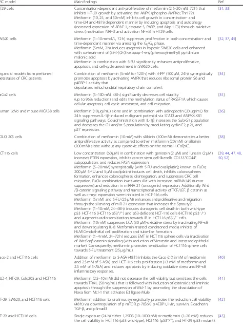
Table 1The summary of preclinical (in vitro) use of metformin in CRC models
CRC model Main findings Ref.
HT29 cells Concentration-dependent anti-proliferative of metformin (2.5–20 mM, 72 h) that inhibits HT-29 growth by activating the AMPK (phospho-AMPKα; Thr172). Metformin (10, 25, and 50 mM) inhibits cell growth in concentration- and time-(24 and 48 h) dependent manner by inducing apoptosis and autophagy (increased expression of APAF-1, caspase-3, PARP, and Map-LC3) through oxidative stress (inactivation NRF-2 and activation NF-κB in HT29 cells.
[31,33]
SW620 cells Metformin (1–10 mmol/L, 72 h) suppresses proliferation in both concentration- and time-dependent manner via arresting the G0/G1phase.
Metformin (5 mM, 2 h) induces apoptosis in hypoxic SW620 cells and enhanced with co-treatment of (E)-4-((2-(3-oxopop-1-enyl)phenoxy)methyl) pyridinium malonic acid
Metformin in combination with 5-FU significantly enhances antiproliferative, apoptosis, and cell-cycle arrestment in SW620 cells.
[32,37,45]
Organoid models from peritoneal metastases of CRC patients
Combination of metformin (5 mM for 120 h) with 4-IPP (100μM, 24 h) synergistically promotes apoptosis by activating AMPK that reduces ribosomal protein S6 and p4EBP-1 activity that
depolarizes mitochondrial respiratory chain complex I.
[34]
CaCo2 cells Metformin (5–100 mM, 48 h) significantly decreases cell viability
(up to 96% reduction) and edits the methylation status of RASSF1A which causes cellular apoptosis, cell cycle arrestment, and cell migration.
[35]
Human LoVo and mouse MCA38 cells Metformin (10μg/mL) alone and in combination with adinopectin (20μg/mL) for 24 h suppresses IL-1β-induced malignant potential via STAT3 and AMPK/LKB1 signaling pathways. Co-administration with IL-1βincreases the Sub-G1 population and decreases the G1 and/or S population by modulating cyclin E2, p21, and p27 expression.
[36]
COLO 205 cells Combination of metformin (10 mM) with silibinin (100 mM) demonstrates a better antiproliferative activity as compared to either metformin (20 mM) or silibinin (200 mM) alone without any cytotoxic effects on the normal HCoEpiC.
[38]
HCT116 cells Low concentration (60μM) in combination with genistein (2μM) and lunasin (2μM) increases PTEN expression, inhibits cancer stem cell-likecells CD133+CD44+
subpopulation, and reduces FASN expression.
Metformin (5–20 mM) synergistically (with 5-FU and oxaliplatin); known as FuOx; 200μM 5-FU and 5μM oxaliplatin) induces cell death, inhibits colonospheres formation, enhances colonospheres disintegration, and suppresses CRC cell migration. FuOx combination inactivates Akt with increased miRNA145 (tumor suppressive) and reduction in miRNA 21 (oncogenic) expression. Additionally Wnt /β-catenin signaling pathway and transcriptional activity of TCF/LEF,β-catenin as well as c-myc expression were inhibited in HCT-116 cells.
Metformin (5 mM) and 5-FU (25μM) enhances antiproliferative and migration through the silencing of miR-21 expression that increases the Sprouty2. Metformin (1–10 mM, 24–48 h) induces clonogenic cell death in both wild-type p53 HCT-116 (HCT116 p53+/+) and p53-deficient HCT-116 cells (HCT116 p53−/−)
and augments radio-sensitization towards IR in HCT116 p53−/−cells.
Metformin (10 mM) suppresses LCA (30μM)-oxidative stress by inactivating NF-κB and downregulating IL-8. Metformin-treated conditioned media inhibits of HUVECendothelial cell proliferation and tube-like formation. .
Metformin (1–4 mM, 24–72 h) reduces EMT in HCT116 sphere cells via inactivation of Wnt3α/β-catenin signaling (with reduction of Vimentin and increased epithelial marker). Consequently, metformin promotes sensitization of HCT116 sphere cells towards 5-FU treatment (25μg/mL).
[39,44,47,48, 50,52]
Caco-2 and HCT116 cells Addition of metformin to 5-ASA (48 h) inhibits the Caco-2 (13 mM of metformin and 2.5 mM of 5-ASA) and HCT-116 cells proliferation (13 mM of metformin and 2.5 mM of 5-ASA) and induces apoptosis by inducing oxidative stress and NF-κB inflammatory responses.
[40]
DLD-1, HT-29, Colo205 and HCT116 Metformin (2.5–10 mM) did not decrease the cell viability but sensitizes the cells towards TRAIL (50 ng/mL) that is followed with induction of extrinsic and intrinsic apoptosis through the suppression of Mcl-1 by promoting the dissociation of Noxa from Mcl-1 that activates E3 ligase Mule.
[41]
HT-29, SW620, and HCT116 cells Metformin addition to sirolimus synergistically promotes the reduction cell viability (48 h) via downregulation of p-mTOR, p-70S6K, p-4EBP1, livin, survivin, E-cadherin, TGF-β, and pSmad3.
[42]
HT-29 and HCT116 cells Single exposure (24 h) either 1,25D3 (10–1000 nM) or metformin (1–20 mM) reduces the cell viability in HCT116 (p53 wild-type), HCT116 (p53−/−), and HT-29 (p53 mutant).
proliferation in both concentration- and time-dependent manner by arresting the G0/G1phase [32]. In a different report, higher concentration of metformin (10, 25, and 50 mM) inhibits HT29 cell growth in concentration- and time-(24 and 48 h) dependent manner and induces cellu-lar apoptosis and autophagy as evident by increased ex-pression of APAF-1, caspase-3, PARP, and Map-LC3 [33]. Moreover, metformin promotes apoptotic and autophagic cell death by suppressing the activation of nuclear factor E2-related factor 2 (NRF-2) and NF-κB in HT29 cells. The combination of metformin (5 mM for 120 h) with 4-iodo-6-phenylpyrimidin (4-IPP, 100μM for 24 h) synergistically promotes apoptotic cell death in two organoid models from peritoneal metastases of CRC patients [34]. While 4-IPP inhibits AMPK, Akt, and JNK signalling, the long term addition of metformin enhances the activation of AMPK that reduces anabolic factors ribosomal protein S6 and p4EBP-1 activities which promotes depolarization of mitochondrial respiratory chain complex I. In CaCo2 cells, metformin (5, 10, 20, 50, and 100 mM, 48 h) significantly decreased the cell viability (up to 96% reduction) [35] even at the lowest concentration of 5 mM. Moreover, metformin alters the methylation status of tumor suppressor gene Ras asscociation domain family 1 isoform A (RASSF1A) which induces apoptosis, cell cycle arrestment, and inhibits cell migration.
Metformin administration alone (10μg/mL) and in com-bination with adinopectin (20μg/mL) for 24 h, suppress IL-1β-induced malignant potential in human (LoVo) and mouse (MCA38) colon cancer cells via STAT3 and AMPK/LKB1 signaling pathways [36]. Furthermore, co-administration of metformin with IL-1βincreases the Sub-G1population and decreases the G1and/or S phase population by modulating cyclin E2, p21, and p27 expression. Additionally, the combin-ation of adinopectin and metformin, co-administered with IL-1βfurther enhances the anticancer effects of metformin.
Metformin (5 mM for 2 h) also induces apoptosis in hypoxic SW620 cells which is further enhanced following co-treatment with cinnamaldehyde derivative, (E)-4-((2-(3-oxo-pop-1-enyl)phenoxy)methyl) pyridinium malonic acid [37]. Combination of metformin (10 mM) with silibinin (100 mM) demonstrates a better antiproliferative activity in COLO 205 cells as compared to either metformin (20 mM) or silibinin (200 mM) alone without any cytotoxic effects on the normal colon cells, HCoEpiC [38]. In another report, low concentra-tion of metformin (60μM) in combination with genistein (2μM) and lunasin (2μM), increased the PTEN expression, inhibited the cancer stem cell-like cells CD133+CD44+ sub-population, and reduced fatty acid synthase (FASN) expres-sion in HCT116 cells [39]. These observations were followed by inhibition of colonosphere formation and cell prolifera-tion. The addition of metformin to 5-aminosalicylic acid (5-ASA) for 48 h significantly inhibits the Caco-2 (13 mM of metformin and 2.5 mM of 5-ASA) and HCT-116 cells prolif-eration (13 mM of metformin and 2.5 mM of 5-ASA) and induces apoptotic cell death via modulation of oxidative stress and NF-κB inflammatory responses [40]. Although the exposure to metformin (2.5–10 mM) in human CRC cells (DLD-1, HT29, Colo205 and HCT116) did not decrease the cell viability to 50%, its exposure (10 mM) sensitized the cells towards TRAIL (50 ng/mL) [41]. This sensitization effect was followed with extrinsic and intrinsic apoptosis through the suppression of myeloid cell leukemia 1 (Mcl-1). Although metformin addition did not influence the Mcl-1, it signifi-cantly enhanced the Mcl-1 protein degradation and polyubi-quitination by promoting the dissociation of Noxa from Mcl-1 that activated E3 ligase Mule. Additionally, metformin is also reported to enhance the anticancer effects of immuno-suppressants in vitro and in vivo CRC models [42]. Metfor-min addition into sirolimus synergistically promotes the reduction of HT29, SW620, and HCT116 cell viability. In HT29 xenografted BALB/c-nude mice, the daily combination Table 1The summary of preclinical (in vitro) use of metformin in CRC models(Continued)
CRC model Main findings Ref.
Both 1,25D3 and metformin synergistically promotes apoptosis, and autophagy irrespective of the p53 status in all of the cells tested via AMPK, intracellular ROS, Bcl-2, and increasing LC3II:LC3I ratio. Additionally, metformin addition in the combination treatment arrests cell cycle in G2/M phase (HCT116 p53−/−) and S
phase (HT-29 cells).
In a different report, metformin at 1 mM (24 h) increases the sensitization of HT29 cells to oxaliplatin (R = 2.66, P < 0.01) but no in HCT116 cells
[46]
DLD-1 cells Metformin (5 mM, 24 h) synergistically promotes oxaliplatin (12.5μM) cytotoxic and anti-proliferative b increasing HMGB1 expression via Akt and ERK1/2.
Metformin activates AMPK signaling at lower concentration and short time exposure (0.5–2μM, 1 h) prior to radiation leads to radioresistance.
[49,54]
SW-480 and HT-29 Pretreatment with metformin (2 mM, 16 h) activates AMPK signaling that inhibits the phosphorylation ofβ-catenin and Akt (Ser473) induced by insulin (10 ng/mL)or IGF-1 (10 ng/mL).
[51]
HCT116, RKO and HT-29 cells Metformin (1 and 5 mM, 24 h) did not inhibit the proliferation and daily treatment (5 mM, 2 weeks) did not suppress the anchorage-independent growth, apoptosis, autophagy, and cell cycle arrest.
administration of metformin (250 mg/kg) with sirolimus (1 mg/kg), tacrolimus (1 mg/kg) or cyclosporin A (5 mg/kg) for 4 weeks significantly suppresses the tumor growth. Further mechanistic study reveals that combination of metformin and sirolimus downregulates the expression of mTOR, p-70S6K, p-4EBP1, livin, survivin, E-cadherin, transforming growth factor (TGF-β), and pSmad3 protein expression in both in vitro and in vivo experiment. In different p53 status CRC cell lines, the single exposure (24 h) to either 1,25D3
(10, 50, 100, 500, and 1000 nM) or metformin (1, 2, 5, 7.5, 10, and 20 mM) reduces the cell viability in HCT116 (p53 wild-type), HCT116 (p53−/−), and HT-29 (p53 mutant) [43]. However, both 1,25D3 and metformin demonstrate the most pronounced effect in wild type 53 HCT116 cells. The com-bination of 1,25D3 (100 nM) and metformin (increasing con-centration) results in synergistic effects, apoptosis, and autophagy irrespective of the p53 status in all of the cells tested. Nevertheless, the combination effect induces AMPK,
Table 2The summary of preclinical (in vivo) use of metformin in CRC models
CRC model Main findings Ref.
HT-29-xenografted BALB/c-nude mice Co-administration of metformin (250 mg/kg) with sirolimus (1 mg/kg), tacrolimus (1 mg/kg) or cyclosporin A (5 mg/kg) for four weeks significantly suppresses the tumor growth in HT-29-xenografted BALB/c-nude mice by downregulating the expression of p-mTOR, p-70S6K, p-4EBP1, livin, survivin, E-cadherin, TGF-β, and pSmad3.
[42]
Apcmutated mice Metformin (250 mg/kg/day, 10 weeks) reduces polyps number (2.0–2.5 mm) but increases polyps ranging from 1.0–1.5 mm in diameter inApcMin/+mice. No
significant reduction in total number of polyps in the small intestine and changes in BrdU index, PCNA index, percentage of apoptotic cells,cyclin D1andc-mycas compared to untreated group.
Metformin (250 mg/kg/day, 6–32 weeks) + basal diet inhibit formation of ACF in azoxymethane-induced mice. Treatment decreased total number of polyp formation (by 20%), polyp expansion (by 11%) and abolished polyps larger than 3 mm.
Metformin suppressed the colonic epithelial cell proliferation (not by apoptosis) in the azoxymethane-induced mice.
[55, 56]
MC38-xenografts mice Metformin mitigates high-energy diet-induced tumor growth in MC38-xenografts mice by reducing FASN expression.
[57]
Organoid peritoneal metastases of CRC patients xenografts
Metformin inhibits DMH-induced ACF formation in diabetic Sprague Dawley rats by reversing the Warburg effect.
[58]
COLO25 and DSS-mice Metformin significantly suppressed TNF-α-stimulated COLO 205 cells and ameliorated DSS-induced acute colitis and colitic cancer in IL-10−/−mice.
[59]
SW48-Mut xenograft nude mice Pre-administration of metformin (one week) reduces tumor volume in a time-dependent manner (maximum inhibition ~ 50%) in SW48-Mut xenograft nude mice.
[60]
HCT116 and HT-29-xenograft SCID mice FuOx mixture (metformin (5 weeks) + 5-FU (IP, 25 mg/kg, once a week for 3 weeks) and oxaliplatin (IP, 2 mg/kg, once a week for 3 weeks)) inhibited tumor volume (50%, day 34) in HCT116-xenografts and in HT-29-xenografts (more than 70%). FuOx downregulatedCD44, upregulatedCK20, and reduced number of stem/ stem like cells
Metformin (IP, 250 mg/kg/day) prior to IR inhibits 59% tumor growth as compared to 4.5% in metformin-treated only and IR-treated only
HCT116 p53−/−xenografts mice. Combination with IR inhibits DNA repair protein that increases radiosensitivity in HCT116 p53−/−xenografts mice.
Metformin (alone, 150 mg/kg body weight) and with rapamycin (intraperitoneal, 0.5 mg/kg body weight) modulates AMPK and mTOR modulation, inhibits tumor volume in HCT116-xenorafted NOD/SCIDs male mice. The addition of probiotic mixture inhibited the intracellular ROS, IL-3, and IL-6 levels which further reduced the tumor volume by 40%.
[44, 48,63]
DMH-induced CRC in diabetic and non-diabetic mice Single (100 or 200 mg/kg) and combination of metformin and/or oxaliplatin inhibited angiogenesis and tumor proliferation in DMH-induced CRC diabetic and non-diabetic mice by suppressing tumor angiogenesis and cell proliferation by reducing serum VEGF level and intratumoral IGFR-I.
[61]
PDX- female SCID mice Metformin (150 mg/kg, 24 days) suppresses tumor growth (by 50%) in PDX CRC-female SCID mice. Combination with 5-FU (IP, 25 mg/kg) inhibited tumor growth (up to 85%). Metformin exposure to ex vivo PDX organoids culture suppresses O2
via activation of AMPK signaling and inhibited culture growth.
[62]
DMH-induced CRC rat and DMH-DSS-induced colitis-associated colon neoplasia mice model
Metformin (medium dose of 120 mg/kg/day) + vitamin D3 (100 IU/kg/day) synergistically enhances the chemopreventive effects against DMH-induced colon cancer rat and DMH-DSS-induced colitis-associated colon neoplasia mice model
intracellular ROS, Bcl-2, and increases LC3II:LC3I ratio which is more pronounced in the wild type p53 cells. Add-itionally, metformin in the combination treatment regime is responsible for arrestment of cell cycle in G2/M phase (HCT116 p53−/−) and S phase (HT-29 cells). These observa-tions suggest that although p53 status does not affect the synergistic anti-proliferative activity of metformin and 1, 25D3, it influences the molecular signaling and cellular re-sponses of the CRC models.
Nangia-Makker et al. [44] demonstrated that metfor-min (5–20 mM) synergistically in combination with 5-fluorouracil (5-FU) and oxaliplatin (FuOx; 200μM 5-FU and 5μM oxaliplatin) induced cell death in HT-29 and HCT-116 cells. The combination treatment (1.25–10 mM of metformin, 50μM of 5-FU and 1.25μM of oxali-platin) significantly inhibited colonospheres formation, enhanced colonospheres disintegration, and suppressed the cell migration by 7–8 folds as compared to untreated cells. The combination of metformin and FuOx inacti-vated the Akt with increased miRNA 145 (tumor suppressive) and dereased in miRNA 21 (oncogenic) expression. Additionally, the combination treatment inactivated the Wnt/β-catenin signaling pathway and inhibited the transcriptional activity of TCF/LEF, de-creased total β-catenin as well as c-myc expression in HCT-116 cells. Zhang et al. [45] demonstrated metfor-min in combination with 5-FU significantly synergized the apoptosis and cell-cycle arrestment in SW620 cells. In a different report, metformin at 1 mM (24 h) increases the sensitization of HT29 cells to oxaliplatin (R = 2.66,
P< 0.01) but not in HCT116 cells [46]. Feng et al. [47] demonstrates that the suppression of HCT-116 cells proliferation and migration by metformin (5 mM) and 5-FU (25μM) can be potentiated by knocking down miR-21 expression which in turn increases the Sprouty2, a tumor suppressor gene expression. In a different study, metformin (1–10 mM, 24–48 h) induces clonogenic cell death in both wild-type p53 HCT-116 (HCT116 p53+/+) and p53-deficient HCT-116 cells (HCT116 p53−/−) [48]. Moreover, metformin augments the radio-sensitization towards ionizing radiation (IR) in the HCT116 p53−/− cells as compared to the wild-type group by suppressing the DNA repair protein expression and prolonging the cell cycle arrestment.
Other than enhancing the effect of chemotherapeutic drugs, metformin also potentiates the adjuvant activity in CRC models. Metformin (5 mM, 24 h) synergistically promotes oxaliplatin (12.5μM) cytotoxic and anti-proliferative effects in DLD-1 cells [49]. The single treat-ment with oxaliplatin (2.5–25μM, 1–24 h) in DLD-1 cells promotes the expression of high-mobility group box 1 protein (HMGB1) via Akt and ERK1/2 that in-duces chemoresistant against chemotherapeutic drugs. Interestingly, metformin reverses this observation by
reducing the HMGB1 expression that promotes the cytotoxic effect of oxaliplatin in DLD-1 cells. The find-ings from this study suggest the incorporation of metfor-min in current CRC adjuvant setting that can reduce the chemoresistant and enhance the cytotoxicity against CRC tumor. Carcinogenesis through angiogenesis can be associated with promotion of inflammation by the aug-mentation of intracellular ROS. Metformin addition (10 mM) significantly suppresses lithocholic acid (LCA, 30μM)-induced intracellular ROS level in HCT116 cells [50] via the inhibition of NADPH oxidase that conse-quently inactivates NF-κB and concomitantly downregu-lates IL-8. Moreover, the metformin-treated conditioned media inhibits HUVEC endothelial cell proliferation and tube-like formation as compared to LCA-treated condi-tioned media, suggesting metformin anti-angiogenic activity. As previously discussed, hyperinsulinemia can lead to insulin-mediated signalling and insulin resistance that promotes CRC progression and metastasis. How-ever, pretreatment with metformin (2 mM, 16 h) in SW-480 and HT-29 activates AMPK signaling that inhibits the phosphorylation of β-catenin and Akt (Ser473) in-duced by insulin (10 ng/mL) or IGF-1 (10 ng/mL) [51]. In a study setting, metformin modulates the stemness of CRC cells by reducing the epithelial–mesenchymal tran-sition (EMT) as observed in HCT116 sphere cells [52]. The cells exposure to metformin (1–4 mM, 24–72 h) re-sults in the inactivation of the Wnt3α/β-catenin signal-ing that leads to reduction of mesenchymal marker Vimentin and increased epithelial marker which further decreases HCT116 sphere cells resistant towards 5-FU treatment (25μg/mL), highlighting metformin capability of suppressing CRC EMT transition while promoting sensitization towards 5-FU.
AMPK signaling by metformin at lower concentration and short time exposure (0.5, 1 and 2μM, 1 h) prior to radiation leads to radioresistance in DLD-1 cells [54]. When the cells were knockdown with AMPK siRNA or treated with compound C, the DLD-1 cells were resensi-tized towards the radiation. Although the report contra-dicts other findings, it is important to note that the pretreatment with metformin at lower dose (below 2μM) at a shorter time duration may be responsible for this contradictory observations.
Metformin inin vivoCRC models
The increased risk of cancer among diabetic patient is postulated to be associated with the hyperglycemic char-acteristic of the cancer cells that require high glucose usage to compensate the high metabolic activity. There-fore, various in vivo studies have investigated the benefi-cial use of metformin as antidiabetic and anticancer agent in CRC. The use of metformin as an anticancer agent against CRC can be associated with the inhibition of polyps growth in the intestine. In Apc mutated mice, metformin treatment (250 mg/kg/day for 10 weeks) sig-nificantly decreases the number of polyps ranging 2.0– 2.5 mm in diameter but increases the number of polyps ranging 1.0–1.5 mm in diameter in ApcMin/+ mice [55]. Moreover, the analysis of BrdU index, PCNA index, per-centage of apoptotic cells, and gene expression ofcyclin D1 and c-myc in tumor tissues of metformin-treated group demonstrates no significant alteration as com-pared to untreated group. The authors reported that metformin treatment did not significantly reduce the total number of polyps in the small intestine as com-pared to the untreated groups (42.11 ± 4.76 vs 38.22 ± 4.53; number of polyp/mouse, respectively). These ob-servations suggest that metformin inhibits the intestinal polyps growth by reducing their size but not by inhibit-ing the total number of intestinal polyps, tumour cell proliferation or activation of apoptosis. In a follow up study, treatment with metformin (250 mg/kg/day) and basal diet combination for 6–32 weeks significantly in-hibits the development of aberrant crypt foci (ACF) per mouse by 68.5 and 58.6%, respectively against azoxy-methane (AZM)-induced mice [56]. Metformin treat-ment for 32 weeks also modestly suppressed the total number of polyp formation (20% reduction) and polyp expansion (11% size reduction) where the appearances of polyps that are larger than 3 mm were abolished in the metformin-treated mice. Additionally, metformin de-creased the BrdU and PCNA indices but did not induce apoptosis in the AZM-induced mice, which indicates that metformin suppresses the ACF formation by sup-pressing the colonic epithelial cell proliferation.
Algire et al., (2010) [57] first demonstrated that metformin possessed the ability to mitigate the effect of high-energy
diet in promoting the growth of tumors in MC38-xenografted mice. The addition of metformin significantly reduced diet induced hyperinsulinemia and FASN which re-duced tumor growth and volume. Additionally, metformin also inhibits DMH-induced formation of colorectal aberrant crypt foci (ACF) in diabetic Sprague Dawley rats by revers-ing the Warburg effect [58]. Metformin is also beneficial in treating inflammatory bowel disease (IBD) and the chronic or long-term IBD can induce the development of colitis-associated colon cancer (CAC). Koh et al., (2014) [59] dem-onstrated that metformin significantly suppressed TNF-α -stimulated COLO 205 cells and ameliorated dextran sulfate sodium (DSS)-induced acute colitis and colitic cancer in IL-10−/− mice. Additionally, in the dietary restriction (DR)-resistant tumors model, 1 week pre-administration of metformin time-dependently reduces the tumor volume (maximum inhibition of approximately 50%) in SW48-Mut xenograft nude mice [60].
oral administration (150 mg/kg body weight) alone or in combination with rapamycin (intraperitoneal, 0.5 mg/kg body weight) only resulted in 20% of tumor volume inhib-ition in HCT116-xenografted NOD/SCIDs male mice [63]. However, addition of probiotic mixture ( Lactobacil-lus rhamnosus, Saccharomyces boulardii, Bifidobacterium breve, Bifidobacterium lactis, Lactobacillus acidophilus, Lactobacillus plantarumandLactobacillus reuteri) inhib-ited the intracellular ROS, IL-3, and IL-6 levels which fur-ther reduced the tumor volume by 40%.
Other than potentiating the chemosensitivity of che-motherapeutic drugs, the addition of metformin (IP, 250 mg/kg, once a day) prior to ionizing radiation (IR) dem-onstrates a better mitigation of the tumor growth up to 59% inhibition as compared to 4.5% in metformin-treated and IR-metformin-treated HCT116 p53−/− xenografts mice [48]. Moreover, metformin addition into IR treatment delays the DNA repair through inhibition of DNA repair protein that leads to an increased radiosensitivity in HCT116 p53−/− xenografts mice model. The combin-ation of metformin (medium dose of 120 mg/kg/day) with vitamin D3 (100 IU/kg/day) enhances the chemo-preventive effects against DMH-induced colon cancer rat and DMH-dextran sodium sulfate (DSS)-induced colitis-associated colon neoplasia mice models [64]. Medium dose of metformin and vitamin D3 demon-strated higher suppression in the total numbers of tu-mors (reduction of 67%), aberrant crypts (reduction of 51%) and total ACF (reduction of 49%) in DMH-induced colon cancer rats at 18 weeks. In addition, the combin-ation of metformin and vitamin D3 further enhanced the inhibition of tumour numbers (more than 50%), tumour volume (up to70%) and incidence of noninvasive adenocarcinoma (100%) as compared to either metfor-min or vitametfor-min D3 alone in DMN + DSS-induced colitis-associated colon neoplasia mice model. Contradictorily to all of the positive findings, metformin did not de-crease the tumor size in HT-29-xenografted mice as compared to 5-Aminoimidazole-4-carboxamide ribonu-cleotide (AICAR), an AMPK activator [53]. The tumor size from the HT-29 xenografts of AICAR group, and not metformin group was smaller compared to control group.
The clinical use of metformin in CRC intervention
In recent years, numerous empirical clinical evidence reported that metformin intervention may prevent and reduce the risk CRC at various stages [65–71]. In a case-control study, Sehdev et al. [72] reported a 12% risk re-duction of CRC in the diabetic patients in USA following the use of metformin over a period of 12 months. Fur-thermore, a large number of meta-analysis comprises of case control and cohort studies demonstrate statistically significant reduction developing CRC in individuals who
were taking metformin compared with non-receiving metformin with mild to moderate heterogeneity [73–75]. In two companion case–control studies conducted in Milan and Pordenone/Udine (Italy) and Barcelona (Spain) between 2007 and 2013, the prevalence of CRC was posi-tively associated with diabetes. Moreover, the use of met-formin was associated with a reduced CRC risk (odd ratio, OR 0.47, 95% and confidence interval, CI 0.24–0.92) risk as compared to increased CRC risk by insulin (OR 2.20, 95% CI 1.12–4.33) [76]. Furthermore, the study found that the long term use of metformin and insulin (over 10 years) either further reduces or strengthens the CRC risk with OR value of 0.36 and 8.18, respectively. The observation demonstrates the safer and beneficial use of metformin than insulin in reducing CRC risk among T2DM patients. Cardel et al., summarizes that from the total of 13 meta-analysis, 12 observational and 1 randomized studies that assessed the association between metformin and CRC, the risk of CRC is decreased by 17% (OR 0.83, 95% CI 0.74– 0.92) among patients treated with metformin compared to those of not using metformin [77]. In another meta-analysis report that includes eight cohort studies and three case-control studies, metformin is associated with 25% re-duction of CRC incidence among T2DM patients [78]. A meta-analysis study reveals that metformin therapy de-creased the risk of all cause of fatality by 44% and the risk of CRC specific fatality by 34% in diabetic CRC patients with an improvement in the overall survival (OS) as com-pared to those in non-metformin patients [79]. In a more recent analysis (12 cohort studies, 7 case-control studies and 1 randomized controlled trial study), metformin intake is associated with 25% reduction of colorectal aden-oma incidence (OR 0.75, 95% CI 0.59–0.97) and 22% decreased of CRC risk (OR 0.78, 95% CI 0.70–0.87) in T2DM metformin users than T2DM non-metformin patients [80]. Metformin is also thought to be beneficial in preventing the incidence of CRC among diabetic patients with previous history of CRC either in T2DM or non-diabetic patients. In a recent systemic review and meta-analysis consisting ten studies (8726 patients), it is concluded that metformin use decreases the risk of aden-oma (OR = 0.76, 95% CI 0.63–0.92) especially in high-risk population (patients with colorectal neoplasia history, OR = 0.61, 95% CI 0.34–1.10) and in high risk population with T2DM (OR = 0.75, 95% CI 0.62–0.91) [81]. In an-other report encompasses 11 studies, although metformin intake did not protect against the risk of the total aden-oma (OR = 0.86, p= 0.274) and adenoma recurrence (OR = 0.89, p= 0.137), its intake significantly reduced the risk advanced adenoma (OR = 0.51,p< 0.001) [82].
found that only 33 patients (28.9%) exhibited adenoma-tous colorectal polyps among the 114 metformin user as compared to 58 (46.0%) patients who developed colorec-tal adenomas among 126 patients non-metformin user [83]. Zhang et al. [45] reported that metformin use in 86 CRC patients with T2DM significantly reduced the pro-portion of patients with poorly differentiated adenocar-cinoma (2.78% vs 16.0%) and distant metastasis rate (5.60% vs 21.6%) than the non-metformin group in Guangzhou, China. Fransgaard et al. reported that, met-formin intake improved the OS among 1962 diabetic CRC patients who underwent surgery and reduced the mortality rate by 15% as compared to patients who were treated with insulin [84]. In a surveillance epidemiology and endpoint research-medicare database study, the combination use of metformin with DPP4 inhibitors fur-ther promoted the survival advantage of CRC patients with hazard ratio (HR) of 0.83 and CI of 0.77–0.90 (P< 0.0001) as compared to the use DPP4 inhibitors alone (HR:0.89; CI: 0.82–0.97, P= 0.007) [85]. The use of DPP4 inhibitors alone further demonstrated positive trend of survival advantage in CRC patients, although it did reach significant statistic threshold with HR value of 0.87 and CI value of 0.75–1.00 (P= 0.055). Likewise, the combination administration of metformin and DPP4 inhibitors resulted in a higher and significant survival advantage with HR value of 0.77 and CI value 0.67–0.89 (P= 0.003). Nevertheless, the encouraging data from this epidemiology study would need to be further strength-ened with a bigger sample size.
The ability of metformin to reduce the CRC incident could be attributed to its capability to intervene the development of colorectal polyps and adenomas either in T2DM or non-diabetic patients as reported in some clinical studies [86–88]. For instance, in a prospective, randomized, placebo clinical trial, metformin intake de-creased the mean number of aberrant cryptic foci in nondiabetic patients after 30 days of treatment with met-formin compared to placebo group of patients [86]. In a phase-3, double-blind, 1-year randomized, placebo con-trolled trial, the safety and chemopreventive effects of metformin (250 mg daily) on sporadic CRC (adenoma and polyp recurrence) in non-diabetic patients with a high risk of adenoma recurrence were assessed [87]. Col-onoscopy examination shows that metformin intake (among 71 patients of metformin group) for 1 year was safe and effective in reducing the occurrence of total polyps (hyperplastic polyps plus adenomas) to 38% (27 out of 71 patients, 95% CI 26.7–49.3%) and of adenomas to 30.6% (22 out of 71 patients, 95% CI 19.9–41.2%) without serious side-effects as compared to the patients [62] who received placebo treatment (56.5 and 51.6%, re-spectively). The data is interesting since metformin is shown to be beneficial in reducing the prevalence of
metachronous adenomas or polyps among non-diabetic patients as compared to most reports among T2DM pa-tients. However, a larger sample and longer-term clinical trials are further required as to ascertain the capability of low dose of metformin in decreasing the total preva-lence metachronous adenomas or polyps after polypect-omy among non-diabetic CRC patients.
and 32.5%, respectively) although the p value is above 0.05 (p= 0.413) [95]. Similarly, the same trend was ob-served for advanced adenoma detection rate (18.2, 15.2 and 10.0%,p= 0.489). Although the reduction rates were not statistically significant, it is noteworthy that the in-take of metformin and metformin in combination with insulin resulted in lower detection rates of adenoma and advanced adenoma among the subjects.
The intake of metformin is also associated with better CRC tumor response towards radiotherapy, especially among diabetic patients treated with neoadjuvant che-moradiotherapy in Korea [96]. In this study, T2DM met-formin patients (n= 42) demonstrated significantly higher N downstaging (p= 0.006) and tumor regression grade 3–4 (p= 0.029) as compared to T2DM non-metformin (n= 29) and non-diabetic (n= 472) patients. However, the intake of metformin did not significantly affect the recurrence-free survival, disease-free survival, and OS rates which further suggest the use of metformin as neoadjuvant for chemotherapy in CRC patients. In a different report, metformin intake significantly improved prognosis among 202 veterans T2DM CRC patients in Tennessee, USA [97]. CRC patients with metformin in-take recorded reduced death percentage (48% versus 76%, P< 0.001), recurrence rate (4% versus 19%, P= 0.002), metastases rate (23% versus 46%, P= 0.001), im-proved 5-year survival rates (57% versus 37%,P= 0.004), OS years (5.7 versus 4.1,P= 0.007), and enhanced reduc-tion of carcinoembryonic antigen (72% versus 47%, P= 0.015) as compared to non-metformin CRC patients. In a population-based cohort study in Taiwan, Tseng, C. H [98]. reported that the longer duration use of metformin (≥ 3 years) in patients showed a significantly lower risk (27%) of CRC as well as chronic obstructive pulmonary disease (COPD) when compared to shorter exposure (< 1 and 1–3 years). In Ireland, the use of metformin among adult patients (207) with stage I - III CRC diag-nosed from 2001 to 2006 showed a nonsignificant reduc-tion in CRC–specific mortality in metformin-exposed patients as compared to metformin [108] and non-diabetic patients (3501) group based on the hazard ratios (HR). Nevertheless, the high-intensity use of metformin significantly demonstrated a reduction in CRC–specific mortality as compared to low-intensity metformin or metformin in combination with other antidiabetic drugs were studied and compared with other antidiabetic drugs only [99]. The high intensity or longer exposure to metformin was also shown to be beneficial in other population groups. In a population-based case-control study among the Danish citizens diagnosed with T2DM, the long term use of metformin (2000 mg within 5 years) only protected and reduced the risk of CRC among women than men [77]. This is by far the only gender specific report on metformin effect on CRC risk in
T2DM patients, and thus, a bigger sample population and studies are required to validate this observation. Additionally, Cardel at al [77]. reported that the intake of metformin dose-dependently and time-dependently (> 250 defined daily dose (DDD) and for the duration > 1 year) decreased CRC risk.
adenoma (low, intermediate and/or high grade intrae-pithelial neoplasia) and prevalence of CRC recurrence at baseline and 12 months after randomization among the patients. Furthermore, the study uses the expression of biomarkers such as NF-κB, pS6K, p53, β-catenin, PI3K, and circulating IL-6, CRP and VEGF as the secondary outcomes. The data to be collected from this study is expected to provide or allow for a better early diagnosis steps of CRC recurrence and potential use of aspirin and metformin synergistic combination with an improved understanding for CRC intervention.
Since obesity is closely related to the onset progression of T2DM, the metformin effects CRC risk among ele-vated BMI patients with colorectal adenoma was studied in a phase IIa clinical trial on Southern California [103]. Patients with BMI above 30 and history of colorectal adenoma within past 3 years (age between 35 and 80, including non-diabetic patients) were enrolled and given metformin at a dose of 1000 mg over 3 weeks (end of study at 12 weeks). They reported that although 4 months intake metformin is safe in a non-diabetic patients, their body weight and glucose level were not significantly differ-ent before the beginning and completion of study. More-over, metformin did not decrease the pS6 levels of biopsies rectal mucosa, although this protein is the main signalling target of LKB1/AMPK/mTOR in CRC models. This observation justifies the need to investigate metfor-min effects on the colorectum tissue itself to determetfor-mine whether metformin may be pursued as an agent that might reduce CRC progression among non-diabetic and elevated BMI patients. Nevertheless, minimizing biases in of metformin intake in decreasing CRC risk remains pertinent among all clinical trials. A cohort study (encom-passes 47, 351 diabetic patients without prior use of met-formin) in Northern California between 1997 until 2012 was conducted to eliminate time-related biases factors (ever use, total duration, recency of use and cumulative dose among patients) [104]. They reported that no clear association between metformin ever use and CRC risk and no significant consistent trend of reduced CRC risk with increase metformin total duration, dose, or recency of use among the diabetic patients. Interestingly, the cumulative and long term use of metformin (more than 5 years) is found to reduce the CRC risk among the total population among current users (HR = 0.78, 95% CIs 0.59–1.04), par-ticularly among the men diabetic patients (HR = 0.65, 95% CI 0.45–0.94). The similar trend was not observed among women diabetic patients, and this further warrant future studies that may explain on the observed gender bias on metformin effect in men. Additionally, patients that switched from sulfonylureas or added metformin intake in their current treatment also reported reduced CRC risk, which strengthen the beneficial use of metformin as T2DM and anticancer CRC agent. Additionally, although
metformin use is safe among diabetic and non-diabetic patients, its intake does not affect the OS or PFS among diabetic patients of advanced (metastatic CRC) treated with first-line chemotherapy FOLFOX6 or FOLFIRI [105]. Moreover, the impact of metformin on non-diabetic CRC patients still remain vague. Currently, a number of com-pleted and ongoing clinical trials (randomized, interven-tion) aim to determine on metformin efficacy in reducing CRC risk among non-diabetic, refractory as well as CRC cancer survivors [106–112]. Most of these studies are using DFS, OS, and PFS as the primary outcome measures between three to 5 year time frame following the intake of metformin (between 500 and 1000 mg/day/oral) in combin-ation with chemotherapy agents, vitamin C, and exercises. Additionally, a MECORA study (phase 2 randomized clin-ical trial) is still ongoing with the aims to determine metfor-min impacts alone on the expression of biomarkers such as Ki67, cleaved caspase-3, and insulin resistance [112]. The results to be obtained from all of this clinical trial would be important in justifying the repurpose use of metformin as potential adjuvant CRC treatment and/or agent in reducing CRC risk among non-diabetic patients.
63.3%), 26 patients demonstrated CRC relapsed (21.7%) and 16 patients died (13.3%) after a median follow-up of 60.4 months. However, since this a sub study of a ran-domized trial, a bigger population studies and a longer median follow-up are required to validate the impacts of metformin on CRC patients’ prognoses. In a different study (Mendelian randomization), the impacts of growth differentiation factor 15 (GDF-15), a potential biomarker for metformin intake was investigated on coronary heart disease, breast cancer, and CRC was evaluated [117]. Interestingly, GDF-15 is associated with reduced risk of coronary heart disease and breast cancer (OR: 0.93 and OR: 0.89, respectively) but not with T2DM and CRC risk factors. Garret et al., (2012) [118] reported in a retro-spective study that metformin prolonged the OS of T2DM with CRC patients by 30% (improvement of 56.9 months to 76.9 months) as compared to other antidia-betic agents in USA. Additionally, a population-based cohort study among 1197 CRC patients between 1998 to 2009 in United Kingdom did not find any relevant or significant CRC protective evidence of metformin as well as other antidiabetic drugs use in CRC-specific death mortality before or after adjustment for potential con-founders [119]. A nested case-control analysis in 920 diabetic patients (age 70 ± 8 years) in U.K. surprisingly revealed that the extensive intake of metformin (≥50 prescriptions) induced an insignificant increased risk of CRC in men [120], which is contradictory to the longer duration use of metformin discussed previously. It was also reported the use of metformin with adjuvant chemotherapy among stage III CRC patients with T2DM resulted in similar disease free survival, OS and time to recurrence with non-diabetic or T2DM patients without metformin [121].
Although these contradictory findings may not support the protective impacts of metformin against CRC, the incre-mental positive outcomes and report suggest otherwise. Col-lectively, these numerous positive observational studies and meta-analysis further strengthen the notion that metformin therapy is beneficial in reducing the risk and improving the survival of diabetic and non-diabetic CRC patients. All of these observations are summarized in Table3.
Molecular mechanisms of metformin in CRC
Metformin targets mTOR through AMPK and insulin/ insulin-like growth factor (IGF) pathways
The pathogenesis of CRC is linked with multiple genetic alterations such as oncogenic Ras activation, hyperactiva-tion of PI3K-Akt, p53 mutahyperactiva-tion, and dysregulahyperactiva-tion of Wnt pathway. Metformin is reported to interfere with the CRC cell growth, proliferation, and angiogenesis by rendering cell death via multifarious signaling pathways (Fig.1) [61]. Various in vitro and in vivo studies have documented that metformin induces anticancer effect mainly by mediating
5’adenosine monophosphate (AMP)-activated protein kin-ase (AMPK)/mammalian target of the rapamycin (mTOR) pathway and insulin/ insulin-like growth factor-related pathways that modulate inflammation and inhibit colon tumor development and growth [31,37,56,57]. Generally, metformin induces its anticancer effect via two main mechanisms: [1] direct mechanism resulting from its sup-pression of adenosine triphosphate (ATP) production due to the inhibition of mitochondrial complex I and [2] indir-ect mechanism involving “endocrine-type effects” related to its insulin-lowering activity which may suppress tumor development in hyperinsulinemic patients.
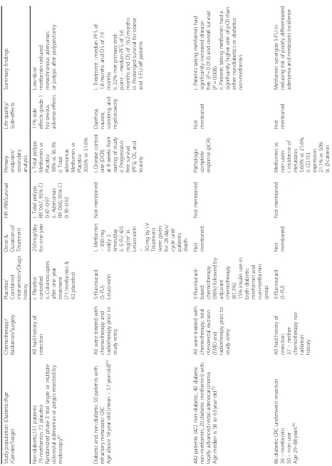
Table 3 The summarization of metformin clinical use for CRC Study population (Di abetic/Age /Gend er/Stag e) Chem otherapy / Rad iation/Surgery Pla cebo/ Comb ined interve ntion/ Drugs hist ory Dose & Duration of Treatmen t HR /RR/ Survival Pri mary end point/ se condary analy sis Life qua lity/ Side-eff ects Summary findings Non-d iabetic ,151 patients 79 metform in, 72 place bo Rand omized phase 3 trial single or mult iple colo rectal ade nomas or po lyps resect ed by end oscopy 87 All had history of res ection i. Pla cebo cont rolled ii. Colono scopies afte r one year treat ment (71 metform in & 62 pla cebo) 250 mg /day for one year i. Tot al polyps RR 0·67, 95% CI 0·47 – 0·97 ii. Aden omas RR 0·60, 95% CI 0·39 – 0·92 i. Total polyps Met formin vs Pla cebo 38% vs 56.5% ii. Total ade nomas Met formin vs Pla cebo 30.6 % vs 51.6% 11% side effect s grad e 1 No se rious adverse eff ects Low-d ose metform in reduced meta chronou s ade nomas or polyps afte r polype ctomy Diab etic an d non-diabetic 50 patie nts with refract ory meta static CRC Age above 18 year-ol d (me an – 57 year-o ld) 92 All we re treat ed with che mothera py and radi otherapy prior to study entry 5-flu orouracil (5-F U) Leu covorin i. Me tformin – 850 mg orally 2
times/day ii.5-FU
425
mg/m
2iii.
Leucovorin – 50
Table 3 The summarization of metformin clinical use for CRC (Co ntinued) Study population (Di abetic/Age /Gend er/Stag e) Chem otherapy / Rad iation/Surgery Pla cebo/ Comb ined interve ntion/ Drugs hist ory Dose & Duration of Treatmen t HR /RR/ Survival Pri mary end point/ se condary analy sis Life qua lity/ Side-eff ects Summary findings 36.8 5 vs 72.2 % iv. Poorl y differ entiat ed ade noma 2.78 % vs 16.0% 240 dia betic without CRC histo ry Age, me tform in 58.8 ± 9. 9 non user 61.6 ± 10.3 90 No i. Asp irin ii. NSAI Ds iii. Sulfon ylurea Not mentio ned Metform in -Adva nced ade noma RR 0.071 Colore ctal ade nocarcinoma – size, occurrence , num ber Not menti oned Metform in lowe r the risk of advanced CRC Adeno mas in new ly dia gnosed patien ts 2088 cases (66 – 80 years) and 9060 cont rol (61 – 77 ye ars) Janu ary 2000 – Dece mber 2009 77 YES i. Asp irin ii. NSAI Ds iii. Sulfon ylurea Cumulative 2000 g (DDD) within 5 years i. Re duced risk of CRC OR 0.83, 95% CI 0.68 – 1.00 ii. Prot ective in women vs me n (OR 0. 66, 95% CI 0.49 – 0.90) vs (OR 0. 96, 95% CI 0.75 – 1.23) Not me ntioned Not menti oned Dose and durati on response -reduc ed risk of CRC metform in > 250 DDD and > 1 year Protect ive effect of long-te rm metform in against CRC in women 424 pat ients Janu ary 2004 – Dece mber 2008 108 YES i. Insu lin ii. ADDs iii. Anti-ch olest erol iv. Asp irin Dose not mentio ned, duration betwee n 2005 and 2008 i. Me tformin vs Non user − 76.9 vs 56.9 mon ths (CI 61.4 – 102. 4) vs CI 44.8 – 68.8) ii. 30% enhancemen t in overall sur vival (OS) HbA1 c level Not menti oned Metform in provi de d 30% improve ment in OS as compared to other ADDs 2066 postm enopa usal Wom en, 50 – 79 years 1854 non-diabetic 84 diabetic(+met formin) 128 dia betic( − metform in) 105 YES i. Insu lin ii. Aspiri n iii. NSAID s Median 4.1
years (3days
-14.4 years ) i. D iabetic Metform in vs Non user -Non-significan t (HR 0.78 , 95% CI 0.38 – 1.55) ii. overa ll survival (HR 0.86 , 95% CI 0.49 – 1.52) i. Tumor size ii. Positive lymph nod es Not menti oned Non-s ignificant differ ence specific sur vival in me tform in compared to non-users 315 pat ients with stage I – III colorect al cance r from 2001 to 20 06 77 YES i. 52% -Met formin + sul fonylurea ii. 72% -no n me tformin + sulf onyl urea iii. Insulin iv. Othe r AD Ds Low an d high intens ity Duration -none Low inten sity HR 0.81, 95% CI 0.41 – 1.58 High intens ity H R 0.44, 95% CI 0.20 – 0.95 Tumor grad e/
Size -Nons
Table 3 The summarization of metformin clinical use for CRC (Co ntinued) Study population (Di abetic/Age /Gend er/Stag e) Chem otherapy / Rad iation/Surgery Pla cebo/ Comb ined interve ntion/ Drugs hist ory Dose & Duration of Treatmen t HR /RR/ Survival Pri mary end point/ se condary analy sis Life qua lity/ Side-eff ects Summary findings A) i. 856 pat ients with CRC from 2003 to 2005 ii. Age: men an d wom en < 40 years B) i. 814 patie nts with CRC from 2003 to 20 05 ii. Age: men an d wom en > 40 years C). D iabetes statu s: ≥ 1 year an d ≥ 3 years 76 YES None i. < 1 year ii. 1 – 3 years iii. ≥ 3 years < 40 years i. < 1 year -0.876 ii. 1 – 3 years
-0.859 iii.≥
3
years
-0.643 >40
years i. < 1 year -0.896 ii. 1 – 3 years
-0.843 iii.≥
3 years -0.646 none Redu ced inciden ce of COP D in metform in users compared to non-users Significantly lower risk CRC by 27 % Longe r use of me tform in inverse ly pro portional to CRC 3775 underwent col onosc opy (May 2001 -March 2013) 912 with me tform in 2193 non users Age > 40 years 88 Colon oscopy Not me ntioned i. < 1 year ii. 1 – 2 years iii. 2 – 3 years iv. ≥ 3 years Not mentio ned i. Colore ctal
polyp -metform
in vs non users (39.4 % vs 62 .4%) ii. aden oma -metform in vs non users (15.2 % vs 20 .5%) iii. Advanced ade noma -metform in vs non users (12.2 % vs 22 %) Not menti oned Metform in is beneficial in prevention of CRC 106 pat ients with stage IV CRC 83 81che moth erapy 25 curat ive resect ion Not me ntioned Not mentio ned Metform in improve d free survival rate for curative group (HR0.024 ,95% CI0.001 – 0.435) i. tumor res ponse, ii. targ et le sion size Chem otherapy – non-sign ificant Lowe r recurrence inciden ce Metform in red uced tumor recurrence afte r curat ive resect ion 8046 patie nt 2682 case group 5364 contro l group (60% male & 40% female each group) Age mean 55 and 57 72 YES i. stati ns ii. insuli n iii. Sulfon ylurea iv. NSAI Ds v.Th iazolidined ione vi. Hea lth care adj ustment i. Inta ke mean and median duration − 218 day s and 240
days ii.Dail
Table 3 The summarization of metformin clinical use for CRC (Co ntinued) Study population (Di abetic/Age /Gend er/Stag e) Chem otherapy / Rad iation/Surgery Pla cebo/ Comb ined interve ntion/ Drugs hist ory Dose & Duration of Treatmen t HR /RR/ Survival Pri mary end point/ se condary analy sis Life qua lity/ Side-eff ects Summary findings 920 dia betic patients with CRC Age 70.2 ± 8.6 years 63.3% mal e, 36.7% fem ale 120 YES i. stati ns ii. insuli n iii. Sulfon ylurea iv. NSAI Ds Not mentio ned i. Exte nsiv e use – increased CRC risk, OR 1.43, 95% CI 1.08 – 1.90 ii. Significant increased ris k in men, OR 1.81, 95% CI: 1.25 – 2.62 Ko0l Not me ntioned Not menti oned Exten sive me tformin inta ke increased risk of CRC i. 675 with Type 2 DM from 1999 to 2009 ii. Age: 5 pat ients < 50, 269 patien ts be tween 50 and 69 an d 401 patients above 70 iii. 437 males and 238 fem ales iv. Stage I – IV of CRC 119 Wi thin the first 6 mont hs of expo sure i. 6 mont hs wit hin CRC dia gnosi s -88 .7% surge ry 28 % che mothera py 15 % radiothe rapy ii.63. 1% sulf onyl ureas iii. 23.1% insu lin Metform in used afte r
CRC diagnosis; Follow
up – i. 6 mont hs
after diagnosis until
death ii. within 5 years up to 14 years i. H R 1.06, 95 % CI
0.80, 1.40 ii.
i. cance r-specif ic mort ality ii. HbA1C levels Lesser inciden ce of cong estive heart disea se, myoc ardial infarc tion and
peripheral vascular disea
In the indirect mechanisms, metformin exerts its an-ticancer effect through the insulin/insulin-like growth factor-1 (IGF-1) pathway. In normal cells, the recep-tors for IGFs and insulin are widely expressed and can be phosphorylated following binding to its ligand which lead to the concomitant activation of down-stream pathways such as PI3K/Akt/mTOR and RAS/ RAF/mitogen-activated protein kinase (MAPK) path-ways. The activation of these pathways via circulated insulin stimulates IGF-1/IGF-1R activation that pro-motes the initial tumor proliferation and growth. How-ever, as an antidiabetic drug, metformin can promote the phosphorylation of IGF-1R that inhibits IGF-1 sig-nalling which increases peripheral insulin sensitivity
and muscle uptake of glucose while reducing plasma insulin levels and hepatic glucose output. As a result, the activation of IGF-1/IGF-1R is further inhibited leading to the indirect anti-proliferative effect of the cancer cell. For example, Cho et al. [37] demonstrated that the combination of metformin and CB-PIC enhances phosphorylation of ACC, AMPK and pERK which sup-presses mTOR and Akt activation in hypoxic SW620 cells. More importantly, the direct and indirect antican-cer mechanisms of metformin are similar, as they both modulate mTOR as a common signaling target. These signaling pathways modulated by metformin are summa-rized in Fig.1in relation with other upstream and down-stream mediators.
Fig. 1The anticancer molecular mechanisms mediated by metformin through the modulation of AMPK and cellular energy homeostasis. Metformin mainly modulates AMPK activation through LKB1 which activates and/or inactivates various downstream signalling targets such as
mTOR, PTEN/PI3K-Akt, MAPKs, transcription factors (NF-κB, FOXO) and p53. The activation of these signalling pathways induce oxidative stress,
Metformin induces apoptosis and autophagy through oxidative stress, inflammation, and metabolic homeostasis via AMPK and mTOR
The activation of AMPK by metformin is a cardinal step that modulates various transcription factors such as NF-κB and FOXO which regulates cellular apoptosis, oxidative stress, inflammation, and neoplastic malig-nancy. Metformin through its anti-inflammatory and anti-oxidant properties targets various cellular mecha-nisms responsible in the development of cancer that is associated with diabetes and obesity. Moreover, metfor-min enhances cellular apoptosis in CRC cells by modu-lating the production of anti- and pro-inflammatory mediators. Metformin inhibits IκBα degradation which suppresses expression of IL-8 and NF-κB activation in TNF-α-stimulated COLO 205 cells [59]. Moreover, metformin induces anti-inflammatory property that in-hibits DSS-induced IκB kinase activation and reduced colitic cancer development in IL-10−/− mice by aug-menting AMPK activation in the intestinal epithelial cells. In addition, co-administration of metformin and DMH in Balb/c female mice effectively reduces the for-mation of AC and ACF (58.3 and 47.4%, respectively) through the modulation of oxidative stress and inflam-mation [126]. Metformin also upregulates p53 and Nrf2 expression while inactivating NF-κB which induces cel-lular apoptosis and modulation of oxidative stress and inflammation. The observations are corroborated by the reduction of malondialdehyde (MDA), inhibition of iNOS expression that decreased NO and nitrotyrosine, suppression of IL-10 and elevation of IL-1β.
Saber et al. [40] demonstrated that metformin in combin-ation with 5-ASA suppresses the pro-inflammatory media-tors such as IL-1β, IL-6, COX-2 and TNF-α, TNF-R1 and TNF-R2 which inactivates of NF-κB and STAT3. These molecular events further decreases MMP-2 and -9 expres-sion and thus, suggests metformin capability to reduce the CRC cell proliferation, migration, and invasiveness. Further-more, the suppression of NF-κB activation enhances apop-tosis by reducing the Bcl-2 protein expression. Exposure to subtoxic concentration of metformin (2.5–10 mM) signifi-cantly potentiated the apoptosis inducing effect of tumor necrosis factor (TNF)-related apoptosis-inducing ligand (TRAIL) through Mcl-1 degradation in HCT116, HT29, DLD-1 and Colo25 cells [127]. Metformin in combination with TRAIL induced the dissociation of Noxa from Mcl-1 followed with an increased E3 ligase Mule activity that pro-moted polyubiquitination of Mcl-1 in the cancer cells. An-other study reported that treatment with metformin alone or in combination with silibinin induced the expression of p-AMPK which suppressed mTOR phosphorylation and induced the activation of PTEN that inactivated PI3K-Akt. Furthermore, modulation of both AMPK/mTOR and PTEN/PI3K-Akt pathways increase the expression of
cleaved caspase-3 and apoptosis inducing factor that pro-moted apoptosis in COLO 25 cells [38]. In a different study, the synergistic anticancer effects of metformin and vitamin D3 activated the AMPK(IGFI)/mTOR pathway that sup-pressed S6P expression and thus, inhibited the formation of early colon neoplasia rats and mice models [64]. It is re-ported that metformin significantly potentiates the vitamin D3 suppression of c-Myc and cyclin D1 mediated through via vitamin D receptor/β-catenin pathway.
Metformin regulates the energy and metabolic homeostasis by regulating the expression of key regulatory lipid enzymes that are associated in metabolic reprogramming of cancer cells through upstream kinase LKB1. Metformin through LKB1 activates AMPK which suppresses the expression of lipogenic transcription factor sterol regulatory element-binding protein-1 (SREBP-1) and its downstream targets such as fatty acid synthase (FAS) and 3-hydroxy-3-methyl glutaryl-CoA reductase [57,128, 129]. Since this process is essential in regulating the metabolic homeostasis and thus, it modulates the plasma concentrations of glucose, insulin, tri-glycerides, and cholesterol. Metformin suppresses the effect of high-energy diet in promoting the growth of tumor in xenografts mice model (MC38 colon carcinoma cells) by re-ducing the insulin level and FASN while inactivating the Akt protein. Additionally, metformin induces apoptosis via the cleavage of poly (ADP-ribose) polymerase (PARP) via AMPK activation, inactivation of acetyl-CoA carboxylase and upreg-ulation of BCL2/Adenovirus E1B 19 kDa Interacting Protein 3 (BNIP3) expression which ultimately suppressed tumor growth and volume [57]. Other than modulating survival and AMPK pathways, metformin also inhibits DMH-induced CRC in diabetic Sprague Dawley rats by reversing the Warburg effect [58] leading to suppression of ACF for-mation and reduction of PCNA expression, proliferation index of colonic tissues which decreases tumors volume. Metformin is also beneficial in treating inflammatory bowel disease (IBD) and the chronic or long-term IBD can induce the development of colitis-associated colon cancer (CAC). Jie et al. [58] suggested that metformin inhibition of the colon cancer cell and produced syner-gistic colon cancer-preventative effect in diabetic patients by modulating the expression of PKM2 and IDH1, two main isoenzymes involved in glycolysis and TCA cycles. The modulation of apoptosis in CRC models by metfor-min through oxidative stress, inflammation and metabolic homeostasis is further exemplified in Fig. 1 in relation with relevant signaling pathways.
Metformin modulates cell cycle and p53 regulation
Moreover, this alteration of cell cycle is also dependent on the status of p53 as a transcription factors that regu-lates cell cycle arrestment, DNA repair, programmed cell death, and senescence [130, 131]. The p53 modulates mTOR by direct modulation AMPK and TSC2 as well as through the regulation PTEN transcription and activa-tion of IGF-1/AKT pathways [132–134]. Cancer cells with a mutated p53 gene that are treated metformin are unable to reprogram their metabolism and therefore, rendered to undergo apoptosis. Metformin can induce cell cycle arrestment following the activation of LKB1/ AMPK that activates p53 and inhibits mTOR. This acti-vation of p53 is regulated by the suppression of cyclin D1 and expression of cyclin- dependent kinase inhibitors p27Kip1 and p21Cip1 [135]. For instance, metformin in-duces arrestment of cell cycle at G0/G1phase via the in-hibition of cyclin D1 expression and telomerase activity [32]. The activation of p53 induces the transcription of p21 which increases the expression of apoptotic genes leading to DNA-damage and fragmentation as well as G0/G1 arrestment. Additionally, metformin in combin-ation with other chemotherapy drug can suppress cancer cell proliferation by regulating cell cycle differently. For example, Zhang et al. [45] demonstrated that pretreat-ment with metformin followed by 5-FU inhibited the proliferation of the SW620 cells by reducing the S phase population without altering the G0/G1 or G2/M phase. Furthermore, metformin can radiosensitize p53-deficient HCT116 cells by arresting the G2/M phase via suppres-sion the DNA repair proteins such as MRE11, BRCA2, Rad51, and ERCC1 [48].
Metformin also inhibits CRC cells proliferation by regulating the expression of microRNAs that further modulate various signaling pathways. Feng et al. [47] demonstrated that suppression of HCT-116 cells pro-liferation and migration by metformin and 5-FU can be potentiated by knocking down miR-21 expression which in turn increased the Sprouty2, tumor suppres-sor expression and PTEN. In a different study, treatment with metformin induced microRNA-34a to inactivate the Sirt1/Pgc-1α/Nrf2 pathway leading to increased suscepti-bility of wild-type p53 cancer cells towards oxidative stress and therapeutic agent in HCT116 cells [136]. Sirtuin 1 (Sirt1), an oncogenic protein promotes resistance against oxidative stress and modulates apoptosis through the deacetylation of its targets such as p53 and FOXO1. The latter can induces a positive-feedback loop through miR-34a that enhances the Sirt1 expression. Sirt1 is found to be overexpressed in human breast, colon, non-small-cell lung, and prostate cancer cells. Sirt1 has been suggested to induce an oncogenic effect in cells expressing wild-type p53 but a tumor-suppressive effect in mutated p53 cells. Although the report by Do et al. found that metformin enhanced apoptosis in the
wild-type p53 HCT116 cells by increasing the p53 expres-sion and miR-34a which downregulates Sirt1 expresexpres-sion and its subsequent downstream effectors, the role of Sirt1 in cancer particularly CRC is still debatable and requires further validation.
Conclusions and future perspectives
The current review depicts the beneficial use of metfor-min from preclinical, epidemiologic, and clinical studies as potential chemotherapeutic and adjuvant agent for CRC with notable association with T2DM. Furthermore, the long history and clinical experience of metformin against various cancer cases simply rebranding it as a potential old drug to be repurposed as cheap and effect-ive chemotherapeutic drug. Metformin use as a chemo-therapeutic agent for CRC also varies but transcendent among gender, age, patients with or without CRC history or resurrection and treatment regimens as sole agent or adjuvant to existing chemotherapeutic drugs. The appli-cation of metformin for various cancer treatment par-ticularly CRC requires further evaluation whether it is effective in preventing the CRC recurrence.