R E S E A R C H
Open Access
Effects of ectopic HER-2/neu gene expression on
the COX-2/PGE
2
/P450arom signaling pathway in
endometrial carcinoma cells: HER-2/neu gene
expression in endometrial carcinoma cells
Shu Li, XiaoXin Ma
*, Li Ma, Cuicui Wang, YuanQi He and ZhiJuan Yu
Abstract
Objectives:To investigate the role of HER-2/neu-mediated COX-2/P450arom signal in estrogen-dependent endometrial carcinoma.
Methods:The recombinant eukaryotic expression vector, pcDNA3.1-HER-2/neu, was constructed and transfect to Ishikawa endometrial carcinoma cells. The expression of COX-2 and P450arom in transfected cells were detected by real-time PCR and western blotting. The levels of estrogen in cell supernatants were detected by ELISA.
Results:Over-expression of HER-2/neu in transfected cells was confirmed by real-time PCR and western blotting. The levels of autocrine estrogen in transfected cells was significantly increased which combination with the enhancement of COX-2 and P450arom expression in transfected cells.
Conclusion:HER-2/neu induced the improvement of autocrine estrogen in endometrial carcinoma cell through triggering the COX-2/P450arom signal.
Keywords:HER-2/neu, COX-2, P450arom, Estradiol, Endometrial carcinoma cell
Introduction
Endometrial carcinoma is a common gynecologic malig-nancy with uncharacterized molecular mechanisms of pathogenesis. A large body of studies has reported that the origin of endometrial carcinoma was associated with long-term estrogen stimulation without counteraction [1]. Long-term stimulation of estrogen can cause endo-metrial hyperplasia, even atypical hyperplasia, and can progress to carcinogenesis. Local synthesis of estrogen may also lead to endometrial carcinoma. A better under-standing of the mechanisms of local estrogen synthesis is important to find the new treatment of endometrial carcinoma.
Aromatase (P450arom), a key enzyme for the bio-synthesis of estrogens, was detectably active and over-expressed in endometrial carcinoma, whereas it was undetectable in normal endometria. P450arom is the
rate-limiting enzyme that catalyzes the final step in the conversion pathway from androgen to estrogen. The quantity and activity of P450arom can directly affect the levels of estrogen in normal or abnormal tissues, in order to maintain estrogen-related physiologic func-tions in normal tissues. Meanwhile, P450arom play a role in the pathogenesis and prognosis of estrogen-dependent diseases. The activity of P450arom is
regu-lated by prostaglandin E2 (PGE2), which is affected by
cyclooxygenase-2 (2). We hypothesize that
COX-2/PGE2/P450arom might be a signaling pathway in
estrogen-dependent diseases to regulate the autocrine activity of estrogen in cancerous tissues. Previous reports indicated that HER-2/neu regulated the expres-sion of COX-2 as the upstream molecular of COX-2-mediated signal pathways [2,3]. In the present paper, our results demonstrated that transfection with HER-2/neu in endometrial cells induced the activation of COX-2/PGE2/ P450arom signal, resulting in the increase of autocrine estrogen from endometrial cells.
* Correspondence:maxiaoxin666@yahoo.com.cn
Department of Gynecology and Obstetrics, Shengjing Hospital of China Medical University, Shenyang 110004, China
Materials and methods Cell culture
The Ishikawa cell line was kindly supplied by the Depart-ment of Pathophysiology, Beijing University. Cells were cul-tured in RIPM1640 with 10% fetal bovine serum, 100 U/ml
penicillin, and 100 μg/ml streptomycin in an incubator
maintained at 37°C and 5% CO2. Celecoxib, a selective
COX-2 inhibitor, was purchased from Santa Cruz Biotech-nology and dissolved in DMSO to generate a 100 mM stock solution that was stored at−20°C. For inhibition
ex-periment, confluence cells were starved by serum
deprivation overnight. Then, cells were treated with 80μM celecoxib and incubated for 48 h.
Construction of pcDNA3.1-HER2/neu
Upstream (50-TGGGAGCCTGGCATTTCTG-30) and
downstream (50-TCCGGCC ATGCTGAGATGTA-30)
primers were designed based on HER-2/neu cDNA
se-quence obtained from GenBank. For cloning, HindIII/ XbaI restriction endonuclease sites were inserted flanking the target gene primers. Primers were synthesized by TaKaRa Biotechnology Co., Ltd. Total RNA was isolated from Ishikawa cells using TRIzol reagent (TaKaRa, China) according to the manufacturer’s instructions. HER-2/neu cDNA was reverse-transcribed using the One Step RNA PCR Kit (TaKaRa) according to the manufacturer’s recom-mendations. PCR conditions included denaturation at 94°C for 5 min, 25 cycles of denaturation at 94°C for 45 s, annealing at 60°C for 1 min, and extension at 72°C for 6 min, with a final extension at 72°C for 10 min. PCR products were separated on 1% agarose gel and eluted. The PCR product was sent to TaKaRa for sequencing.
PcDNA3.1 plasmid and HER2cDNA were digested with
HindIII/XbaI double endonucleases. The digested pro-ducts were separated by agarose gel electrophoresis and
purified. PureHER2cDNA and vector were mixed at a 4:1
ratio and were ligated at 16°C for 20 h. Ligation products were transformed intoE. coli. After culturing at 37°C over-night,E. colicells were screened. DNA was isolated from positive colonies (named pcDNA3.1 (+)-HER2), and were sequence-verified.
Gene transfection and G418 screening
pcDNA3.1 (+)-HER2 was isolated using a plasmid extrac-tion kit (Invitrogen, USA) according to the manufacturer’s protocol. Ishikawa cells in logarithmic growth were trans-ferred to 6-well culture plates at 1 × 10⁶ cells/well and were cultured for 1 d prior to transfection. Ishikawa cells then were transfected with pcDNA3.1-HER-2/neu or pcDNA3.1 (control) using Lipofectamine2000 (Invitrogen,
USA) according to the manufacturer’s protocol.
Non-transfected cells were cultured as the negative control. The original culture media was discarded 3 d after trans-fection, and cells were cultured in complete media
containing 10% fetal bovine serum and 700μg/ml of G418
(Invitrogen, USA). G418-resistant clones were selected and transferred to culture media containing 350μg/ml of G418 for scale-up culture.
RNA interference
To knock down the HER2/neu in Ishikawa cells, the siRNA transient transfection experiment was performed according to the previous publication [4]. Briefly, Ishikawa cells were transfected with 25 nM COX-2 siRNAs, respectively.
Non-targeting siRNA was used as negative control. Transfections were carried out according to the guidelines
for the DharmaFECTW siRNA Transfection Reagents
(Dharmacon). Ishikawa cells were collected at 72 hours post-siRNA addition for protein western blotting analysis.
Real-time RT-PCR analysis ofHER-2/neu
Total RNA was extracted from Ishikawa cells stably transfected with pcDNA3.1 (+)-HER2 using TRIzol, as
above. The same HER-2/neuprimers were used as in the
construction of pcDNA3.1 (+)-HER2. GAPDH was used as
the internal control (upstream 50-CATCCATGACAACT
TTGGTATC-30; downstream 50-CCATCACGCCACAGTT
TC-30). cDNA was synthesized from 1 μg of total RNA
using oligo(dT) primers in the presence of reverse tran-scriptase. Gene amplification was performed on a real-time
PCR instrument (TaKaRa, China) using 1 μl of cDNA
as template in a 25μl volume. PCR was started at 95°C for 5 min, followed by 30 cycles of denaturation at 95°C for 10 s, annealing at 59°C for 15 s, and extension at 72°C for 20 s, with a final extension at 72°C for 10 min. Fluores-cence intensity was monitored and recorded in real time. A melting curve analysis was performed after amplification
was complete. The ΔΔCt value was used to evaluate
ex-pression levels of HER2 and COX-2 mRNA. By this
method, a higher expression level is related to a lower
ΔΔCt value.
Western blotting for HER-2/neu, COX-2, and P450arom
Cells collected from non-transfected, pcDNA3.1-transfec-ted, and pcDNA3.1-HER2-transfected groups were lysed with 250 μl protein extracting fluid (RIPA lysis buffer: 50 mM Tris [pH 7.4], 150 mM NaCl, 1% Triton X-100, 1% sodium deoxycholate, 0.1% SDS), homogenized for 10 min, incubated in an ice-bath for 1 h, and centrifuged
at 12,000 × g for 30 min at 4°C. Supernatants were
col-lected, and protein concentrations were determined using the BCA protein assay system (Pierce, USA). Proteins were separated by 12% SDS-PAGE and were transferred to PVDF membranes. After blocking overnight at 4°C in 1 × PBS, 0.1% Tween 20, and 5% non-fat milk, membranes were incubated with anti-HER-2/neu (1:800), COX-2 (1:400), P450arom (1:400) and β-actin (1:800) polyclonal antibodies (Santa Cruz Biotechnology, USA) for 3 h at room temperature. Membranes were washed twice and in-cubated with horseradish peroxidase-conjugated goat anti-rabbit secondary antibody (ZhongShan, China, 1:1,500) for 2 h at room temperature. Immunodetection was performed by chemiluminescence (ECL reagent, Beyotime, China) and membranes were exposed to film. Images were captured using a transmission scanner. For quantification, target pro-teins were normalized toβ-actin (the internal standard) by comparing the gray-scale values of proteins to
correspond-ing β-actin values. Quantification was performed using
UVP Gelworks ID Advanced v2.5 software (Bio-Rad, USA).
ELISA for PGE2and E2detection
Supernatants were collected from non-transfected,
pcDNA3.1-transfected, and pcDNA3.1-HER2-transfected Table 1 HER-2/neu mRNA expression
Group ΔCt -ΔΔCt 2-ΔΔCt
HER-2 transfected 97.16 ± 0.71 2.62 ± 0.71 6.15 (3.75–10.06)*
pcDNA3.1 transfected 9.88 ± 1.10 0.1 ± 0.10 1.07 (1.06–1.08)
Non-transfected 9.78 ± 1.09 0 ± 1.09 1 (0.47-2.13)
*P< 0.05.
groups for ELISA. Supernatant PGE2and E2 concentra-tions were measured using an ELISA kit (R&D Systems,
Minneapolis, MN, USA) according to the manufacturer’s
instructions. Each sample was examined in triple and averaged for data analysis.
Statistical methods
SPSS v10.0 software was used for all statistical analyses. Data were expressed as mean ± standard error of the mean (SEM). One-factor analysis of variance was used for pairwise comparison. Statistical significance was defined asP< 0.05.
Results
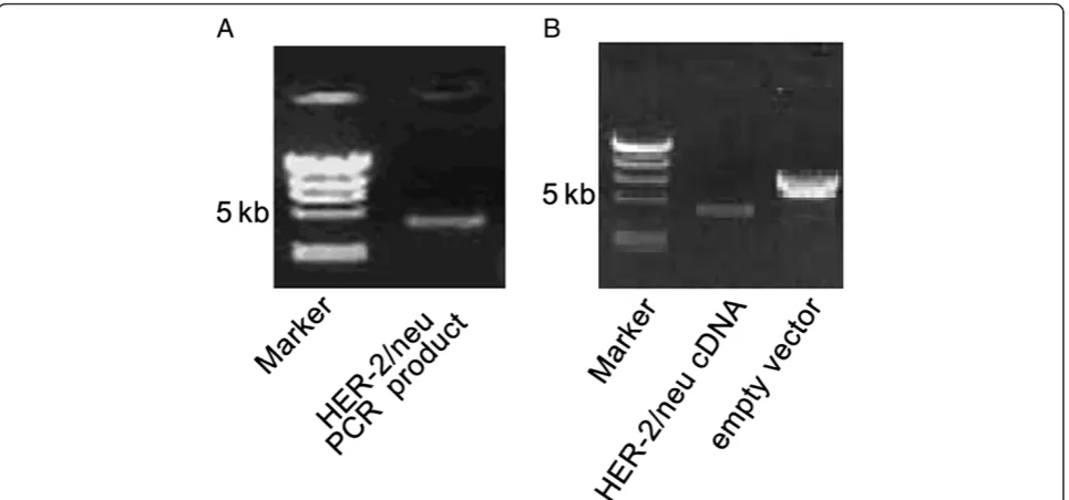
Construction of pcDNA3.1-HER2
RT-PCR ofHER-2/neuyielded a specific band of
approxi-mately 4.4 kb (Figure 1A). The DNA fragment sizes from
HER-2/neu cDNA and pcDNA3.1 plasmid digested with HindIII and XbaI were as predicted from the sequence (Figure 1B). DNA sequencing confirmed the absence of point or frameshift mutations in HER-2/neu cDNA.
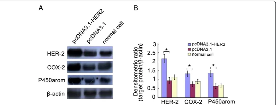
Expression of HER-2/neu in Ishikawa cells stably transfected with pcDNA3.1-HER2
Real-time RT-PCR demonstrated significantly higher HER-2/neu mRNA expression in pcDNA3.1-HER2-transfected cells compared with empty plasmid-transfected or
non-transfected cells (Table 1). Western blotting indicated a sig-nificant increase in HER-2/neu protein levels of cells transfected with pcDNA3.1-HER2 compared with empty plasmid-transfected or non-transfected cells (Figure 2). These results imply that the transfection was effective, and that the cells were appropriate for subsequent analyses.
Transfected with pcDNA3.1-HER2 in Ishikawa cells induced the increase of COX-2, PGE2and P450arom
expression
Western blotting demonstrated that levels of COX-2 and P450arom in Ishikawa cells stably transfected with pcDNA3.1-HER2 were significantly higher compared to those in empty plasmid-transfected or non-transfected cells (Figure 2). In additionally, ELISA analysis showed
that the supernatant level of PEG2in
pcDNA3.1-HER2-transfected group was significant higher than that of the empty plasmid-transfected group, and the normal cell group.
Transfected with pcDNA3.1-HER2 induced the increase of autocrine E2from Ishikawa cells
ELISA indicated was there were statistically significant dif-ferences in the cell supernatants of E2 levels among the pcDNA3.1-HER2-transfected group, the empty plasmid-transfected group, and the normal cell group (Table 2).
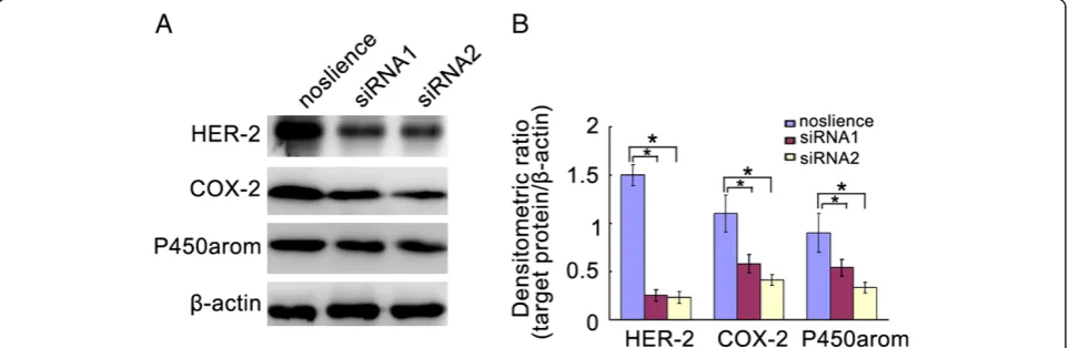
Inhibition of HER2 in Ishikawa cells induced the decrease of COX-2 and P450arom expression
RNA interference technology was used for the down-regulation of HER2 expression in Ishikawa cells. As shown in Figure 3, HER2 siRNAs were effectively able to knockdown the levels of HER2 in Ishikawa cells. Inter-estingly, down-regulation of HER2 expression induced significantly the reduction of COX-2 and P450arom levels in Ishikawa cells (Figure 3).
Figure 3The levels of COX-2, and P450armo in the ishikawa cells transfencted with HER2 siRNA. A. Represent image for western blot.B. Analysis of protein levels in each group and quantification of band density was done using Image J. *P< 0.05.
Table 2 ELISA analyses for PGE2and E2in the
supernatants of endometrial carcinoma cells
Group PGE2(pg/ml) E2(pg/ml)
Transfected 41.69 ± 0.87* 31.49 ± 2.14* pcDNA3.1 transfected 31.35 ± 1.06 21.16 ± 2.37 Non-transfected 27.67 ± 1.20 20.56 ± 3.27
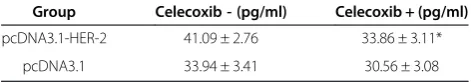
Inhibition of COX-2 in the over-expressed HER2 Ishikawa cells led to the decrease of PGE2 and P450arom
expression
To further investigate the relationship between the COX-2/
PGE2/P450arom signal and HER2, celecoxib, a selective
COX-2 inhibitor, was used for inhibition experiment. The results showed that inhibition of COX-2 in the over-expressed HER2 Ishikawa cells led to the obvious decrease of PGE2 and P450arom expression (Figure 4; Table 3).
Discussion
An important member of the epidermal growth factor re-ceptor (EGFR) family, the proto-oncogene HER-2/neu en-codes a 185-kD transmembrane glycoprotein with tyrosine kinase activity [5]. HER-2/neu over-expression typically oc-curs in the placenta, embryonic epithelial tissue, and several types of tumor cells. In contrast, HER-2/neu is absent or minimally expressed in normal tissues [6]. The positive ex-pression rate of the HER-2/neu protein in endometrial car-cinoma is associated with clinical staging, a lower degree of tissue differentiation, and lymph node metastasis [7]. We have applied RT-PCR and ELISA to detect the expression of HER-2/neu, COX-2, p450arom and PGE2 in normal endometrium, hyperplasia endometrium and endometrial carcinoma respectively. The results showed that the expres-sion of HER-2/neu was significantly correlated with patho-logic grading, FIGO staging, and lymph node metastasis. But it has no correlation with menopausal status [8]. There are some studies also shows that the HER-2/neu gene con-tributes to the progression of carcinomas and tumor resist-ance to chemotherapy [9-11]. A better characterization of this proto-oncogene can lend insight to the pathogenesis and molecular mechanisms involved in the development of endometrial carcinoma.
We have preciously made nude mice transplanted with Ishikawa cells, which were stably transfected with HER2/ neu plasmid and empty plasmid,respectively. The tumor volume and weight were measured.It showed that the tumor formation rate and tumor size in HER2/neu
plasmid transfection group were significantly higher than those of the control group, which suggested that HER2 could promoted the growth of Ishikawa cells. In the present study, we confirmed that HER-2/neu mRNA and protein levels were significantly elevated in cells stably transfected with pcDNA3.1-HER2/neu compared with non-transfected cells or those transfected with empty vec-tor. Using these cells, we identified the significant in-creases in the levels of COX-2 and P450arom. In addition, the E2 concentration was also significantly increased in cells stably transfected with pcDNA3.1-HER2/neu com-pared with non-transfected or empty vector-transfected groups. As an alternative approach, RNA interference technology was used for the down-regulation of HER2 ex-pression in Ishikawa cells. The results showed that inhib-ition of HER2 in Ishikawa cells significantly induced the decrease of COX-2 and P450arom expression. Mean-while, celecoxib, a selective COX-2 inhibitor, inhibited the expression of PGE2 and P450arom in the over-expressed HER2 Ishikawa cells. These results indicated that HER-2/neu induced the upregulation of COX-2, PGE2 and P450arom to promote the autocrine of E2 in endometrial carcinoma cells.
As a transmembrane glycoprotein, the cell membrane portion of HER-2/neu is the primary contributor to trans-duction of cell proliferation signals [12,13]. The tyrosine kinase activity of HER-2/neu is essential for COX-2 tran-scriptional activation [14] and regulates the expression of COX-2 via multiple pathways [15]. Over-expression of COX-2, which was detected in endometrial carcinoma, stimulated the proliferation and angiogenesis of cancer cell
Table 3 ELISA analysis for PGE2in the supernatants of
tranfected endometrial carcinoma cells treated with Celecoxib
Group Celecoxib - (pg/ml) Celecoxib + (pg/ml)
pcDNA3.1-HER-2 41.09 ± 2.76 33.86 ± 3.11*
pcDNA3.1 33.94 ± 3.41 30.56 ± 3.08
*P< 0.05.
[16]. COX-2 also is an important rate-limiting enzyme in prostaglandin synthesis [13]. The endometrial
prostaglan-din E2 induced the activity of aromatase (P450arom) by
up-regulating intracellular cAMP levels in endometrial stromal cells. COX-2 indirectly regulated the expression of
P450arom by influencing the synthesis of PGE2 [17].
P450arom is the rate-limiting enzyme catalyzing the final step in the conversion from androgen to estrogen. P450arom determined the levels of estrogen in normal and abnormal tissues directly, which maintained the estrogen-related physiologic functions and impacted the pathogenesis and prognosis of estrogen-dependent dis-eases [18]. High levels of HER-2/neu have been detected in endometrial carcinoma tissues and were found to cor-relate with tumor malignancy [19-21]. Our results sug-gested that HER-2/neu, as a potential upstream regulatory
molecule in the COX-2/PGE2/P450arom signaling
path-way, could play a critical role in estrogen-dependent endo-metrial carcinoma. These findings provided an improved understanding of the molecular mechanisms of estrogen-dependent endometrial carcinoma, and might instruct to screen the targets for hormone-dependent gynecologic tumors related to HER-2/neu.
Competing interest
The authors declare that they have no competing interests.
Authors’contributions
XXM and SL, conception, experimental design and performance, data analysis and interpretation, manuscript writing; CW conception and design, data analysis and interpretation; LM, YQH and ZJY performed research; SL conception and design, financial support, provision of study material, final approval of manuscript. All the authors read and approved the final manuscript.
Acknowledgement
This study was supported by grants from the National Natural Science Foundation of China (No. 81272874), the Project from Educational Department of Liaoning Province (No. L2010642), and the Science and Technology Project of Shenyang City (No. F10-205-1-58).
Received: 7 December 2012 Accepted: 23 February 2013 Published: 2 March 2013
References
1. Le J:Obstetrics and Gynecology [M].6th edition. China: Beijing: Beijing People’s Medical Publishing House; 2005:300.
2. Simeone AM, Li YJ, Broemeling LD,et al:Cyclooxygenase-2 is essential for HER2/neu tosuppress N-(4-hydroxyphenyl) retinamide apoptotic effects in breast cancer cells.Cancer Res2004,64(4):1224–1228.
3. Wang SC, Lien HC, Xia W,et al:Binding at and transactivation of the COX-2 promoter by nuclear tyrosine kinase receptor ErbB-COX-2.Cancer Cell2004,
6(3):251–261.
4. Faltus T, Yuan J, Zimmer, Krämer A, Loibl S, Kaufmann M, Strebhardt K:
Silencing of the HER2/neu gene by siRNA inhibits proliferation and induces apoptosis in HER2/neu-overexpressing breast cancer cells.
Neoplasia2004,6(6):786–795.
5. Tiseo M, Loprevite M, Ardizzoni A,et al:Epidermalgrowth factor receptor inhibitors: a new prospective in the treatment of lung cancer.
CurrMed Chem Anti-Canc Agents2004,4(2):139–148.
6. Kokay Y, Cohen JA,et al:Stage-and tissue-specific expression of neu oncogene in rat development.Proc Natl Acad Sci USA1987,84:8498.
7. Xiao-xin M, Qing M:Expression of protein c-erbb-2 in endometrial diseases and clinical significance testis neoplasms.China Journal of Modern Medicine2006,16(4):550–552.
8. Yuan-qi HE, Xiao-xin MA, Shu LI:Effects of HER2 on COX-2/PGE2 /P450arom Signal Pathway in Nude Mice Endometial Model.The Journal of China Medical University2010,39(10):823–826.
9. Anastasi S, SALA G, Huiping C,et al:Loss of R ALT/M IG -6expression in ER BB2-amplified breastcarcinom as enhances ErbB-2 oncogenic potency and favors resistance to Herceptin.Oncogene2005,24:4540–4548. 10. Jager R, Friedrichs N, Heim I,et al: Dual role of A P-2 gamma in ErbB-2
-induced mammary tumorigenesis.Breast Cancer Res Treat2005,90(3):273–280. 11. Goncalves A, BRA DAC, Vire F,et al:High-dose alkylating agents with
autologous hem atopoietic stem cell support and trastuzumab in ERBB2 overexpressing metastatic breast cancer: afeasibility study.A nticancer Res2005,25(1B):663–667.
12. Essapen S, Thomas H, Green M,et al:The expression and prognostic significance of HER-2 in colorectal cancer and its relationship with clinicopathological parameters.Int J On-col2004,24(2):241–248. 13. Half E, Broaddus R, Danenberg KD,et al:HER−2 receptor expression,
localization, and activation in colorectal cancer cell lines and human tumors.Int J Cancer2004,108(4):540–548.
14. Ratna V, Mahitosh M, Liana A,et al:Regulation of cyclooxygenase-2 pathway by HER-2 receptor.Oncogene1999,18(2):305–314.
15. Wang KH, Kao AP, Chang CC,et al:Increasing CD44+/CD24(-) tumor stem cells, and upregulation of COX-2 and HDAC6, as major functions of HER2 in breast tumorigenesis.Mole Cancer2010,9:288.
16. Ohno S, Ohno Y, Suzuki N,et al:Multiple roles of cy-clooxygenase-2 inendometrial cancer.Anticancer Res2005,25(6A):3679–3687. 17. Milczarek R, Klimek J:Aromatase–key enzyme of estrogen biosynthesis.
Postepy Biochem2005,51(4):430–439.
18. Zeitoun KM, Takayama K, Michael MD,et al:Stimulation of aromatase P450 promoter (II)activity in endometriosis and its inhibition in endometrium are regulated by competitive binding of SF-1 and COUP-TF to the same cis-acting element.Mol Endocrinol1999,13:239–253.
19. Chan SK, Hill ME, Gullick WJ: The role of the epidermal growth factor receptor in breast cancer.J Mammary Gland Biol Neoplasia2006,11(1):3–11. 20. Livasy CA, Reading FC, Moore DT,et al:EGFR expression and HER2/neu
overexpression/amplification in endometrial carcinosarcoma.
Gynecol Oncol2006,100(1):101–106.
21. Ejskjaer K, Sorensen BS, Poulsen SS,et al:Expression of the epidermal growth factor system in endometrioid endometrial cancer.Gynecol Oncol 2007,104(1):158–167.
doi:10.1186/1756-9966-32-11
Cite this article as:Liet al.:Effects of ectopic HER-2/neu gene expression on the COX-2/PGE2/P450arom signaling pathway in endometrial carcinoma cells: HER-2/neu gene expression in endometrial carcinoma cells.Journal of Experimental & Clinical Cancer Research2013 32:11.
Submit your next manuscript to BioMed Central and take full advantage of:
• Convenient online submission
• Thorough peer review
• No space constraints or color figure charges
• Immediate publication on acceptance
• Inclusion in PubMed, CAS, Scopus and Google Scholar
• Research which is freely available for redistribution