Biolinguistics 6.3–4: 383–392, 2012 ISSN 1450–341 http://www.biolinguistics.eu
Harry J. Jerison
This report is based on 3D digital scans of endocasts of 110 species of fossil mammals and 35 species of living mammals. It presents direct evidence of the last 60 million years of brain evolution. Endocasts are casts of the cranial cavity. They are brainlike in size and shape, and their surface features can be named as if they were brain structures. Although endocast data are restricted to outer surfaces of brains, a few inferences about inner structure are possible. Neocortex in the forebrain, for example, is identifiable and measurable as cerebral forebrain on the endocast dorsal to the rhinal fissure. An important result in this report is that surface area of neocortex as identified on endocasts appears to have reached a maximum of about 80% of the total endocast surface area in anthropoid primates including humans. This may be a fundamental limitation in brain size. The average neocorticalization percentage for mammals as a whole rose from about 20% to about 50% of the surface area during the 60 million years covered by this analysis. Neocorticalization is associated with the evolution of higher mental processes, including the evolution of language as a hominin specialization. The limitation of the increase in relative amount of neocortex is similar in all anthropoids. Neocortex is greater in absolute area in living humans because the total size of the hominin brain is so much larger than in other primates.
Keywords: cerebral cortex; encephalization; neuroimaging; primates
1. Introduction
The measurements of surface area, as I will review them, show that progressive neocorticalization occurred in mammalian evolution. It would be fruitful if comparative neuroanatomists would routinely add information on the outer surface areas to reports on micro and molecular anatomy of brains they study to improve the correlation between fossil data on endocasts with their data on living brains.
Endocasts are not brains. They are usually rock, plaster, or latex casts molded by the cranial cavity. In birds and mammals they are remarkably brainlike in size and shape. The literature on the evolution of the brain is usually on the comparative neuroanatomy of living brains with living species arranged as if they represent a phylogenetic tree. Fossil endocasts represent a more truly evolutionary pattern of brain evolution, which can help anchor the data of comparative neuroanatomy. Endocasts, however, are limited to data on external surfaces. In this report I present measurements of external surface area in endocasts in the centimeter-gram-second (cgs) system and present an illustrative example of the use of such measurements to analyze the evolution of neocorticalization.
Endocasts are not always perfect pictures of brains, but the natural endocast in Figure 1 shows just how good an image they provide. That endocast is of the 37 Ma (million years ago) artiodactyl Bathygenys reevesi, one of the many fossil nonprimate brains in my sample. My conclusions are based on measurements of endocasts of many species of mammals including those of primates. This report is on the evolution of neocortex in mammals during the past 60 million years. Cerebral cortex is a uniquely mammalian trait, and Bathygenys is a fine example of the identification of that trait in a fossil mammal’s endocast. For biolinguistics as a trait that evolved I will emphasize primate neocortex, of course, but all of the mammals that I have worked with provide as good an example of the identification of neocortex. My judgments are about behavioral capacities controlled by neocortex in all mammals and can be applied in particular to neocortical control of language in the human species.
2. Methods and Results
The utility of one’s measurements depends on the quality of the endocast as an image of the brain. Figure 1 of the endocast of Bathygenys reevesi shows how good the image can be. Laser scans in turn provide accurate information on volume, length, and surface area of any endocast, but because olfactory bulbs are often distorted in endocasts I exclude them from my computations. My scanning software enables me to mark surface area for measurement. Those for Bathygenys are in the legend to Figure 2, which shows how the scanned image is actually clearer than the natural endocast in illustrating the important presence of the rhinal fissure. An endocast is a picture only of the brain’s outer surface, but there is a good relationship between the area hidden in the convolutions and the total area. Endocast surface area is a good base for reasoning about total cortical surface and its implications for the evolution of higher mental processes. The surface area of the cerebral cortex is recognized as estimating the total number of neurons, of information processing units.
Figure 2: Laser scan of Bathygenys reevesi. Top is lateral view of 3D image; bottom is dorsal view with marked half of the neocortical surface area in green. Rhinal Fissure is very evident. Endocast length = 5.2 cm; volume = 11.9 ml; total surface area = 34 cm2; olfactory bulb surface area = 2.5 cm2;
The measurement of neocorticalization depends partly on one's ability to mark the limits of neocortex. This region is visible on the lateral surface of the brain and endocast. Its ventral border is the rhinal fissure; anterior border is at the entry of the olfactory tract; a dorsal border is at the midline, and posterior border is at the end of forebrain. None of these borders are perfectly visible and depend on the judgment of the investigator. Because of artifacts introduced by measurements of olfactory bulbs in fossils I always subtracted their surface area from total endocast surface area. Neocorticalization was then measured as the surface area of neocortex in cm2 relative to the entire endocast surface area reduced by olfactory bulbs. In the case of Bathygenys it was 30%. Slightly less than 1/3rd of the endocast/brain of Bathygenys was devoted to neocortex.
In addition to the measurement of surface area of neocortex relative to the rest of the brain one determines the geological age of one’s fossils. Bathygenys marks the Chadronian at the end of the Eocene, which was about 37 million years ago (37 Ma).
A word now about the use of the Rhinal Fissure as a ventral margin of neocor-tex. Examining many slides in the Wisconsin brain collection this fissure is evident in all 275 of their species of mammals. Figure 3 shows additional evidence in the Arma-dillo.Rhinal fissure is marked on the lateral surface of the brain.A section about mid-way through the neocortex of this species (Wisc 60-465) shows the heavily stained layer of nerve cells in the cerebral cortex ventral to the rhinal fissure. This is Lamina II of the cortex and is a landmark identifying paleocortex as opposed to neocortex.
Acknowledging our interest in primate evolution my scan of an Eocene prosi-mian primate endocast, Adapis parisiensis, is shown in Figure 4. Its neocortex is as I marked it. The living galago brain beside it happens to be about the same size, 10 ml or so. The images of the fossil endocast and of the living brain are remarkably simi-lar, but differences are also evident. Galago neocortex covers a larger portion of the surface area. Rhinal fissure is partially masked in both specimens by ventral neocor-tex. An important point can be made about the apparent size of the medulla. It is much thicker on the endocast than on the brain. This reflects the fact that the ‘medulla’ of the endocast is actually a cast of the foramen magnum. This entry of the spinal cord as medulla into the cranial cavity is enlarged to contain blood vessels and the venous cisterna magnum that surround medulla. (I have argued that this cis-tern is part of a cushioning system that protects medulla from ballistic brain move-ments within the cranial cavity. It is, therefore related to both brain size and body size. The dimensions of the foramen magnum have been used to estimate body size for analysis of encephalization. Although dimensions of medulla are excellent for this purpose those of the foramen magnum as an independent variable to replace bo-dy size in allometric analysis ‘confound’ a brain size effect with the bobo-dy size effect.) In Adapis the percentage of neocortex area relative to whole brain area was 51% as I measured it. This degree of neocorticalization is obviously greater than in its contemporary the artiodactyl, Bathygenys. I have no data on neocorticalization in the brain of Galago which was prepared at the University of Wisconsin, but it is clearly greater than my measure on the Adapis endocast. This pattern in differential brain evolution is evident in most of my data. With respect to encephalization, Adapis weighed about 1700 grams. Galago weighs about 250 grams. The similarity of the two brains in size as well as shape is striking, reflecting the much greater encephalization of the living lemuroid compared to its Eocene relative. The comparisons of brain and endocast in these prosimians support our treatment of endocasts as brains.
Figure 4: Endocast of late Eocene prosimian, Adapis parisiensis (Field Museum specimen 59259) and living Galago senegalensis (U Wisc 61-686), both about the same size, about 10 ml. (by permission)
photograph of lateral views of the human brain in the Wisconsin Brain Collection. Left hemisphere is at the left and the right hemisphere at the right. Those familiar with living brains at autopsy will immediately recognize that as in a large majority of human brains the Sylvian Fissure of the left hemisphere is longer than the right. The lengthened Sylvian allows space for Heschl’s gyrus buried in the region of the insula. Hemispheric asymmetry in this respect has frequently been measured and analyzed in the human brain. Neither of the endocasts in the lower half of Figure 6 provides an image of the Sylvian fissure. The Falk endocast is significant for other reasons (Falk 2012), showing the layout of cerebral arteries, which are not visible in the endocast at the lower right, prepared from MRI. For our analysis of the evolution of the capacity for language I regret the absence of surface features in endocasts that represent measurable language areas in the brain. Unlike the Sylvian Fissure the Rhinal Fissure and other boundaries of neocortex as present in endocasts enable one to measure and analyze neocorticalization. The new data in this report are on neocor-ticalization, which is also important for the evolution of language in hominins.
3. Results: Neocorticalization
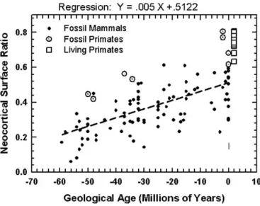
Figure 6 graphs a quantitative analysis of neocorticalization as measured on digitized images of mammalian endocasts. I am in the midst of a monograph for the Field Museum in Chicago with more details on my collection of these images: about 150 species of mammals. Digitization was with laser scans (see http://hjerison.bol. ucla.edu). Many of my scans are of specimens collected by the late Professor Len Radinsky of the University of Chicago who deposited them at the Field Museum to form its Radinsky Collection.
A general point about the data of Figure 6. We see how primates are all ‘above average’. I do not label the data on other groups in this graph. I could have identified a dozen species of equids, for example, which are first known in the fossil record of about 55 Ma. A side issue for us, they have been ‘average’ (near the regression line) for most of their evolutionary history. Marsupials as a group have always been ‘below average’ as have most of the fossil Neotropical species of South America. These are secondary matters for biolinguistics but appropriate for a broad picture. For this report I present the data most relevant for the evolution of language, in particular in the human species. I place my statistical words in quotation marks to acknowledge the questionable theoretical status of my regression analysis.
The graph in Figure 6 shows a regression equation fitted to all of the fossil data. The unit X is in millions of years before the present (Ma). For example the pre-sent geological age has Ma = X = 0 and the ‘average’ neocorticalization is now 0.51, or 51%. That means that at present an average mammal has 51% of its brain’s surface area devoted to neocortex. The earliest graphed geological age is 60 Ma. At X = –60 the average amount of neocorticalization was (.005)(–60) + .51 = 0.51–0.3 = 0.21. 21% of the surface area of the brain of the average mammals of 60 Ma was devoted to neocortex. The linear regression equation fitted to the data may be interpreted as showing an average of 5% increase in neocortex in mammals per 10 million years.
If we are not carried away by the use of averages this is a fine way of sum-marizing what we observe. Regression analysis is based on working with a sample taken randomly from its population, and it is well to keep that in mind. The primates in Figure 6 form a fine cohort for our purpose. The four Eocene species were prosimians (Infraorder Strepsirrhini) related to living lemuroids. The four Plio-Pleistocene species were two australopithecines in the hominin lineage and two pro-simians. Two points of interest: Both groups of primates were ‘above average’ in neocorticalization that is described by the regression line. With respect to the theory of regression analysis they do not represent a random selection from the mammals as a class. But it is helpful to recognize that there is something about brain size in pri-mates that makes them a specialized order of mammals with respect to enlarged brains.
4. Discussion
The most unusual feature of the graph for me was the comparison between living and fossil primates. Here are a few of the numbers. The two australopithecines are Taung and Sk 1585 (Swartkrans) and were respectively 80.5% and 77.2% neocorti-calized. I have two human skulls collected by Falk (2012) in my living primate sample, and these are 80.0% and 77.7% neocorticalized. I have one chimpanzee and it’s 80.7% neocorticalized, which makes it my top performer by 0.2%. I have a Patas monkey from Africa, and it is 78% neocorticalized. I have two New World Saki monkeys and these are 78.0% and 79.0% neocorticalized. The point is that neocorti-calization in primates has topped out at about 80%. I have guessed that the growth of mammalian brains is limited with respect to the amount of neocortex that is developed. This is presumably genetically determined.
Brai-tenburg & Schuez 1992) that display the remarkably extensive neocortical neural net-works involved with language. Broca’s and Wernicke’s areas are only a fraction of those revealed by brain scans of living humans performing language tasks. My spec-ulation is straightforward. Assuming a selective advantage for the early adaptations and the later developments of language in our hominin lineage there would have to be space for the language network in the neocortex. To enlarge our neocortex there remains the 80% barrier. Hence, to produce an appropriately enlarged mass of neo-cortex the whole brain would have to be enlarged. My speculation is that the remarkable extent of human evolutionary encephalization resulted from selection to provide additional neocortex in the face of the 80% barrier.
References
Braitenberg, V. & A. Schuez. 1992. Basic features of cortical connectivity and some considerations on language. In J. Wind, B. Chiarelli, B. H. Bichakjian, A. Nocentini & A. Jonker (eds.), Language Origin: A Multidisciplinary Approach, 89– 102. Dordrecht: Kluwer.
Falk, D. 1992. Braindance. New York: Holt.
Falk, D. 2012. Hominin paleoneurology: Where are we now? In M. A. Hofman & D. Falk (eds.), Progress in Brain Research, vol. 195, 255–272. Amsterdam & New York: Elsevier.
Hofman, M. A. 2012. Design principles of the human brain: An evolutionary perspective. In M. A. Hofman & D. Falk (eds.), Progress in Brain Research, vol. 195, 373–390. Amsterdam & New York: Elsevier.
Jerison, H. J. 1979. The evolution of diversity in brain size. In M. E. Hahn, C. Jensen & B. C. Dudek (eds.). Development and Evolution of Brain Size: Behavioral Implications, 29–57. New York: Academic Press.
Jerison, H. J. 2001. Adaptation and preadaptation in hominid evolution. In P. V. Tobias, M. A. Raath, J. Moggi-Cecchi & G. A. Doyle (eds.), Humanity from African Naissance to Coming Millennia, 373–378. Florence: Firenze University Press and Johannesburg: Witwatersrand University Press.
Jerison, H. J. 2007. Fossils, brains, and behavior. In S. Watanabe & M. A. Hofman (eds), Integration of Comparative Neuroanatomy and Cognition, 13–31. Tokyo: Keio University Press.
Kaas, J. H. 2012. The evolution of neocortex in primates. In M. A. Hofman & D. Falk (eds.), Progress in Brain Research, vol. 195, 91–102. Amsterdam: Elsevier.
Pulvermüller, F. 2010. Brain-language research: Where is the progress? Biolinguistics
4, 255–266.
Wilson, J. A. 1971. Early Tertiary vertebrate faunas, Vieja Group. Trans- Pecos Texas: Agriochoeridae and Merycoidodontidae. Texas Memorial Museum Bulletin 18, 1– 83.
Harry J. Jerison University of California
Department of Psychiatry and Biobehavioral Sciences 760 Westwood Plaza
Los Angeles, CA 90095 USA