Anna Czeczuk
1, A–F, Elżbieta Huk-Wieliczuk
1, C–F, Agnieszka Michalska
2, C–F,
Dorota Bylina
2, C–F, Jarosław Sołtan
3, C–F, Zofia Dzięcioł
4, D–FThe Effect of Menopause on Bone Tissue in Former
Swimmers and in Non-Athletes
Wpływ menopauzy na jakość tkanki kostnej u byłych pływaczek
i kobiet nieuprawiających sportu w przeszłości
1 Physical Education Department, Faculty of Physical Education in Biala Podlaska, Poland
2 Department of Anatomy and Physiology, Faculty of Physical Education in Biala Podlaska, Poland 3 Foreign Languages Department, Faculty of Physical Education in Biala Podlaska, Poland
4 Rehabilitation Department, Medical University in Bialystok, Poland
A – research concept and design; B – collection and/or assembly of data; C – data analysis and interpretation;
D – writing the article; E – critical revision of the article; F – final approval of article; G – other
Abstract
Background. An increased loss of bone density during the first years after menopause induces osteoporosis.
Objectives. The aim of the research presented in this paper was to ascertain the difference in the rate of involu-tional changes in bone tissue in former athletes and in non-athletes of the same age.
Material and Methods. The research involved 18 former swimmers and 18 females of similar age who had never practiced sports. The subjects were subdivided into two subgroups: Subgroup I had been post-menopausal for ≤ 5 years, and Subgroup II for > 5 years; this was done to assess bone mineral contentrelative to the length of the post-menopausal time period. Bone mineral content (BMC) and bone mineral density (BMD) were measured in lumbar vertebrae by dual-emission X-ray absorptiometry (DXA). Bone strength was measured in the heel using the bone stiffness index. Each subject was examined twice, with a one-year period in between. A diagnostic questionnaire was used to compile date on the subjects’ physical activity and their gonad functioning. Dietary habits (calcium intake) were established by three interviews and the Dieta 4.0 computer program.
Results. Anthropometric features did not differentiate the subjects in the subgroups. Former athletes in both sub-groups spent off-work time on physical activities significantly more frequently. In both sub-groups, calcium intake was sufficient and did not exceed ¾ of the daily norm. A higher calcium intake was found in former athletes compared to non-athletes. The subjects in Subgroup I had significantly greater BMC and BMD than those in Subgroup II. In Subgroup I, the second examination showed somewhat lower reductions in BMC and BMD among the former athletes than among the non-athletes. In Subgroup II, BMC and BMD increased somewhat among the former athletes, while non-significant reductions were observed in the BMC and BMD of the non-athletes. All the subjects undertook pharmacologic treatment after the first examination, which caused improvement of bone parameters in the second examination.
Conclusions. The rate of bone mass loss in former athletes proved to be consistent with the involutional process and similar to that of non-athletes. The reduced BMD in the lumbar vertebrae of 22% of the women in the study demonstrates the need for regular densitometric examinations in postmenopausal females (Adv Clin Exp Med 2012, 21, 5, 645–652).
Key words: female athletes, bone mineral content, bone mineral density, bone tissue involution, postmenopausal osteoporosis.
Streszczenie
Wprowadzenie. W czasie wczesnej menopauzy następuje gwałtowna utrata wapnia w kościach, co wywołuje oste-oporozę.
Cel pracy. Ocena tempa zmian inwolucyjnych tkanki kostnej u byłych sportsmenek, będących we wczesnym i póź-nym okresie postmenopauzalpóź-nym.
Adv Clin Exp Med 2012, 21, 5, 645–652 ISSN 1899–5276
ORIgINAL PAPERS
Materiał i metody. Badaniami objęto 18 byłych pływaczek w wieku 50–62 lat oraz grupę 18 kobiet w analogicznym wieku nieuprawiających sportu w przeszłości. W celu analizy parametrów tkanki kostnej w zależności od okresu po menopauzie grupę zbadanych kobiet podzielono na dwie podgrupy: podgrupę I – okres po menopauzie ≤ 5 lat i podgrupę II – okres po menopauzalny > 5 lat. Zawartość minerału w kości (BMC) i średnią gęstość kości (BMD) oznaczono w odcinku lędźwiowym kręgosłupa metodą DEXA. Wytrzymałość kości oszacowano współczynnikiem Stiffness w kości piętowej metodą ultrasonograficzną. Dane dotyczące aktywności fizycznej i funkcji gonad w prze-szłości oraz stosowania hormonalnej terapii zastępczej, zbierano metodą sondażu diagnostycznego. Obecny sposób żywienia (podaż wapnia) oszacowano za pomocą programu Dieta 4.0.
Wyniki. Parametry antropometryczne nie różnicowały kobiet w obu podgrupach. Byłe pływaczki znacznie więcej czasu poświęcały w ciągu tygodnia na rekreację fizyczną. Średnie spożycie wapnia nie przekraczało 75% dzienne-go zapotrzebowania (AI). Kobiety, u których od ostatniej miesiączki upłynęło mniej niż 5 lat, miały znamiennie większą mineralizację i gęstość kości (p < 0,05) w porównaniu do kobiet, u których upłynęło więcej niż 5 lat. Po upływie roku u pań będących 5 lub mniej lat po menopauzie nastąpił spadek BMC i BMD. U kobiet powyżej 5 lat po menopauzie wzrosły średnie wartości BMC i BMD w granicach 1,8% – byłe pływaczki i 0,4% – grupa odniesie-nia. U 4 byłych pływaczek i 4 kobiet z grupy odniesienia w obu badaniach wykazano zmniejszoną w stosunku do normy gęstość mineralną kości.
Wnioski. Tempo utraty masy kostnej byłych pływaczek nie odbiega od tempa stwierdzonego w grupie odniesienia. Stwierdzenie zmniejszonej gęstości kości u 22% byłych zawodniczek i 22% kobiet z grupy odniesienia wskazuje na potrzebę systematycznych badań densytometrycznych po menopauzie (Adv Clin Exp Med 2012, 21, 5, 645–652).
Słowa kluczowe: sportsmenki, mineralizacja kości, gęstość kości, inwolucja tkanki kostnej, osteoporoza pomeno-pauzalna.
Bone tissue is one of the basic structures whose correct metabolism depends on estrogens. Those hormones multilaterally inhibit calcium resorption, restrain osteoclasts, influence the maturing and activity of osteoblasts by means of transforming growth factor beta (TgF-β) and insulin-like growth factor 1 (IgF-I) [1]. Estrogens also inhibit the secre-tion of osteoclast stimulating factors IL-1 and IL-6 from osteoblasts, regulate vitamin D metabolism and increase both the production of calcitonin and the absorption of calcium in the small intestine [1].
One of the features of menopause is disable-ment of the hypothalamic-pituitary-gonadal axis (HPg axis) circular activity caused by atrophy in production of cyclic stimuli of hypothalamus by the centers located above it (the limbic system, the midbrain reticular formation and certain areas of the cerebral cortex [20].
In menopause, the hypothalamus produces more gonadotropin-releasing hormone (gnRH), whereas the pituitary gland produces more gonad-otropins, especially follitropins (FSH). Cyclic se-cretion of lutropin (LH) fades away, which causes the regular functions of the ovaries to become ex-tinct and the ovaries gradually stop reacting to the stimuli of gonadotropins. As a result the produc-tion of 17β-estradiol is reduced [20].
In menopause estradiol content in plasma is reduced from 30–350 pg/ml before menopause to 5–20 pg/ml, which, along with the reduced secre-tion of progesterone by the ovaries, causes meta-bolic disorders. Osteoporosis is one of the conse-quences [2].
Postmenopausal osteoporosis (primary type 1) is currently one of the greatest health problems in
women after menopause. It appears 3 to 5 years after the last menstruation and is diagnosed in its clinical form in around 25–44% of females [3, 4].
In the last century, the average life span has significantly increased in females. In the 19th
cen-tury fewer than 30% of females lived to meno-pausal age; nowadays, almost 90% of females live to menopause, and 60% of them live to be 70 years old [5]. This is why the number of females with osteoporosis symptoms has risen greatly.
In humans, after peak bone mass is attained, it remains at the same level until the age of around 40 years, after which it drops. There are two stages in the involution of compact and spongy types of osseous tissue: slow and fast.
The first stage is found in both males and males, whereas the second stage is found only in fe-males just after menopause, and lasts an average of 8 to 10 years. Numerous clinical investigations have reported that the greatest reduction of bone mass takes place within the first five years after menopause. During that time period, cancellous bone mass could be lost at a rate of 5% to 8% annually [6, 7].
Females lose around 60 mg of calcium a day during the first five years after menopause, where-as calcium loss before menopause is around 20 mg a day [8]. The increased loss is a reflection of in-tensified bone resorption, which happens as a sult of the activation of new elements of bone re-modelling. At the same time, calcium regulation changes in the whole body: Calcium absorption in the intestine is reduced, and calcium excretion is enhanced, probably due to blocking of parathyroid hormone (PTH) secretion. This results in a loss of 1000 g (about 13%) of primary calcium mass in the whole body, and brings about a change of 1 standard deviation in bone mass density (BMD), increasing the risk of bone fracture twofold or threefold [9, 10]. Fractures of the femoral neck, vertebrae, ribs, pelvis, and distal radius are the most common [11].
It should be emphasized that (apart from the normal processes of aging) postmenopausal os-teoporosis is caused 60% to 80% by genetic fac-tors, which determine the amount of bone mass, and 20% to 40% by lifestyle. Unhealthy diet and insufficient physical activity are common causes of postmenopausal osteoporosis [12].
Prevention of osteoporosis is based on precau-tions taken long before menopause. One of the best precautions to prevent osteoporosis is physical ac-tivity. A prolonged active lifestyle before, during and after menopause, along with regular physical exercises that strenghtens the skeleton can signifi-cantly reduce the risk of bone fractures that hap-pen as a result of bone mass loss. Physical exercise is also beneficial because it reduces susceptibility to fracture and a predisposition to accidents by increasing muscle strength and improving coordi-nation [13]. However, there are numerous clinical reports that beneficial effect of physical exercise is achieved only when it is combined with sufficient intake of calcium in the daily diet [14, 15].
The aim of the research presented in this paper was to establish the difference in the rate of invo-lutional changes in bone tissue in former athletes and non-athletes in the same age group.
Material and Methods
The study involved 36 women: 18 former swim-mers aged 50–62 years, who had ended their profes-sional careers around 20 years prior to the research and who had regularly performed intense physical exercise in the past; 18 females aged 49–63 years who had never practiced sports constituted the control group. The selection criterion for the con-trol group was menopausal age similar to the for-mer athletes. All the subjects were in good general
health. All the subjects participated in the research voluntarily, and submitted written consent.
The research was approved by the Józef Piłsudski Academy of Physical Education Review Board.
The subjects’ body height and weight were measured by conventional methods with a preci-sion of 0.1 cm and 0.1 kg. The method suggested by Durnin andWomersley was employed to de-termine body fat: Four skin fold measurements were taken, at the bicep, tricep, subscapular and suprailiac [16].
A diagnostic questionnaire was employed to compile data on the subjects’ current physical ac-tivity and their gonad functioning in past.
Current dietary habits were ascertained by three interviews that covered the period 24 hours prior (two interviews covered workdays and one interview covered a day off) and by the comput-er program Dieta 4.0 (developed by the National Food and Nutrition Institute, Poland) based on Polish nutrition norm charts [17].
Bone mineral content (BMC, g) and bone mineral density (BMD, g/cm2) were measured in
lumbar vertebrae (L2–L4) by dual-emission X-ray
absorptiometry (DXA) Luna DPX-L (USA). Luna Achilles (USA) was used to measure the ultrasonographic stiffness index in the heel. In ac-cordance with World Health Organization (WHO) requirements, densitometric and ultrasonographic measurements were performed twice, with a pe-riod of 12 months in between. The measurements were performed at the Department of Biochem-istry of Experimental Medicine at the Children’s Memorial Health Institute in Warsaw, Poland. All the measurements were taken by the same techni-cian using the same equipment.
In cases of reduced BMD, the T-score was used to indicate how much the subject’s BMD departed from the mean. The results were interpreted ac-cording to WHO criteria.
All the data has been presented as arithmetic means and standard deviations. The results have been analyzed using both single-factor variation analysis and Tukey’s test of unequal samples to de-fine the range of differences in the average values of the investigated features in particular groups. The Shapiro-Wilk test [18] was used to verify the hypothesis of normal data distribution.
The Brown-Forsythe test was used to verify variations of bone parameters in particular groups. Significance of the statistical tests was defined as a p
Results
In order to assess bone mineral content in relation to the time period that had elapsed since menopause, all the subjects from both groups were further divided into two subgroups: Subgroup I, consisting of women ≤ 5 years from menopause (average age 51 years), and Subgroup II, consisting of women > 5 years from menopause (average age 59 years). The subjects' anthropometric features are presented in Table 1. The average means of the anthropometric features did not differentiate the subjects in the two subgroups. At the time of the examination, themajority of the former athletes were professionally active (81%). Some worked as physical education teachers (21%) and sports coaches (12%); the professions of the rest of them did not require physical effort.
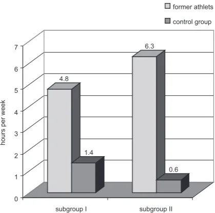
Analyzing the data regarding current physical activity in off-work time (Figure 1) indicated that former athletes in both subgroups more frequently spent their off-work time on physical activities – i.e. 4.8 and 6.3 hours per week in Subgroups I and II, respectively, compared to non-athletes, who spent 1.4 and 0.6 hours per week respectively on
physi-Table 1. Anthropometric features of former athletes and non-athletes (control group)
Tabela 1. Cechy antropometryczne byłych sportsmenek i kobiet nieuprawiających sportu (grupa kontrolna)
Years after menopause (Lata po menopauzie)
group (grupa) (n) Age – years
(Wiek – lata) Body mass – kg (Masa ciała – kg)
Body height – cm (Wzrost – cm)
BMI
– kg/m2 Adipose tissue – %
(Tkanka tłuszczowa – %)
≤ 5 former athletes 11 52.1 ± 3.3 72.1 ± 11.3 167.6 ± 4.5 25.7 ± 3.5 31.1 ± 5.2
control group 11 50.7 ± 2.2 68.3 ± 8.2 162.7 ± 4.5 25.9 ± 3.5 33.4 ± 8.0
> 5 former athletes 7 63.3 ± 4.3 65.5 ± 11.4 164.2 ± 4.3 22.9 ± 152 29.8 ± 3.5
control group 7 60.6 ± 2.3 67.7 ± 6.9 162.4 ± 3.8 24.9 ± 2.7 34.2 ± 9.4
Table 2. Current average daily calcium intake
Tabela 2. Bieżąca średnia dzienna dawka wapnia
Years after menopause
(Lata po menopauzie) group (grupa) (n) Calcium (Wapń)
mg %*
≤ 5 former athletes 11 683.61± 145.7 52.6
control group 11 612.5± 2992.0 47.1
> 5 former athletes 7 780.5± 294.2 60.1
control group 7 673.7± 208.6 51.8
* Percentage of adequate intake (AI). * Odsetek dziennego zapotrzebowania (AI).
4.8
1.4
6.3
0.6
0 1 2 3 4 5 6 7
subgroup I
hours per week
subgroup II
former athlets
control group
Fig. 1. Subjects’ current physical activity per week Subgroup I ≤ 5 years from menopause.
Subgroup II > 5 years from menopause.
Ryc. 1. Bieżąca aktywność fizyczna badanych kobiet w ciągu tygodnia
cal activities. Swimming, bicycle riding and walking were the most frequent kinds of physical activities.
Current calcium intake was sufficient and did not exceed ¾ of the daily norm among both the former athletes and the controls [17]. However, higher intake of calcium was noted among the for-mer athletes (in both subgroups) compared to the controls (Table 2).
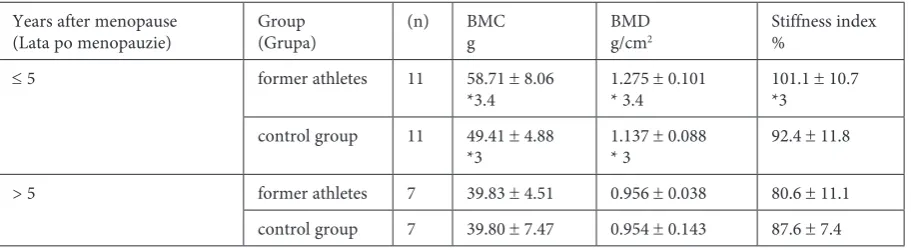
The analysis of the bone tissue data compiled in the first examination (Table 3) showed that Subgroup I of both the former athletes and non-athletes had significantly greater bone mineral content and bone mineral density (p < 0.05) than the women in Subgroup II, who had gone through menopause more than 5 years prior to the exami-nation. The average means of the stiffness index proved to be the greatest in former athletes during the first five years after menopause (10.1 ± 10.7%). The differences in the average means of the stiff-ness index between former athletes in Subgroup I and former athletes in Subgroup II were found to be significant. There was no statistically signifi-cant difference between the stiffness index of non-athletes in Subgroup I and that of the non-non-athletes in Subgroup II. However, a tendency was observed
for the stiffness index to increase in subjects who had gone through menopause 5 years or less prior to the examination.
The analysis of individual examination results of all subjects showed that BMD in lumbar verte-brae was insufficient in four former athletes and four non-athletes. Interpreting the results of the densitometric examinations according to WHO criteria [19] in each separate subject indicated osteoporosis (T-score below –2.5) in one former athlete and two non-athletes, and osteopenia (T-score within the range –1 to –2.5) in three former athletes and two non-athletes. These pathologi-cally low BMD scores were found only in subjects in Subgroup II, who had undergone menopause more than 5 years prior to the examination. The average means of BMD in subjects with osteopo-rosis and osteopenia were lower than those of the rest of the subjects with normal T-scores (28% lower in former sportswomen, and 18% lower in the control group).
After the first examination, all the subjects un-derwent pharmacological treatment as a result of the medical consultations. One former athlete and one non-athlete were being treated with Fosamax
Table 3. Bone features of former athletes and non-athletes (control group) (examination I)
Tabela 3. Cechy kości byłych sportsmenek i kobiet nieuprawiających sportu (grupa kontrolna) (badanie I)
Years after menopause
(Lata po menopauzie) group (grupa) (n) BMCg BMDg/cm2 Stiffness index%
≤ 5 former athletes 11 58.71 ± 8.06
*3.4 1.275 * 3.4 ± 0.101 101.1 *3 ± 10.7
control group 11 49.41 ± 4.88
*3 1.137 * 3 ± 0.088 92.4 ± 11.8
> 5 former athletes 7 39.83 ± 4.51 0.956 ± 0.038 80.6 ± 11.1
control group 7 39.80 ± 7.47 0.954 ± 0.143 87.6 ± 7.4
* Significant differences at p < 0.05 between groups denoted by ordinal numbers.
* Różnice istotne statystycznie dla p < 0,05 między grupami oznaczonymi liczbami porządkowymi.
Table 4. Differences in bone features between examinations I and II
Tabela 4. Różnice cech kości między badaniem I i II Years after
menopause (Lata po menopauzie)
group (grupa) (n) BMC BMD Stiffness index
g % g/cm2 % %
≤ 5 former athletes 11 –1.45 –2.7 –0.021 –2 –2.5
control group 11 –1.40 –2.9 –0.031 –2.8 –3.4
> 5 former athletes 7 +0.63 +1.6 +0.021 +2 +0.1
(bisphosphonate) at a dosage of 10 mg a day, with simultaneous supplementation of calcium and vi-tamin D. The rest of the subjects were on hormone replacement therapy with calcium supplementa-tion; those subjects declared that they altered their life style, increasing their calcium intake and active leisure activities. Three former athletes and two non-athletes from Subgroup I had been on hor-mone replacement therapy for three years.
Changes in the subjects’ bone tissue param-eters after one year are presented in Table 4. Evi-dent reductions both in bone mineral density and bone mineral content were found in subjects who had gone through menopause 5 years or less pri-or to the examination (Subgroup I). The average means of BMC and BMD reduced by 1.45 g (2.7%) and 0.021 g/cm (2.0%) respectively in former ath-letes; in non-athletes the average means of BMC and BMD reduced by 1.4 g (2.9%) and 0.031 g/ cm (2.8%) respectively. In addition, there was an evident drop in the stiffness index among both the former athletes and non-athletes in Subgroup I.
After one year, in subjects who had gone through menopause more than 5 years prior to the study (Subgroup II), improvements in the average means of BMC and BMD of 1.6% and 2%, respec-tively, were noted in the former athletes, while non-significant reductions (0.5% and 0.1%) were found in the non-athletes. The average means of the stiffness index dropped by 2.4% in the non-athletes and increased by 0.1% in the former ath-letes after a year in Subgroup II.
Discussion
Reproductive activity declines in women during menopause. Intense hormonal changes take place not only in the hypothalamic-pituitary-gonadal axis but in other endocrine glands as well [20].
Metabolic changes take place along with hor-monal changes in target organs and tissues, includ-ing bone tissue. Rapid bone mass loss is observed in females after menopause, which takes place at the rate of 1–3% of all cortical (compound) bone mass annually and 3–5% of all cancellous bone mass annually [7]. This rapid process of bone mass loss lasts for around 8–10 years and after that pe-riod slows down.
The quality of bone tissue after menopause and in the later years of life depends upon the peak bone mass (PBM) attained during growth and maturity, as well as the ability to maintain optimal PBM during adulthood. The rate of bone mass loss in adulthood depends on many factors, among which lifestyle tends to be the most promi-nent. Unhealthy dietary habits, insufficient
move-ment, smoking and excessive alcohol consumption contribute to bone mass loss, which consequently leads to osteoporosis.
In the current study, during the year between the first and second examinations, the average means of the reduction in the subjects’ BMC and BMD was found to be the greatest in Subgroup I, comprised of women who had gone through menopause ≤ 5 prior to the first examination (in former athletes the loss of BMC was −2.7% and of BMD −2%; in non-athletes the reductions were −2.9% and −2.8% respectively).
Numerous clinical trials have proven that the most intense bone mass loss takes place during the first five years after menopause – up to 5–8% an-nually, especially in cancellous bone [7]. The fact that magnitudes of involutional bone mass loss in the subjects of this study were rather low when compared to the rest of the population could be explained by the preventive hormone replacement therapy that three of the former athletes and two of the non-athletes had undergone; their average BMD values were 2–4% greater in the second test than in the first test. Preventive hormone replace-ment therapy (HRT) is considered a successful way to prevent postmenopausal osteoporosis. Since 1978a number of scientific papers have been pub-lished that undeniably prove the positive effects of HRT on the human skeleton [21, 22].
The current study found that the annual re-duction in the average means of BMC and BMD in Subgroup I was greater in the non-athletes than in the former athletes. Former athletes spent signifi-cantly more time on physical activities, including swimming. Most scientific papers suggest swim-ming to be of minimal positive influence on bone tissue, because this form of exercise does not cause bone mass growth, unlike such sports as gymnas-tics or volleyball [23]. Despite those other opin-ions, the authors of the current paper consider swimming to have a positive influence on bone tissue.
It should be also emphasized that the diet of the former athletes in Subgroup I was richer in calcium than the control group’s diets. Numerous investigations have proved the positive influence of a high-calcium diet on bone tissue in postmeno-pausal females [24, 14].
diagnosed with osteoporosis and osteopenia after the first examination. Other factors which could have increased the mean BMC and BMD in the lumber vertebrae of former athletes could include greater intake of calcium in the diet and a greater level of physical activity compared to the control group. Moreover, according to numerous clinical investigations, bone mass loss in women who went through menopause more than five years earlier is not as drastic as it is during the first five years after the gonads stop functioning [6].
The National Health and Nutrition Examina-tion Survey (NHANES) diagnosed 13% to 18% of American females aged over 50 years with osteo-porosis and 37% with low bone mass, according to WHO standards [25]. Similarly, the National Os-teoporosis Risk Assessment (NORA), the largest American osteoporosis risk registry, diagnosed al-most half of the female population over age 50 with reduced bone density [26]. Similar results were re-ported by Hoszowski et al., [27] who screened fe-males of a similar age in Warsaw.
The current research found dangerously re-duced bone density, compared to population norms, in 22% of the former swimmers and 22% of the non-athletes; osteoporosis T-scores in 5.5% of the former swimmers and 11% of the non-athletes; and osteopenia T-scores in 16.6% of the former swimmers and 11% of the non-athletes. This cor-roborates the scale of the problem as reported by many authors. Whereas educational information about the prevention of circulatory system diseases is widespread nowadays and reaches a wide audi-ence, there is a great shortage of knowledge about osteoporosis and its management in the general population [28].The problem is aggravated by the
lack of clinical symptoms of osteoporosis. It should be emphasized that none of the subjects of the cur-rent study who were diagnosed with reduced bone density had ever experienced any clinical symp-toms of osteoporosis or bone fracture. The women included in this study were chosen at random, therefore the discovery of significantly reduced bone density in their lumber vertebrae during the densitometric examination was accidental. On the basis of thorough medical examinations and interviews, it could be assumed that the reduced bone density in 7 subjects could have resulted from postmenopausal changes and previous lifestyle. In one of the subjects, reduced bone density could have been a side effect of medication (cytostatic therapy ten years prior to the examination). One of the side effects of cancer treatment is secondary osteoporosis, which develops as a result of either hypogonadism or the direct influence of drugs or medical procedures on bone tissue [28]. The afore-mentioned subject was treated with 10mg doses of Fosamax daily, coupled with supplementation of calcium and vitamin D (as noted in the research results). The second examination showed a 13% increase in bone density in the lumber vertebrae compared with the first examination. An increase of 2–6% of BMD was found in another subject af-ter a year of the same treatment. The average BMD means in subjects who had supplemented their diet with calcium and vitamin D for a year (osteopenia T-score) was 2–4% greater in second examination compared with first examination.
The present research results suggest that in-creased physical activity and normal dietary con-sumption of calcium have positive effect on pre-serving age optimal bone mass.
References
[1] Riggs B, Spelsberg T: Oestrogen and bone. Raport of Fourth International Symposium. Hong Kong 1993, 8–9.
[2] Uhrynowska-Tyszkiewicz J, Kamińska A: Mechanizm działania niektórych hormonów na proces przebudowy tkanki kostnej. Terapia 2002, 6, 5–12.
[3] Czerwiński E, Kukiełka R, Strzępek J: Patogeneza, diagnostyka osteoporozy. Med. Sport 1999, 3 (Suppl. 2), 9–17.
[4] Warenik-Szymankiewicz A, Słopień A, Męczekalski B: Menopauza i jej wpływ na osteoporozę. Twój Mag Med 2004, 2(137), 14–24.
[5] Krasomski G: Współczesne poglądy na stosowanie terapii zastępczej u kobiet po menopauzie. gin Pol 1995, 66(1), 28–31.
[6] Dębski R: Substytucja hormonalna w okresie pomenopauzalnym. Nowa Med 1995, 2(8), 64–66.
[7] Leszczyński P: Zasady postępowania diagnostycznego I terapeutycznego w osteoporozie u progu XXI wieku. Osteoporoza – Biuletyn Informacyjny Wielkopolskiego Kolegium Osteoporozy 2001, 1, 3–4.
[8] Rosen CJ: Osteoproza. Zasady rozpoznania i leczenia. Springer PWN, Warszawa 1998.
[9] Melton LJ, Atkinson EJ, O’Fallon WM, Wahner HW, Riggs BL: Long-term fracture production by bone mineral assessed at different skeletal sites. J Bone Miner Res 1993, 8, 1227–1234.
[10] Olszyński WP: Wyniki stosowania aldronianu disodowego w leczeniu osteoporozy. gerontol Pol 2000, 8(4), 1–6.
[11] Horst-Sikorska W, Marcinkowska M: Osteoporotyczne złamania kostne – profilaktyka, leczenie, rehabilitacja. Terapia 2005, 2(162), 37–40.
[13] Chmielewski D: Zapobieganie osteoporozie. Twój Mag Med 2004, 2(137), 25–32.
[14] Czeczuk A, Dmitruk A, Popławska H: Effect of calcium intake on bone parameters in women training and not-training in the past being in the postmenopausal period. Med Sport 2006, 10(3), 81–84.
[15] Melendez-Ortega A: Osteoporosis, falls and exercise. Eur Rev Aging Phys Act 2007, 4, 61–70.
[16] Durnin JVGA, Womersley J: Body fat assessed from total body density and its estimation from skinfold thickness: measurements on 481 men and women aged from 16 to 72 years. Br J Nutr 1974, 32, 77–97.
[17] Jarosz M, Bułhak-Jachymczyk B: Normy żywienia człowieka. PZWL, Warszawa 2008.
[18] Ferguson GF, Takane Y: Analiza statystyczna w psychologii i pedagogice. PWN, Warszawa 1993.
[19] Badurski JE: Zasady diagnostyki osteoporozy i ryzyka złamań oraz leczenia farmakologicznego. Post Osteoartrol 2001, 12, Supl. 1, 146.
[20] Skałba P: Endokrynologia ginekologiczna. PZWL, Warszawa 1993.
[21] Sawicki A: Charakterystyka szczegółowa leków i metod leczenia osteoporozy. In: Osteoporoza. Eds.: Badurski J, Sawicki A, Boczoń S, Osteoprint, Białystok 1994.
[22] Cauley JA, Seeley DG, Enstrud K, Ettinger B, Black D, Cummings SR: For the study of osteoporosis fractures research group. Estrogen replacement therapy and fractures in older women. Ann Int Med 1995, 122, 9–16.
[23] Fehling PC, Alekel L, Clasey J, Rector A, Stillman RJ: A comparison of bone mineral densities among female athletes in impact loading and active loading sorts. Bone 1995, 17(3), 205–210.
[24] Kanis JA: The use of calcium in management of osteoporosis. Bone 1999, 24, 274–290.
[25] Goddard D, Kleerekoper M: Epidemiologia osteoporozy. Med Dypl 1999, 8(3), 22–30.
[26] Siris ES, Miler PD, Barret-Connor E, Faulkner KG, Wehren LE, Abbot TA, Berger ML, Santora AC, Sherwood LM: Występowanie nierozpoznanej małej gęstości mineralnej tkanki kostnej oraz związanych z nią złamań kości u kobiet po menopauzie. JAMA 2002, 4(2), 119–127.
[27] Hoszowski K, Gawron J, Korczyk P, Grabski T, Jędrzejewska-Korczyk J, Markiewicz J, Lorenc RS: Analiza czyn-ników ryzyka i częstości występowania osteoporozy kręgosłupa w próbie populacyjnej mieszkańców Warszawy powyżej 50. roku życia. Pol Tyg Lek 1993, 48 (Suppl. 3), 31–35.
[28] Spaczyński M: Osteoporoza jako powikłanie leczenia w różnych chorobach nowotworowych. Osteoporoza – Biuletyn Informacyjny Wielkopolskiego Kolegium Osteoporozy 2003, 5, 11–12.
Address for correspondence:
Anna Czeczuk
Faculty of Physical Education in Biala Podlaska Akademicka 2
21-500 Biala Podlaska Poland
Tel.: +48 502 538 405, +48 83 342 87 34 E-mail: [email protected]
Conflict of interest: None declared