This is an open access journal, and articles are distributed under the terms of the Creative Commons Attribution-Non Commercial-ShareAlike 4.0 License, which allows others to remix, tweak, and build upon the work non-commercially, as long as appropriate credit is given and the new creations are licensed under the identical terms.
© 2017 Journal of Advanced Pharmacy Education & Research | Published by SPER Publication
486
Effect of transcranial direct current stimulation on upper
extremity functional recovery in stroke patients
Abdaleem Ateia
1, Waleed Talat
1, Amani Nawito
2, Noura Elkafrawy
1*1Departments of Neuromuscular Disorders and Its Surgery, Faculty of Physical Therapy, Cairo University, Egypt , 2Clinical Neurophysiology, Faculty of Medicine, Cairo
University, Egypt.
Correspondence: Noura Elkafrawy, Departments of Neuromuscular Disorders and Its Surgery, Faculty of Physical Therapy, Cairo University, Egypt. E_mail: [email protected]
ABSTRACT
Background: Upper extremity impairment as a deficit, affects the daily lives’ of the sufferers. Objective: To investigate the effect of bihemispheric tDCS on functional recovery of the affected upper extremity in chronic stroke patients. Patients and methods: Forty chronic stroke patients were allocated randomly to receive 10 consecutive sessions of either 1) simultaneous bihemispheric transcranial direct current stimulation (tDCS) and constraint induced movement therapy (CIMT) (study group) or 2) simultaneous sham stimulation and CIMT (control group). Outcome measures include changes in affected upper extremity motor impairment (Motricity Index (MI), Fugl-Meyer Upper Extremity Motor Assessment (UE-FM)) and motor activity (Action Research Arm Test (ARAT)) assessments. Results: The improvement of motor function (MI, ARAT and UE-FM scores) were significantly greater in the real stimulation group than the sham group (p = 0.002, 0.01 and <0.001 respectively). Conclusions: Bihemispheric tDCS can improve the recovery of motor functions in chronic stroke.
Keywords:Stoke, constraint induced movement therapy, transcranial direct current stimulation.
Introduction
Upper extremity impairment is a common motor deficit after a stroke, and 30–60% of stroke sufferers have disability and inability to use their affected upper extremity in their daily lives
[1]. Stroke is a common neurological condition that causes
long-term disability in adults [2]. Stroke often survives with
neurological and functional deficits [3], upper limb impairment is
one of the most common motor deficits after stroke and about 33% to 66% of patients with upper limb impairments report a little recovery 6 months after stroke [4]. Excessive using of
unaffected arms in daily living activities causes "non-use" and decreased movements of the affected one [5].
To overwhelm non-use, "constraint-induced movement therapy" (CIMT) was recommended by Taub et al.; it is a
profound, functional task specific training with paretic upper extremity while restraining the unaffected one [6].
Transcranial direct current stimulation (tDCS) is a non-invasive brain excitation that can be applied during motor training. By transmitting a weak continuous current on the scalp, the tDCS depolarize or hyperpolarize the neurons and cause changes cortical excitability [7]. A motor impairment can occur from
reduced output of the damaged hemisphere and unopposed inhibition from the unaffected hemisphere according to the model of interhemispheric rivalry between affected hemisphere and intact one [8]. Therefore, recovery of motor functions could
be induced by increasing excitability of the affected motor cortex by anodal tDCS or by decreasing the excitability of cathodal tDCS-mediated motor cortex (unaffected motor cortex) [9].
In this study, we assessed the effects of bihemispheric tDCS combined with intensive peripheral sensorimotor training on upper extremity functional recovery. This combined treatment was compared with benefits achieved by the peripheral training alone (plus shame tDCS).
Patients and Methods
Forty chronic stroke patients from outpatient clinic of the Department of Neurology, faculty of physical therapy, Cairo University were included in this study. Inclusion criteria
Access this article online
Website: www.japer.in E-ISSN: 2249-3379
How to cite this article: Abdaleem Ateia, Waleed Talat, Amani Nawito,
Noura Elkafrawy.
Effect of transcranial direct current stimulation on upper extremity functional recovery in stroke patients. J Adv Pharm Edu Res 2017;7(4):486-490.
consisted of occurrence of ischemic stroke in the territory of the middle cerebral artery at least 6 months prior to enrollment; age ranged from 45 to 60 years old; no previous or subsequent strokes; with at least 10 degrees’ active wrist dorsiflexion and extension of themetacarpophalangealand interphalangeal joints of the thumb and at least 2 additional digits 10 degrees at enrollment. Exclusion criteria were as follows: spasticity of grade 3 or more; other neurologic or orthopedic disorders that could influence upper extremity function; history of neurosurgery or epilepsy; metallic implants within the brain and cardiac pacemaker. The study was approved by the ethical committee of faculty of physical therapy and informed consent was obtained from all participants. Patients were randomly assigned to one of two groups—real tDCS with CIMT (study group) or sham tDCS with CIMT (control group).
The following measures for motor function assessment were used. The Action Research Arm Test (ARAT) [10], a standardized
measure of arm motor function after stroke. It is divided into four subscales: grasp, grip, pinch and gross movement. It has high intra- and interrater reliability and validity [11]. Motricity
Index(MI) [12], a valid instrument for characterizing the strength
of the paretic upper extremity following stroke [13]. Upper
Extremity Fugl-Meyer Motor Score (UE-FM) [14], a valid
measure of upper extremity motor impairment. It has excellent internal consistency, intrarater and inter-rater reliability. In addition, it does not have any significant floor and ceiling effects
[15]. These measures were obtained at baseline and at the end of
the treatment. All the evaluations were conducted at the baseline and at the end of the treatment.
Constraint-induced movement therapy. Each subject underwent 10-day CIMT. Participants had to wear a hand mitt on the nonparetic hand which hindered hand and finger activity. The mitt should be worn for at least 90% of waking hours [16]. During
the treatment period, all patients received up to 6 hours training per day of the affected arm in the outpatient clinic. Training tasks were designed according to a behavioral “shaping” technique and were designed to force an intensive use of the paretic extremity, while requiring a progressive improvement of the quality of movement [17].
Transcranial direct current stimulation. Direct current was transferred through pair of saline-soaked surface sponge electrodes (7.62x7.62 cm) and delivered by a battery-driven, adjustable direct current generator (Apex type A, USA). Simultaneous bilateral real stimulation consisted of 20 minutes of 2 mA direct current with the anode placed over the ipsilesional and the cathode over the contralesional motor area. For sham stimulation, the same electrode positions were used, but the stimulator was turned off after 30 seconds of stimulation. This ensured that patients could feel the initial itching sensation at the beginning of tDCS required for successful masking [18]. The
patients were blinded as to whether they received real or sham tDCS.
Statistical analysis
In this study, outcome was the motor function measured by the ARAT, MI and UE-FM. The data were analyzed using the statistical package SPSS (Statistical Package for the Social Sciences) version 24. Data was summarized using mean, standard deviation, median, minimum and maximum in quantitative data and using frequency (count) and relative frequency (percentage) for categorical data. Comparisons between quantitative variables in the 2 groups were done using the non-parametric Mann-Whitney test. For comparison of serial measurements within each patient the non-parametric
Wilcoxon signed rank test was used [19]. For comparing
categorical data, Chi square (𝜒𝜒2) test was performed. Exact test was used instead when the expected frequency is less than 5 [20].
Correlations between quantitative variables were done using Spearman correlation coefficient [21]. P-values less than 0.05
were considered as statistically significant.
Results
Demographic data.
There is no significant difference between the two groups regarding age, duration of illness, gender and affected side distribution (all p > 0.089; table 1,2).
Table 1.The duration of illness in both groups
study group control group value P
Me an SD Me di an M in im um Max imu m Me an SD Me di an M in im um Max imu m
Age 53. 05 69 5. 54. 00 45. 00 60. 00 54. 30 03 5. 56. 00 45. 00 60. 00 529 0.
dur ati on of illne ss (m onth ) 8.
37 22 2. 00 8. 00 6. 12. 00 60 9. 44 2. 11. 00 00 6. 12. 00 084 0.
Table 2. The Sex and paralysis distribution in both groups
Real stimulation
group (GII) group (GI) Sham P value
Count % Count %
Sex FEMALE MALE 15 5 75.0% 25.0% 14 6 70.0% 30.0% 0.723 hemiplegic
side RT LT 14 6 70.0% 30.0% 12 8 60.0% 40.0% 0.507
Motor assessments
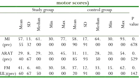
The two groups did not differ with respect to baseline motor score (all p ≥ 0.142; table 3).
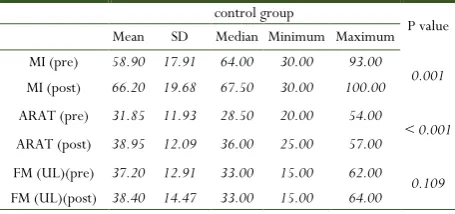
Study group gained greater improvement than control group in both MI and ARAT scores. There was a significant improvement in FM(UE) scores in the study group only post intervention; instead the sham group showed insignificant improvement. (Table 4, 5). Comparison of clinical motor scores post intervention in (GI) and (GII) revealed a statistical significant difference (Table 6).
Table 3. Difference between groups (Baseline motor scores)
Study group control group
P value Me an SD Me di an M in
Max Mean SD Medi
an
M
in
Max
MI
(pre) 57. 55 13. 32 61. 00 30. 00 77. 00 58. 90 17. 91 64. 00 30. 00 93. 00 678 0. ARAT
(pre) 29. 40 67 8. 29. 00 20. 00 45. 00 31. 85 11. 93 28. 50 20. 00 54. 00 529 0. FM
Abdaleem Ateia, et al.: Effect of transcranial direct current
Table4. comparison between pre and post intervention in study group
patient group P value
Mean SD Median Minimum Maximum MI (pre) 57.55 13.32 61.00 30.00 77.00
< 0.001 MI (post) 86.20 18.01 91.00 40.00 100.00
ARAT (pre) 29.40 8.67 29.00 20.00 45.00
< 0.001 ARAT (post) 48.95 7.59 53.00 37.00 57.00
FM (UL)(pre) 41.60 6.67 40.50 30.00 58.00 < 0.001 FM (UL)(post) 58.20 6.77 61.00 40.00 64.00
Table 5. comparison between pre and post intervention in control group
control group
P value Mean SD Median Minimum Maximum MI (pre) 58.90 17.91 64.00 30.00 93.00
0.001 MI (post) 66.20 19.68 67.50 30.00 100.00 ARAT (pre) 31.85 11.93 28.50 20.00 54.00
< 0.001 ARAT (post) 38.95 12.09 36.00 25.00 57.00
FM (UL)(pre) 37.20 12.91 33.00 15.00 62.00 0.109 FM (UL)(post) 38.40 14.47 33.00 15.00 64.00
Table 6. Difference between groups (Post intervention motor scores)
study group control group
P value
Me
an SD
Me
di
an
M
in
Max Mean SD Medi
an
M
in
Max
MI
(post) 86. 20 18. 01 91. 00 40. 00 100. 00 66. 20 .68 19 67. 50 30. 00 100. 00 002 0.
ARAT
(post) .95 48 59 7. 53. 00 37. 00 57. 00 38. 95 12. 09 36. 00 25. 00 57. 00 01 0. FM
(UL) (post)
58
.20 77 6. 61. 00 40. 00 64. 00 38. 40 .47 14 .00 33 15. 00 64. 00 < 0. 001
Discussion
Bihemispheric tDCS improves the functional gain caused by CIMT, as measured by MI, ARAT and UE-FM. This is the finding of our study.
The present study focused on examining patients whose age ranged from 45 to 60 years old, as 95% of strokes occur in people aged 45 and older [22]. Older people have a lower capacity
for functional recovery. There is a 7% reduction in gain on the Barthel Index following rehabilitation for each 10-year rise in life after age of 60 [23]. Khan et al. (2012) reported that old age is one
of the independent predictors of poor functional outcome post stroke [24]. Old age is normally associated with stereotypical
structural and physiological changes in the brain that are caused by deterioration in elementary cognitive, sensory, and sensorimotor functions as well as increased susceptibility to stress. These changes are connected with falls, especially among patients with neurological diseases [25].
Spontaneous recovery in the chronic phase of stroke (>6 months) is improbable [26], so any improvement is attributed to
intervention. About 60% of the post stroke individuals suffer from residual motor dysfunction as a long-term disability after
the first year [27]. Upper extremity chronic motor problems seen
from the first year after stroke could lead to learned nonuse as individuals stop trying to voluntarily move their affected upper extremity. Accordingly, the present study comprised patients whose stroke onset ranged from 6 to 12 months prior to study enrollment.
There is evidence which suggests that CIMT is an effective therapeutic modality for the treatment of patients with hemiparesis [28]. Thus, we chose CIMT as a protocol for physical
rehabilitation of this study. Also, we decided using bihemispheric tDCS as several previous studies showed that dual-hemispheric tDCS provide more effective cortical stimulation than single-hemispheric tDCS [29, 30].
The current study revealed that, despite CIMT-dependent motor recovery, bihemispheric tDCS significantly promote the effectiveness of CIMT. This finding is in agreement with previous studies [9, 31-33]. Also, bihemispheric tDCS produced
significant improvements in the precision grip and digital dexterity motor control after the stimulation time [34]. It
improves synergy learning, causing rapid and more coincided performance [35]. Consequently, bihemispheric tDCS may be a
hopeful assistant to regimes of neurorehabilitation training. Contrary to our results was a study investigated the impact of electrode arrangement on tDCS efficacy on upper extremity function in stroke survivors. This study clarified significant increase in hand function after unilateral tDCS compared with sham, but not after bihemispheric stimulation [36]. The reason
why bihemispheric tDCS was ineffective is unknown and couldn’t be explained. There is a contrast between this study and all previously mentioned studies.
Despite the previously mentioned promising findings, Future studies involving larger number of patients resembled in site and lesion size are needed to clarify our results.
References
1. Mercier L, Audet T, Hébert R, Rochette A, Dubois MF. Impact of motor, cognitive, and perceptual disorders on ability to perform activities of daily living after stroke. Stroke. 2001; 32: 2602–2608.
2. Monaco M, Trucco M, Monaco R, Tappero R, Cavanna A. The relationship between initial trunk control or postural balance and inpatient rehabilitation outcome after stroke: a prospective comparative study. Clinical Rehabilitation. 2010;24(6):543–554.
3. Pollock A, Durward B, Rowe P, Paul J. The effect of independent practice of motor tasks by stroke patients: A pilot randomized controlled trial. Clinical Rehabilitation. 2002;16(5):473–80.
4. Mercier L, Audet T, Hébert R, Rochette A, Dubois MF. Impact of motor, cognitive, and perceptual disorders on ability to perform activities of daily living after stroke. Stroke. 2001; 32:2602–2608.
6. Taub E1, Miller NE, Novack TA, Cook EW 3rd, Fleming WC, Nepomuceno CS et al. Technique to improve chronic motor deficit after stroke. Arch Phys Med Rehabil. 1993; 74:347–354.
7. Dimyan, M. A. & Cohen, L. G. Contribution of transcranial magnetic stimulation to the understanding of functional recovery mechanisms after stroke. Neurorehabil Neural Repair. 2010; 24,125–135.
8. Nowak DA, Grefkes C, Ameli M, Fink GR. Interhemispheric competition after stroke: brain stimulation to enhance recovery of function of the affected hand. Neurorehabil Neural Repair. 2009; 23:641-656. 9. Lindenberg R, Renga V, Zhu L, Nair D, Schlaug G.
Bihemispheric brain stimulation facilitates motor recovery in chronic stroke patients. Neurology. 2010;14;75(24):2176-2184.
10. De Weerdt WJG, Harrison MA. Measuring recovery of arm-hand function in stroke patients: a comparison of the Brunnstrom-Fugl-Meyer test and the Action Research Arm test. Physiotherapy Canada. 1985; 37:65-70.
11. Van der Lee J, De Groot V, Beckerman H, Wagenaar R, Lankhorst G, Bouter L. The intra- and interrater reliability of the action Research Arm Test: a practical test of upper extremity function in patients with stroke. Arch Phys Med Rehabil. 2001; 82:14-9.
12. Collin C Wade D. Assessing motor impairment after stroke: a pilot reliability study. J Neurology Neurosurg Psychiatry. 1990; 53: 576-579.
13. Lindow J, Domin M, Grothe M, Horn U, Eickhoff SB and Lotze M. Connectivity-Based Predictions of Hand Motor Outcome for Patients at the Subacute Stage After Stroke. Frontiers in Human Neuroscience. 2016; 10:101. 14. Fugl-Meyer AR, Jaasko L, Leyman I, Olsson S, Steglind S.
The post-stroke hemiplegic patient. 1. A method for evaluation of physical performance. Scand J Rehabil Med. 1975; 7:13-31.
15. Lin JH, Hsu MJ, Sheu CF, Wu TS, Lin RT, Chen CH et al. Psychometric Comparisons of 4 Measures for Assessing Upper- Extremity Function in People with Stroke. Phys Ther. 2009; 89: 840–850.
16. Andrade SM, Batista LM, Nogueira LL, Oliveira EA, Carvalho AG, Lima SS et al. Constraint-Induced Movement Therapy Combined with Transcranial Direct Current Stimulation over Premotor Cortex. Rehabil Res Pract.2017.
17. Taub E, Uswatte G, King DK, Morris D, Crago JE, Chatterjee A. A placebocontrolled trial of constraint-induced movement therapy for upper extremity after stroke. Stroke. 2006; 37:1045–1049.
18. Gandiga PC, Hummel FC, Cohen LG. Transcranial DC stimulation (tDCS): a tool for double-blind sham-controlled clinical studies in brain stimulation. ClinNeurophysiol. 2006;117: 845-850.
19. Chan YH. Biostatistics102: Quantitative Data – Parametric & Non-parametric Tests. Singapore Med J. 2003a; 44(8):391-396.
20. Chan YH. Biostatistics 103: Qualitative Data –Tests of Independence. Singapore Med J. 2003b ;44(10):498-503. 21. Chan YH. Biostatistics 104: Correlational Analysis.
Singapore Med J. 2003c;44(12): 614-619.
22. Hatem AK. Prognosis of Stroke Patients who need Mechanical Ventilator. Int.J.Curr.Res.Aca.Rev.2017; 5(1): 98-103.
23. Ergeletzis, D.; Kevorkian, C.G.; Rintala, D. Rehabilitation of the older stroke patient. Am. J. Phys. Med. Rehabil. 2002;81: 881–889.
24. Khan M, Ahmed B, Ahmed M, Najeeb M, Raza Eet al. Functional, cognitive and psychological outcomes, and recurrent vascular events in Pakistani stroke survivors: a cross sectional study. BMC Research Notes. 2012; 5(89). 25. Pratali L, Mastorci F, Vitiello N, Sironi A, Gastaldelli A Et
al. Motor Activity in Aging: An Integrated Approach for Better Quality of Life. International Scholarly Research Notices. 2014.
26. Li S. Spasticity, Motor Recovery and Neural Plasticity after Stroke. Front Neurol. 2017; 8: 120.
27. Cauraugh J, Light K, Kim S, Thigpen M, Behrman A. Chronic Motor Dysfunction After Stroke Recovering Wrist and Finger Extension by Electromyography-Triggered Neuromuscular Stimulation. Stroke. 2000; 31:1360-1364 28. Nanji LS, Cardoso AT, Costa J, and Vaz-Carneiro A.
Analysis of the cochrane review: interventions for improving upper limb function after stroke. cochrane database syst rev. ActaMedica Portuguesa. 2015;28(5): 551–553.
29. Kwon YH and Jang SH. Onsite-effects of dual-hemisphere versus conventional single-hemisphere transcranial direct current stimulation. A functional MRI study. Neural Regen Res. 2012;7(24):1889-1894.
30. Sehm B, Kipping J, Schäfer A, Villringer A; Ragert P. A comparison between uni- and bilateral tDCS effects on functional connectivity of the human motor cortex. Front Hum Neurosci. 2013; 7:183.
31. Goodwill AM, Teo WP, Morgan P, Daly RM, Kidgell DJ. Bihemispheric-tDCS and Upper Limb Rehabilitation Improves Retention of Motor Function in Chronic Stroke: A Pilot Study. Front Hum Neurosci. 2016; 10: 258. 32. Lefebvre S, Dricot L, Laloux P, Gradkowski W,
Desfontaines P, Evrard F Et al. Neural substrates underlying stimulation-enhanced motor skill learning after stroke. Brain. 2015;138(1):149–163.
33. Bolognini N, Vallar G, Casati C, Abdul Latif L, El-NazeRr E, Williams J Et al. Neurophysiological and Behavioral Effects of tDCS Combined with Constraint-Induced Movement Therapy in Poststroke Patients. Neurorehabil Neural Repair. 2011;25(9):819 –829.
Abdaleem Ateia, et al.: Effect of transcranial direct current
improves precision grip and dexterity of the paretic and after stroke. Neurorehabil Neural Repair. 2014; 28:100– 110.
35. Waters-Metenier S, Husain M, Wiestler T, Diedrichsen J. Bihemispherictranscranial direct current stimulation enhances effector-independent representations of motor synergy and sequence learning. J. Neurosci. 2014; 34:1037–1050.