© 2017 Aziza B. Shalby et al. This is an open access article distributed under the terms of the Creative Commons Attribution License -NonCommercial-ShareAlikeUnported License (http://creativecommons.org/licenses/by-nc-sa/3.0/).
Journal of Applied Pharmaceutical Science Vol. 7 (09), pp. 191-198, September, 2017 Available online at http://www.japsonline.com
DOI: 10.7324/JAPS.2017.70926 ISSN 2231-3354
Antifibrotic candidates of Selenium nanoparticles and selenium in
the experimental model
Aziza B. Shalby1*, Mohamed Diaa Abd El-Maksoud2, Ahmed E. Abdel Moneim3, Hanaa H. Ahmed1
1
Hormones Department, National Research Centre, 33 El Bohous Street, 12622 Dokki, Giza, Egypt; Affiliation ID 60014618.
2
Biochemistry Department, National Research Centre, Giza, Egypt.
3
Department of Zoology and Entomology, Faculty of Science, Helwan University, Cairo, Egypt.
ARTICLE INFO ABSTRACT
Article history: Received on: 25/04/2017 Accepted on: 22/06/2017 Available online: 30/09/2017
This research was designed to compare the efficacy of selenium in nanoscale (SeNPs) with its free form (Se) against liver fibrosis induced by thioacetamide (TAA) in rats. In a completely randomized design, 60 adult female rats were distributed as: Group (1) control (received saline) and other three groups received TAA to induce liver fibrosis (100 mg/kg b.wt of three times a week for 6 weeks). Fifteen rats were termed TAA (Group 2). Rats in group (3) were simultaneously administered SeNPs (0.48 mg/kg/b.wt) orally (TAA+SeNPs). Rats in group (4) were simultaneously administered Se (0.48 mg/kg/b.wt) orally (TAA+Se). TAA injection enhanced liver enzymes activity, oxidative stress markers and inflammatory mediators, while suppressed the activity of the antioxidant enzymes activity versus the control group. SeNPs as well as Se supplementation blunted liver enzymes activity, oxidative stress indicators and inflammatory intermediates, while potentiated the activity of the antioxidant enzymes relative to TAA group. Histological investigation of liver tissue appreciated the biochemical findings. Aforementioned data clearly indicate that the mitigation of oxidative stress and inflammation may be the probable mechanisms by which SeNPs or Se can offer their antifibrotic action. Worth mentioning, SeNPs showed superior effect above Se in its free form in this respect.
Key words:
Selenium nanoparticles; Liver fibrosis; Inflammation; Antifibrotic action; Rats.
INTRODUCTION
Liver fibrosis, an exacerbated wound-healing response to a variety chronic stimuli, is manifested by the immoderate production of extracellular matrix (ECM) proteins particularly type I collagen. It is a character of the most of chronic liver diseases involving non-alcoholic steatohepatitis (NASH), chronic viral hepatitis and alcohol abuse (Scott et al., 2015). This chronic process distorts hepatic architecture, disturbs normal function, and may lead to the formation of cirrhosis associated with morbidity and mortality (Xiaoling et al., 2015). The risk of hepatic fibrosis and complications associated with liver cirrhosis; including ascites, portal hypertension, encephalopathy, liver
* Corresponding Author
Aziza B. Shalby, Hormones Department, National Research Centre, 33 El Bohous Street, 12622 Dokki, Giza, Egypt.
Email: drazizanrc @ yahoo.com
failure, and hepatocellular carcinoma, evolve a substantial burden on individual, society, and health care system (He et al., 2015). Activated hepatic stellate cell (HSC) is a key effector cell in the hepatic fibrosis progression. HSCs are activated from quiescent cells to myofibroblast-like cells, which is accompanied with
obvious phenotypic alterations, including increased cell
The pro-inflammatory cytokines; TNFα is a potent cytokine that exerts pleiotropic inflammatory function via
triggering downstream signaling cascade leading to hepatic fibrosis (Osawa et al., 2013). Levels of circulating TNFα are increased in patients with liver fibrosis and are associated with poor prognosis (Amara et al., 2015). Thus, any approaches that attenuate its production, receptor activation, or downstream signal transduction should inhibit HSC activation and its subsidiary events (Wu and Zern, 2000). Selenium (Se) is an essential micronutrient with powerful antioxidant properties as it is an essential ingredient of some oxido-reductase enzymes (Ding et al., 2010). Most notable is Se-dependent glutathione peroxidase enzyme (Se-GSH-PX) which catalyses the reduction of hydrogen peroxide (H2O2) to water (H2O) by transformation of reduced glutathione (GSH) to its oxidized form (GSSG). Se and Se-GSH-PX deficiency are associated with higher levels of ROS and lipid peroxidation, which can be reversed by Se supplementation (Ding
et al., 2010). Noteworthy, the Se, like all biologically essential
trace elements, might be toxic when provided at excess levels of Se might be toxic, and the maximum safe levels for selenium have been set at 0.5 mg/Kg in the European Union (2004) and China (Ministry of Agriculture, 2010) and 2.0 mg/kg for the United States (AAFCO, 2011).
Selenium nanoparticles (SeNPs) have attracted
widespread attention because of nanometer particulates display novel features as large surface area, high surface activity, high catalytic potential, powerful adsorbing ability, and low toxicity (Zhang et al., 2008) paralleled by strong antioxidant activity (Zheng et al., 2012). It has been mentioned that SeNPs possess
physiological properties similar to selenite and
Se-methylselenocysteine in activation but with minimal toxicity (Zheng et al., 2012).Therefore, the objective of this research study was to estimate the antifribrotic activity of SeNPs in comparison with free form of Se in an experimental model of liver fibrosis.
MATERIALS AND METHODS
Preparation of SeNPs
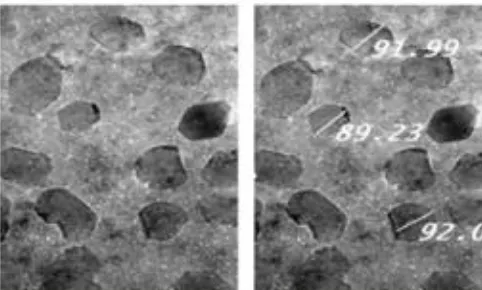
SeNPs was prepared according to Zhang et al. (2001) and Wang et al. (2007). By using bovine albumin protein as a disperser, elemental selenium ions were produced via reducing 25 mM sodium selenite by 25 mM glutathione (GSH). This leds to an aggregation of Se into particles 88.23-92.00 nm in size (SeNPs), forming nano red elemental Se and oxidized glutathione (GSSG). The red solution was dialyzed to separate GSSG from the SeNPs. The final solution containing SeNPs was lyophilized and stored at room temperature.
Characterization of SeNPs
The produced SeNPs were characterized by TEM. According to Chen et al. (2008), TEM samples were prepared by dispersing the powder particles onto holey carbon film on copper grids. Then, the micrographs were obtained on TEM (Philips CM-10, FEI Inc., Hillsboro, OR, USA) as shown in Fig.(1).
Rats
Sixty adult female albino rats of Wistar strain (150 ± 10 g) were obtained from a breeding stock preserved in the Animal House of the National Research Centre, Egypt. They were kept in a group of 5 in polypropylene cage, housed in animal facility in an environmentally conditioned room with respect to light, temperature and air humidity and fed with standard rodent chow
ad libitum and water. All rats were accommodated with these
laboratory conditions for at least two weeks before the commencement of the experiment and they were maintained under the same conditions all over the experiment. All procedures were done with proper approval of Animal care and Ethics Committee of Medical Research of the National Research Centre, Giza, Egypt.
Experimental Setting
Rats were randomly classified into four groups (15 rats for each). The first group received 0.5 ml saline solution intraperitoneally three times a week for 6 weeks and served as normal control group (control). The rats in the second group were injected intraperitoneally (i.p) with 100mg/Kg b.wt of TAA dissolved in saline three times a week for 6 weeks (Strand et al., 2008) (TAA group). The rats in the third group were injected i.p with TAA and simultaneously administered orally with SeNPs 3 mg/Kg b.wt three times/week for 6 weeks (TAA+ SeNPs group). The rats in the forth group were injected i.p with TAA and simultaneously administered orally with 3 mg/ Kg b.wt Se three times/week for 6 weeks (Heikal et al., 2012) (TAA + Se group).
After completion of the round, the diets were withheld from the experimental rats for 12 hours and then blood samples were immediately withdrawn from the retro-orbital venous plexus under diethyl ether anesthesia, left for 15 min., centrifuged at 1899 xg for separation of seram. After that, the rats were sacrificed by cervical dislocation and the liver was dissected carefully weighted and blotted dry. Then, each liver was divided sagittally into two portions; the first one was immediately homogenized in phosphate buffer (pH 7.4), centrifuged at 1800 xg and the supernatant was obtained for biochemical analysis. The second portion was used for histopathological examination and stained by hematoxylin & eosin stain for examination through the light microscope (Banchroft et al, 1996).
Biochemical analyses
Determination of liver enzymes
Determination of oxidative stress parameters and glutathione content
Homogenate liver was prepared in 50 mM Tris-HCl and 300 mM sucrose, pH 7.4 using homogenizer to give 10% homogenate. This homogenate was used for determination of malondialdehyde (MDA) by reaction with thiobarbituric acid (Ohkawa et al., 1979), nitric oxide (NO) by optimized acid reduction method (Green et al., 1982) and glutathione contents by the reduction of Elman's reagent (5,5` dithiobis (2-nitrobenzoic acid) "DTNB") (Ellman, 1959).
Determination of enzymatic antioxidants
Homogenate liver was used for assessment of superoxide dismutase (SOD) by inhibiting phenazine methosulphate-mediated reduction of nitroblue tetrazolium (NBT) dye (Nishikimi et al.,1972), catalase (CAT) by reaction with known quantity of H2O2 (Aebi, 1984), glutathione peroxidase (GPx) by the recycling of oxidized glutathione (GSSG) to its reduced state (Paglia and Valentine, 1967) and glutathione reductase (GR) by catalyzing the reduction of glutathione in the presence of NADPH (Factor et al., 1998).
Determination of cytokines
Prostaglandin E2 (PGE2), prostaglandin F2α (PGF2α), tumor necrosis factor-alpha (TNF-α) and angiogenin (Ang) were quantified in liver homogenate using ELISA kits obtained from
Abcam Company (Cambridge, UK) according to the
manufacturer’s instructions.
Histopathological method
Autopsy samples from the second portion of liver of each rat were taken in the different groups and fixed in 10% formalin saline for 24 h. Washing was performed by tap water, then serial dilutions of alcohol (methyl, ethyl and absolute ethyl)
were used for dehydration. Specimens were cleared in xylene and embedded in paraffin at 56˚ in hot air for 24 h. Paraffin bees wax tissue blocks were prepared for sectioning at 4 microns thickness by sledge microtome. The obtained tissue sections were collected on glass slides, deparaffinized,
stained by hematoxylin & eosin stain then examination was done through the light microscope (Banchroft et al., 1996).
Statistical analysis
All data were subjected to a one-way analysis of variance ANOVA, and the significance of the differences between means was tested using Tukey's honestly significant difference test (p<0.05). The software used was SAS, version 9.1 (Statsoft Inc., Tulsa, OK). Values are expressed as means ± standard error.
RESULT AND DISCUSSION Biochemical results
The current data show significant elevation in serum AST, ALT, ALP and GGT activities in rats upon TAA administration versus the controls. Whereas, the enzyme activities of AST, ALT, ALP and GGT revealed significant reduction as a result of the treatment with SeNPs (-27.74%, -43.13%, -31.96% and -30.18%, respectively) and Se (-23.66%, -38.63%, -28.55% and -22.86%, respectively) in comparison with TAA-challenged group (Table 1). The tabulated results in Table (2) show significant rise in hepatic MDA and NO levels paralleled by significant drop in hepatic GSH level in TAA administered group relative to the control group. Meanwhile, hepatic MDA and NO levels revealed significant decline in the group treated with SeNPs 13.70% and – 49.71% respectively) and that treated with Se (-7.31% and -43.88% respectively) as compared with TAA-challenged group. Treatment with SeNPs or Se also revealed significant elevation in hepatic GSH level (77.02% and 59.39% respectively) as compared to TAA-challenged group. In the present study, TAA administration evoked significant reduction in liver antioxidant enzymes (CAT, SOD, GRd and GPx) activity versus the control group. Whereas, the activities of the antioxidant enzymes revealed significant up regulation in the group treated with SeNPs ( 63.41%, 13.43%, 42.26 % and 37.56% respectively) and that treated with Se (56.1%, 11.81%, 24.62% and 21.09% respectively) as compared with TAA-challenged group (Table 3). The existing results show significant increase in liver prostaglandin F2α, prostaglandin E2, angiogenin (Ang) and TNF-α levels in TAA administered group in respect to the corresponding values in the control group. However, treatment with SeNPs or Se evoked significant downregulation in liver prostaglandin F2α (7.96% and 2.78% respectively), prostaglandin E2 (15.13% and -7.94% respectively), Ang (-42.26% and -32.7% respectively) and TNF-α (-29.78% and -23.8% respectively) relative to the corresponding values in TAA-challenged group (Table 4).
Table 1: Influence of treatment with SeNPs and Se on TAA-induced alterations in liver enzymes of rats.
Groups ALT (U/L) AST (U/L) ALP (U/L) GGT (U/L)
Control 64.60±3.36 85.50±2.33 129.70±4.23 3.75±0.34
TAA 139.03±5.87a 127.46±5.04a 199.58±3.33a 5.60 ±0.45a
TAA + SeNPs 79.07±4.00b 92.10±2.06b 135.80±5.10b 3.91±0.22b
TAA + Se 85.32±3.62b 97.30±4.76b 142.60±3.10b 4.32±0.40b
Table. 2: Influence of treatment with SeNPs and Se on TAA-induced devaststing effect on the oxidant/antioxidant markers in liver tissue of rats.
Groups GSH (mmol/g tissue) MDA (nmol/g tissue) NO (μmol/g tissue)
Control 0.0957±0.02 275.11±13.32 205.89±5.22
TAA 0.0692±0.01a 321.20±12.73a 495.08±11.00a
TAA + SeNPs 0.1225±0.04b 277.18± 4.73b 248.96±9.51b
Histological findings
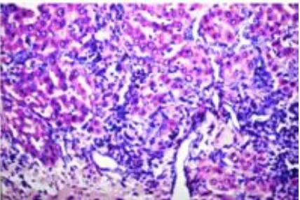
Microscopic examination of liver tissue section of control rat shows normal histological structure of the central vein and the surrounding hepatocytes in the parenchyma as well (Fig-1). Fig. (2) represents photomicrograph of liver tissue section of TAA-administered rat showing fibrosis with inflammatory cells infiltration in between the multiple numbers of newly formed bile duct in portal area that extended between the hepatocytes to form lobules with congesting portal vein. Also, microscopic investigation of liver tissue section of rat administered TAA shows oval cells hyperplasia (Fig. 3). Fig.(4) records photomicrograph of liver tissue section of rat treated with SeNPs showing inflammatory cells infiltration in portal area. Fig.(5) illustrats the photomicrograph of liver tissue section of rat treated with Se showing inflammatory cells infiltration in portal area and between the hepatocytes with congestion of central and portal veins.
Fig. 1: High-resolution transmission electron microscopy (TEM) image of SeNPs.
TAA experimental model represents liver fibrosis mimic to human nonbiliary liver diseases. From this pre-clinical animal model, various essential pathological processes and the contributed mechanisms of liver fibrosis can be cleared and participated in the development of novel diagnostic and therapeutic strategies for this disease. TAA is metabolically transformed into thioacetamide-S-oxide (TASO), acetamide and sulfate through microsomal oxidase
system. TASO has been found to cause centrilobular hepatic necrosis. A further metabolism causes biotransformation of TASO to thioacetamide-S, S-dioxide (TASO2), which covalently binds to proteins forming acetylimidolysine derivatives that act as hepatotoxic compounds (Chen et al., 2015). Metabolism of TAA generates reactive compounds that increase the oxidative stress which is responsible for the damage of liver cells, in parallel with the leakage of AST, ALT, ALP and GGT from the destroyed liver cells (Czechowska et al., 2015). The amount of these cellular enzymes present in the blood reflects the alteration in hepatic cells plasma membrane integrity and/or permeability (Chen et al., 2008).
Fig. 2: Optical micrograph of a cross-sectioned liver tissue of rat in the control group showing normal histological structure of the central vein and the surrounding hepatocytes in the parenchyma (H. E x40).
Reddy et al. (2004) cited that the hepatotoxic compound yields from the binding of TASO2 with tissue macromolecules is responsible for the excessive production of oxidative stress in association with considerable reduction in the glutathione (GSH) pool in the liver. The reduction of intracellular GSH due to TAA administration as shown in the present study has been explained by Fontana et al. (1996) as they reported that the active metabolite of TAA can combine with the sulphahydryl groups of proteins resulting in rapid depletion of intracellular GSH concentration. Therefore, the decline of GSH becomes one of the most important Table 3: Influence of treatment with SeNPs and Se on TAA–induced detrimental impact on hepatic antioxidant enzymes activity of rats.
Groups SOD
(U/g tissue)
CAT (U/g tissue)
GPx (U/g tissue)
GRd (μmol/g tissue)
Control 2213.54±35.81 0.52±0.008 980.15±13.19 2089.88±71.89
TAA 1913.21±7.38a 0.41±0.004a 870.78±29.96a 1488.52±74.43a
TAA + SeNPs 2170.10±17.82b 0.67±0.004b 1238.74±50.36b 2047.65±54.34b
TAA + Se 2139.26±2.30b 0.64±0.008b 1085.18±33.02b 1802.46±81.52b
GRd : glutathione reductase; GPx : glutathione peroxidase
Table 4: Influence of treatment with SeNPs and Se on TAA –induced overshotting of liver inflammatory mediators of rats.
Groups Prostaglandin F2α (pg/g tissue) PGE2
(pg/g tissue)
Ang (pg/g tissue)
TNFα (pg/g tissue)
Control 57.12±1.75 719.53±16.19 20.59±0.98 74.28±3.24
TAA 70.39±1.25a 1018.15±23.88a 38.90±1.53a 282.27±12.29a
TAA + SeNPs 64.79±1.85b 864.11±17.83b 22.46±0.49b 198.21±7.10b
TAA + Se 68.45±1.47b 937.33±19.22b 26.18±1.21b 215.10±22.76b
toxic effects of TAA which motivates excessive generation of ROS at the mitochondrial level leading to damage of cellular components.
Depletion of endogenous GSH leads to overproduction of H2O2 which is very toxic compound and in the presence of transition metal such as iron; it can generate a highly toxic hydroxyl ions that induce lipid peroxidation. This mechanism explains the burden of TAA on hepatic cells following its metabolism into highly reactive metabolites which elicit the denaturation of cellular biomolecules like lipids resulting in lipid peroxidation and its by product (Cheng-Haung et al., 2004).
Overproduction of lipid peroxidation product leads to
destabilization in cellular lipid substances and motivation of oxidative damage especially of membrane structures.
Several lines of evidences indicated that exposure of cells to H2O2 upregulates eNOS expression and NO production (Drummond et al., 2000). This represents the underlying mechanism by which TAA could elevate hepatic NO level in the present work. The major components of the antioxidant protective system in the mammalian cells are the following enzymes; CAT, SOD, GPx and GRd. These enzymes act as a mutually supportive team for defense against ROS (Salama et al., 2013). The suppressed activity of SOD and CAT in liver upon TAA administration might be due to the enhanced lipid peroxidation and/or structural and functional inactivation of these antioxidant enzymes due to overproduction of free radicals (Georgieva et al., 2004). The observed drop in hepatic GSH-Px and GRd activity in TAA administered rats could be attributed to the decreased availability of intracellular GSH. The detectable inhibition in the activity of hepatic SOD, CAT, GPx and GRd indicate hepatic damage in rats following administration of TAA.
NF-κB acts as a key candidate of fibrosis in HSCs and/or
hepatic myofibroblasts (HMF). Luedde and Schwabe (2011)
suggested that NF-κB regulates three key aspects of HSC and/or
hepatic myofibroblast biology; activation, survival and
inflammatory responses. NF-κB potentiates hepatic fibrosis due to several actions; direct fibrogenic action, antiapoptotic action and the secretion of macrophage-recruiting chemokines. The intensified activation of NF-κB in HSC/HMF may also implicate to a tumor-friendly microenvironment in the fibrotic liver (Appel
et al., 2015). A wide range of proinflammatory mediators can
promote NF-κB in HSC/HMF including LPS, TNF-α, IL-1β, angiotensin II and CD40L (Seki et al., 2007). Also, NF-κB can be activated by the generation of ROS in the liver (Hyoudou et al., 2007). The induction of NFκB in association with the activation of HSC often relates to liver damage because it imposes a constraint on HSC apoptosis, further leading to aggregating hepatic fibrosis (Czechowska et al., 2015). Wen et al. (2014) stated that tissue damage occurred after TAA intoxication is accompanied by significant rise in the proinflammatory modulators. These investigators suggested that such changes are due to activation of NF-Kβ. Therefore, the current data of increasing PGF2α, PGE2, ang and TNF-α in liver tissue upon TAA administration could be derived from the motivation of NF-Kβ signaling. Selenium is an
essential trace element that is presented in the body as
Se-containing proteins (selenoproteins), which contain a
selenocysteine group instead of the sulfur-containing cysteine. The well-characterized selenoproteins are GPx and thioredoxin reductase (Min-Chang et al., 2014). Selenoproteins perform variety of important physiological tasks. The current biochemical analysis and histological examination indicated that selenium in its two forms could suppress hepatic fibrogenesis and restore liver functions. These findings are in harmony with those of He et al. (2004) who stated that selenium has powerful influences on hepatic fibrosis in rats by improving immunity and inhibiting NF-Kβ and TGF-β1 expression. Se posses anti-inflammatory effects through regression of the proinflammatory mediators, likes TNF-α and IL-1β and retraction of NF-Kβ which has a positive correlation with the other proinflammatory mediators (Min-Chang et al., 2014). In addition, selenium is able to protect hepatic cells from oxidative damage via its free radical scavenging activity (Shafik and El Batsh, 2016) plus its ability to enhance the antioxidant protective system (Newairy et al., 2007; Jihen et al., 2009). Through this way, selenium could reverse liver enzymes activity (AST, AST, ALP and GGT) in serum (Messarah et al., 2012). The inhibition of the liver enzymes activity in the blood confirms that selenium can maintain the normal structural and architectural integrity of hepatocytes by restricting the leakage of these enzymes. This evidenced the membrane-stabilizing property of selenium.
recover the liver structural organization of the treated rats as shown in biochemical and histological findings of the current study. This is the third document for the strong ability of SeNPs to mitigate liver fibrosis. On the histological point of view TAA
administration showed necrosis with inflammatory cells
infiltration and fibrosis (Fig. 2&3). These findings agree with the previous report of Anbarasu et al. (2012) who observed that TAA
supplementation caused liver toxicity distinguished by
centrilobular necrosis along with various gradations of fatty changes comprising of tiny to large sized vacuoles.
Fig. 3: Optical micrograph of a cross-sectioned liver tissue of rat in TAA group showing fibrosis (f) with inflammatory cells infiltration (m) in between the multiple numbers of newly formed bile duct (bd) in portal area and extended between the hepatocytes forming a lobules with congestion in portal vein (pv) (H. E x10).
Treatment with SeNPs resulted in the disappearance of liver fibrosis but the inflammatory cells infiltration in portal area as shown in Fig. (4). Examination of liver tissue sections of rats Se –treated group are still present showed inflammatory cells infiltration in portal area and between the hepatocytes with congestion of central and portal veins (Fig. 5). Bhattacharjee et al. (2014) confirmed these findings as they mentioned that SeNPs and Se can effectively decrease the degree of hepatic fibrosis and support the recovery process (Ding et al., 2010).
Fig. 4: The magnification of (Figure 3.) showing oval cells hyperplasia (c) (H. E x40).
In conclusion, the outcomes of the current research study encourage the use of selenium either in free form or in
nanoformulation as antifibrotic candidate. The underlying mechanisms for this effect include: 1) membrane stabilizing capacity, 2) free radical scavenging activity, 3) antioxidative potential and anti-inflammatory action. Notably, SeNPs showed superior potency than Se in our study and the main cause could be attributed to the small sized particles, large surface area and increased bioavailabilty.
Fig. 5: Optical micrograph of a cross-sectioned liver tissue of rat in TAA + SeNPs group showing inflammatory cells infiltration (m) in portal area (H. E x40).
Fig. 6: Optical micrograph of a cross-sectioned liver tissue of rat in TAA + Se group showing inflammatory cells infiltration (m) in portal area and between the hepatocytes with congestion in central (cv) and portal veins (pv)(H. E x10).
Financial support and sponsorship: Nil.
Conflict of Interests: There are no conflicts of interest.
REFERENCES
AAFCO, AAFCO Model Guidance Document. Official guidelines suggested for contaminants in individual mineral feed ingredients. Official publication. Association of American Feed Control Officials Inc., Olympia, WA. 2011; 304.
Aebi H, Catalase in vitro. Methods Enzymol. 1984; 105:121-126.
Amara S, Lopez K, Banan B, Brown SK, Whalen M, Myles E, Ivy MT, Johnson T, Schey KL, and Tiriveedhi V, Synergistic effect of proinflammatory TNFα and IL-17 in periostin mediated collagen deposition: Potential role in liver fibrosis. Molecular Immunology. 2015; 64: 26-35.
induced hepatotoxicity in rats. Asian Pac J Trop Biomed, 2012; 2: 511-515.
Appel K, Meiser P, Millán E, Collado JA, Rose T, Gras CC, Carle R, and Muñoz E, Chokeberry (Aronia melanocarpa (Michx.) Elliot) concentrate inhibits NF-κB and synergizes with selenium to inhibit the release of pro-inflammatory mediators in macrophages. Fitoterapia, 2015; 105:73-82.
Banchroft JD, Stevens A, and Turner DR, Theory and practice of histological techniques 4th ed. New York, London, San Francisco, Tokyo, Churchil Livingstone, 1996.
Bhattacharjee A, Basu A, Ghosh P, Biswas J, and Bhattacharya S, Protective effect of Selenium nanoparticle against cyclophosphamide induced hepatotoxicity and genotoxicity in Swiss albino mice. J Biomater Appl, 2014; 29: 303-317.
Bowers GN, and McComb RB. A continuous spectrophotometric method for measuring the activity of serum alkaline phosphatase. Clin Chem, 1966; 12:70-89.
Chen PJ, Chiu CH, Tseng JK, Yang KT and Chen YC, Ameliorative effects of D-glucuronolactone on oxidative stress and inflammatory/fibrogenic responses in livers of thioacetamide-treated rats. Journal of Functional Foods, 2015; 14:154-162.
Chen TF, Wong YS, Zheng WJ, Bai Y, and Huang L, Selenium nanoparticles fabricated in Undaria pinnatifida polysaccharide solutions induce mitochondria-mediated apoptosis in A375 human melanoma cells. Colloids Surf B, 2008; 67: 26–31.
Czechowska G, Celinski K, Korolczuk A, Wojcicka G, Dudka J, Bojarska A, and Reiter RJ, Protective effects of melatonin against thioacetamide-induced liver fibrosis in rats. J Physiol Pharmacol, 2015; 6: 567-579.
Ding M, Potter JJ, Liu X, Torbenson MS, and Mezey E, Selenium supplementation decreases hepatic fibrosis in mice after chronic carbon tetrachloride administration. Biol Trace Elem Res, 2010; 133: 83-97.
Drummond GR, Cai H, Davis ME, Ramasamy S, and Harrison DG, Transcriptional and Posttranscriptional Regulation of Endothelial Nitric Oxide Synthase Expression by Hydrogen Peroxide. Circ. Res, 2000; 86: 347-354.
Ellman GL, Tissue sulfhydryl groups. Arch Biochem Biophys, 1959; 82:70-77.
European Union, List of the authorized additives in feeding-stuffs published in application of Article 9t (b) of Council Directive 70/524/EEC concerning additives in feeding stuffs. Off. J. Eur. Union, 2004; C/50:1–144.
Factor VM, Kiss A, Woitach JT, Wirth PJ, and Thorgeirsson SS, Disruption of redox homeostasis in the transforming growth factoralpha/c-myc transgenic mouse model of accelerated hepatocarcinogenesis. J Biol Chem, 1998; 273: 15846–15853.
Fontana L, Moreira E, Torres MI, Fernández MI, Ríos A, Sánchez de Medina F, and Gil A, Serum amino acid changes in rats with thioacetamide-induced liver cirrhosis. Toxicology, 1996; 106: 197-206.
Georgieva N, Gadjeva V, and Tolekova A, New isonicotinoylhydrazones with SSA protect against oxidative-hepatic injury of isoniazid. TJS, 2004; 2: 37-43.
Green LC, Wagner DA, Glogowski J, Skipper PL, Wishnok JS, and Tannenbaum SR, Analysis of nitrate, nitrite, and [15N] nitrate in biological fluids. Anal Biochem, 1982; 126: 131-138.
He W, Shi F, Zhou ZW, Li B, Zhang K, Zhang X, Ouyang C, Zhou SF, and Zhu X, A bioinformatic and mechanistic study elicits the antifibrotic effect of ursolic acid through the attenuation of oxidative stress with the involvement of ERK, PI3K/Akt, and p38 MAPK signaling pathways in human hepatic stellate cells and rat liver. Drug Des Devel Ther, 2015; 9: 3989-4104.
He YT, Liu DW, Ding LY, Li Q, and Xiao YH, Therapeutic effects and molecular mechanisms of anti-fibrosis herbs and selenium on rats with hepatic fibrosis. World J Gastroenterol, 2004, 10: 703-706.
Heikal TM, EL-Sherbiny M, Hassan SA, Arafa A, and Ghanem HZ, Antioxidant effect of selenium on hepatotoxicity induced by chlorpyrifos in male rats. Int J Pharm Pharm Sci, 2012; 4: 603–609.
Hyoudou K, Nishikawa M, Kobayashi Y, Kuramoto Y, Yamashita F, and Hashida M, Analysis of in vivo nuclear factor-kappaB activation during liver inflammation in mice: prevention by catalase delivery. Mol Pharmacol, 2007; 71: 446-453.
Jihen H, Imed M, Fatima H, and Abdelhamid K, Protective effects of selenium (Se) and zinc (Zn) on cadmium (Cd) toxicity in the liver of the rat: effects on the oxidative stress. Ecotoxicol Environ Saf, 2009; 72: 1559-1564.
Luedde T, and Schwabe RF, NF-κB in the liver--linking injury, fibrosis and hepatocellular carcinoma. Nat Rev Gastroenterol Hepatol, 2011; 8: 108-118.
Messarah M, Klibet F, Boumendjel A, Abdennour C, Bouzerna N, Boulakoud MS, El Feki A, Hepatoprotective role and antioxidant capacity of selenium on arsenic-induced liver injury in rats. Exp Toxicol Pathol, 2012; 64: 167-174.
Min-Chang G, Wei-Hong T, Zhen X, and Jie S, Effects of Selenium-Enriched Protein from Ganoderma lucidum on the Levels of IL-1 β and TNF- α , Oxidative Stress, and NF- κ B Activation in Ovalbumin-Induced Asthmatic Mice. Evid Based Complement Alternat Med, 2014; 2014:182817.
Ministry of Agriculture, Ministry of Agriculture of People’s Republic of China Bulletin No. 1224–2010. The safety use standard of feed additives. Beijing, China, 2010.
Newairy AA, El-Sharaky AS, Badreldeen MM, Eweda SM, and Sheweita SA, The hepatoprotective effects of selenium against cadmium toxicity in rats. Toxicology, 2007; 242: 23-30.
Nishikimi M, Appaji N, and Yagi K, The occurrence of superoxide anion in the reaction of reduced phenazine methosulfate and molecular oxygen. Biochem Biophys Res Commun, 1972; 46: 849-854.
Novo E, Cannito S, Paternostro C, Bocca C, Miglietta A, and Parola M, Cellular and molecular mechanisms in liver fibrogenesis. Arch Biochem Biophys, 2014; 548: 20–37.
Ohkawa H, Ohishi N, and Yagi K, Assay for lipid peroxides in animal tissues by thiobarbituric acid reaction. Anal Biochem, 1979; 95: 351-358.
Osawa Y, Hoshi M, Yasuda I, Saibara T, Moriwaki H, and Kozawa O, Tumor necrosis factor-alpha promotes cholestasis-induced liver fibrosis in the mouse through tissue inhibitor of metalloproteinase-1 production in hepatic stellate cells. PLoS One, 2013; e65251.
Paglia DE, and Valentin WN, Studies on the quantitative and qualitative characterization of erythrocyte glutathione peroxidase. J. Lab. Clin. Med, 1967; 70:158-169.
Pelyhe C, and Mézes M, Myths and facts about the effects of nanoselenium in farm animals-mini review. Eur Chem Bull, 2013; 2: 1049–1052.
Reddy PV, Murthy Ch R, and Reddanna P, Fulminant hepatic failure induced oxidative stress in nonsynaptic mitochondria of cerebral cortex in rats. Neurosci Lett, 2004; 368: 15-20.
Reitman S, and Frankel SA, A colorimetric method for the determination of serum glutamic oxaloacetic and pyruvic transaminases. Am. J. Clin. Pathol, 1957; 28: 56-63.
Rosenbloom J, Mendoza FA, and Jimenez SA, Strategies for anti-fibrotic therapies. Biochim Biophys Acta, 2013; 1832: 1088–1103.
Salama SM, Abdulla MA, Alrashdi AS, and Hadi AH, Mechanism of Hepatoprotective Effect of Boesenbergia rotunda in Thioacetamide-Induced Liver Damage in Rats. Evid Based Complement Alternat Med, 2013; 2013:157456.
Sarkar B, Bhattacharjee S, Daware A, Tribedi P, Krishnani KK, and Minhas PS, Selenium Nanoparticles for Stress-Resilient Fish and Livestock. Nanoscale Res Lett, 2015; 10: 371.
Scott C, Cha K, Rao R, Liddle C, George J, and Gunton JE, Hepatocyte-specific deletion of ARNT (aryl hydrocarbon Receptor Nuclear Translocator) results in altered fibrotic gene expression in the thioacetamide model of liver injury. PLoS One, 2015; 10: e0121650.
Seki E, De Minicis S, Osterreicher CH, Kluwe J, Osawa Y, Brenner DA, and Schwabe RF, TLR4 enhances TGF-beta signaling and hepatic fibrosis. Nat Med, 2007; 13: 1324-1332.
Oxidative Stress Markers in Arsenic-Induced Hepatotoxicity in Rats. Biol Trace Elem Res, 2016; 169: 121-128.
Strand P, Tao GZ, Zhou Q, Harada M, Toivola DM, Brunt EM, and Omary MB, Keratin mutation predisposes to mouse liver fibrosis and unmasks differential effects of the CCl4 and thioacetamide models. Gastroenterology, 2008; 134: 1169-1179.
Tietz NW, Clinical Guide to Laboratory Tests, 3rd ed. W.B. Sanuders Co. Philadelphia PA, 1995.
Wang CH, Chen YJ, Lee TH, Chen YS., Jawan B, Hung KS, Lu CN, and Liu JK, Protective effect of MDL 28170 against thioacetamide induced acute liver failure in mice". J. Biomed. Sci, 2004; 11: 571-578.
Wang HL, Zhang JS, and Yu HQ, Elemental selenium at nano size possesses lower toxicity without compromising the fundamental effect on selenoenzymes: comparison with selenomethionine in mice. Free Radic Biol Med, 2007; 42: 1524–1533.
Wang J, Zhang Y, Yuan Y, and Yue T, Immunomodulatory of selenium nano-particles decorated by sulfated Ganoderma lucidum polysaccharides. Food Chem Toxicol, 2014; 68: 183-189.
Wang Y, Yan X, and Fu L, Effect of selenium nanoparticles with different sizes in primary cultured intestinal epithelial cells of crucian carp, Carassius auratus gibelio. Int J Nanomedicine, 2013; 8: 4007–4013.
Wen SL, Gao JH, Yang WJ, Lu YY, Tong H, Huang ZY, Liu ZX, and Tang CW, Celecoxib attenuates hepatic cirrhosis through inhibition of epithelial-to-mesenchymal transition of hepatocytes. J Gastroenterol Hepatol, 2014; 29(11):1932-1942.
Wu J, and Zern MA, Hepatic stellate cells: A target for the treatment of liver fibrosis. Journal of Gastroenterology, 2000; 35: 665– 672.
Zhang J, Wang X, and Xu T, Elemental selenium at nano size (Nano-Se) as a potential chemopreventive agent with reduced risk of selenium toxicity: comparison with se-methylselenocysteine in mice. Toxicol Sci, 2008; 101: 22-31.
Zhang JS, Gao XY, Zhang LD, and Bao YP, Biological effects of a nano red elemental selenium. Biofactors, 2001; 15: 27-38.
Zhang X, Xu Y, Qi Y, Han X, Yin L, Xu L, Liu K, and Peng J, Potent effects of dioscin against thioacetamide-induced liver fibrosis through attenuating oxidative stress in turn inhibiting inflammation, TGF-β/Smad and MAPK signaling pathways. Journal of Functional Foods, 2015; 16: 436-447.
Zheng S, Li X, Zhang Y, Xie Q, Wong YS, Zheng W, and Chen T, PEG-nanolized ultrasmall selenium nanoparticles overcome drug resistance in hepatocellular carcinoma HepG2 cells through induction of mitochondria dysfunction. Int J Nanomedicine, 2012; 7: 3939-3949.
How to cite this article: