consequences of CVD, all attempts to identify oth-er modifiable risk factors are extremely important, as they would contribute to more effective preven-tion of CVD.
The amount of evidence confirming the role of chronic inflammatory processes in initiation and progression of CVD continues to increase [2]. However, it is not conclusively clarified wheth-er local inflammation in the pwheth-eriodontium is suf-ficient to trigger a systemic inflammatory cascade and initiate atherosclerosis [3]. In view of conflict-Cardiovascular diseases (CVD) constitute the
most frequent cause of death worldwide. Accord-ing to the National Institute of Public Health, in Poland in 2010, 456 individuals per 100,000 died of CVD, which accounted for 46.0% of all deaths [1]. MI was the cause of 17,800 deaths, which repre-sented 10.0% of deaths from cardiovascular causes. Etiopathogenesis of CVD as social diseases is mul-tifactorial, but the coexistence of traditional CVD risk factors does not fully explain the overall car-diovascular risk in all patients. Due to the fatal
Bartłomiej Górski
1, B, D, Ewa Nargiełło
2, B, D, Ewa Grabowska
1, C,
Grzegorz Opolski
2, A, E, Renata Górska
1, A, E, FThe Association Between Dental Status
and Risk of Acute Myocardial Infarction Among Poles:
Case-control Study
1 Department of Periodontology and Oral Mucous Membrane Diseases, Medical University of Warsaw, Poland 2 First Department of Cardiology, Medical University of Warsaw, Poland
A – research concept and design; B – collection and/or assembly of data; C – data analysis and interpretation;
D – writing the article; E – critical revision of the article; F – final approval of article
Abstract
Background. Results of scientific research on the effects of periodontitis on the incidence of myocardial infarction (MI) are ambiguous.
Objectives. The aim of this study was to investigate the association of the severity and extent of periodontitis with acute MI in Poles.
Material and Methods. This case-control study included 134 cases hospitalized with acute MI under the age of 70 years and 155 controls drawn from the general population with no MI history. Sociodemographic, cardiologic and periodontal variables were assessed. Three periodontal indicators were evaluated: (1) the severity of periodon-titis classified in accordance with Page and Eke definition, (2) the extent of periodonperiodon-titis determined on the basis of the percentage of sites with CAL ≥ 3 mm (Arbes Index) and (3) tooth loss (> 10 teeth). In a logistic regression model, the association of periodontal parameters with MI occurrence was evaluated after adjusting for well-known cardiovascular risk factors.
Results. The extent of periodontitis was significantly associated with the risk of acute MI even after adjusting for age, sex, tobacco smoking, hypertension, diabetes, BMI, education and income (odds ratio [OR] = 2.4; 95% con-fidence interval [CI] = 1.1 to 5.2; p = 0.0203). However, the severity of periodontitis was associated with MI after adjusting for age and sex (OR = 2.0; 95% CI = 1.2–3.5; p = 0.0109), but not after adjusting for the other above-men-tioned risk factors. The association between the number of lost teeth and acute MI was significant after adjusting for age, sex, tobacco smoking, arterial hypertension and diabetes mellitus (OR = 2.1; 95% CI = 1.2–5.9; p = 0.0151).
Conclusions. This study proves the positive association between periodontitis and acute MI in Poles. This associa-tion seems to be stronger with regard to the extent rather than to the severity of periodontitis (Adv Clin Exp Med 2016, 25, 5, 861–870).
Key words: periodontitis, acute myocardial infarction, risk factor.
ORIGINAL PAPERS
Adv Clin Exp Med 2016, 25, 5, 861–870
ing results of studies, two meta-analyzes on the association between periodontitis and CVD were conducted [4, 5]. Both demonstrated that peri-odontitis independently increases the risk of cor-onary heart disease (CHD). Most recently, a com-prehensive review performed by the American Heart Association (AHA) working group, as well as a report by the Joint Workshop of Europe-an Federation of Periodontology Europe-and the Ameri-can Academy of Periodontology (EFP/AAP) high-lighted that periodontitis is associated with CVD independently of known confounders, but there is no evidence for a causal link [3, 6]. What is more, the Fifth Joint Task Force of the European Society of Cardiology (ESC) and Other Societies on Car-diovascular Disease Prevention in Clinical Practice stated that “periodontitis can be considered a risk indicator for a generally decreased cardiovascular health status” [7]. However, all of the statements concluded that more studies are needed to draw further conclusions.
This is of great importance due to the wide-spread occurrence of periodontal disease in the Polish population. Among Poles periodontitis oc-curred in 57.2% of those aged 35–44 years and even more frequently among older people in contrast to 37.2% of adults between the age of 35–44 years in the United States [8, 9].
To date, few studies have assessed the associa-tion between periodontitis and MI, but the results of conducted research are inconclusive. Some of them demonstrated that periodontitis constitutes an independent MI risk factor [10–17], where-as other studies did not confirm these relation-ships [18–20]. Therefore, the aim of this study was to answer the question whether the periodontitis severity and extent is an independent and signifi-cant risk factor for acute MI in adult Poles under the age of 70 years.
Material and Methods
Study Sample
This study was conducted in the Department of Oral Medicine and Periodontal Diseases, Medi-cal University of Warsaw (MUW) and in the First Clinic and Department of Cardiology, Faculty of Medicine, MUW, in 2011–2013 in accordance with the STROBE guidelines. All participants were Polish Caucasians.
The study group included 134 patients (29 women, 105 men) hospitalized in the First Clinic and Department of Cardiology due to acute MI on the second or third day of hospitalization. The mean age of this group was 54.3 years (± 8.1).
The inclusion criteria were: 1) MI history and 2) age under 70 years. MI (STEMI and NSTEMI) was diagnosed in accordance with the criteria of Guidelines of ESC. The exclusion criteria included: 1) edentulousness; 2) cancer; 3) rheumatic disease; 4) autoimmune disease; 5) chronic liver disease; 6) chronic renal disease stage 4 and 5; 7) stroke his-tory. Recruitment and inclusion of patients in the study group was conducted by a single cardiolo-gist (EN).
The control group consisted of people select-ed by the Ministry of the Interior and Adminis-tration from the general population of adult Poles who reported to Department of Oral Medicine and Periodontal Diseases. These individuals had to meet the following criteria: 1) age under 70 years; 2) present natural teeth; 3) no cancer history; 4) no rheumatic disease history; 5) no autoim-mune disease history; 6) no chronic liver disease; 7) no chronic renal disease; 8) no stroke history, and 9) no MI history. The control group included 155 individuals (94 women, 61 men). The average age of this group was 54.9 (± 10.0) years.
Social and medical history of the participants was taken. Physical examinations were performed by a single cardiologist (EN). Similarly, a single dentist (BG) conducted dental and periodontal ex-aminations. The study was carried out taking into account the ethical standards resulting from the Declaration of Helsinki of 1975, as revised in 2000. The research was approved by the Bioethics Com-mittee, MUW (approval number KB-145/2011). All individuals involved in the study declared a conscious approval to participate in the project by signing the informed consent form.
Data Collection
Sociodemographic and general medical his-tory of the participants were taken using a tailor-made questionnaire. Information about education, income, and nicotinism was obtained from inter-views. Education was defined as primary, second-ary and higher. Income was determined on the basis of income per family member per month: < €200, €200–350, > €350. The participants were classified as current smokers if they declared smoking 10 or more cigarettes a day continually for at least five years, past smokers and never smokers.
Arterial hypertension was defined as systolic blood pressure ≥ 140 mm Hg or diastolic blood pressure ≥ 90 mm Hg (mean of three consecutive measurements taken at five-minute intervals us-ing a certified sphygmomanometer), or as previ-ous use of antihypertensive drugs.
consecutive tests, random blood sugar was over 200 mg/dL or if the patient was taking drugs due to diagnosed diabetes.
Measurements of height and weight were per-formed using the physician scale with height rod. Measurements of body height in the standing posi-tion were made with 1 cm accuracy and body weight was assessed in the standing position with 0.1 kg accuracy. Waist circumference (in cm) and hip cir-cumference (in cm) were determined using a body measuring tape. Body mass index (BMI) was cal-culated by dividing body weight (kg) by height (in m2). Overweight was diagnosed in case of BMI
25–29.9 kg/m2, and obesity when BMI ≥ 30 kg/m2.
Waist-hip ratio (WHR) was calculated by dividing waist circumference by hip circumference.
Dental Examination
and Periodontal Measurement
Dental and periodontal examinations were performed by one examiner (BG). A calibration exercise was carried out on 10 patients probed twice before conducting a periodontal examina-tion to reach intraexaminer reproducibility. Cal-ibration was accepted if ≥ 90% of the recordings (pocket depth, clinical attachment level) could be reproduced within a difference of 1.0 mm.Dental and periodontal examinations were performed in artificial light, using a dental mir-ror and a periodontal probe with 1 mm scale (HU-FRIEDY PCPUNC 15). The examinations did not include third molars.
Dichotomous plaque index (PI) by O’Leary was established on four surfaces of all teeth (mesi-al, dist(mesi-al, lingual and buccal). The index was eval-uated as the ratio of the surfaces with plaque to all tested surfaces. Bleeding on probing index (BoP) by Ainamo and Bay was evaluated at 4 points around all teeth: mesial-buccal (MB), buccal (B), distal buccal (DB) and lingual (L). BoP was calcu-lated by dividing the sum of bleeding points by the sum of all the test points. Pocket depth (PD) and clinical attachment level (CAL) were evaluated at four sites per tooth: MB, B, DB, L. PD was defined as the distance from the gingival margin to the bot-tom of the pocket established by probing (in mm). CAL was defined as the distance between the bot-tom of the pocket determined by probing and the cementoenamel junction (in mm). Measurements were rounded down to the nearest mm.
Clinical diagnosis of periodontitis and its se-verity was made in accordance with the case defi-nitions for surveillance of periodontitis proposed by the Centers for Disease Control and AAP (Page and Eke definition) as follows:
– mild periodontitis as ≥ 2 interproximal sites with CAL ≥ 3 mm and ≥ 2 interproximal sites with PD ≥ 4 mm (not on the same tooth),
– moderate periodontitis as ≥ 2 interproximal sites with CAL ≥ 4 mm or ≥ 2 interproximal sites with PD ≥ 5 mm (not on the same tooth),
– severe periodontitis as ≥ 2 interproximal sites with CAL ≥ 6 mm (not on the same tooth) and ≥ 1 interproximal site with PD ≥ 5 mm [21].
The extent of periodontitis was classified by the percentage of sites with CAL ≥ 3 mm in line with Arbes Index: 0% = absent; > 0 – ≤ 33% = mild; > 33 – ≤ 67% = moderate; and > 67% = extensive perio-dontitis [10].
Data Analyses
Statistical analysis of the collected data was performed using the PQStat v. 1.4.4 software. Data was analyzed using the χ2 test for categorical
vari-ables and Mann-Whitney U test for continuous variables. The threshold for statistical significance was assumed at p = 0.05, odds ratio (OR) of MI or severe periodontitis was determined with 95% confidence interval (95% CI).
Because a correlation between CVD risk fac-tors and the occurrence of MI, as well as peri-odontitis was confirmed, some models were con-structed taking into account the CVD risk factors and periodontal parameters as potential indepen-dent variables while the case status was a depen-dent variable.
Multivariate analysis was performed by for-ward selection. The first two parameters included in the model were non-modifiable factors associ-ated with CVD and periodontitis: age and gender of the patients. The next step was adding classical CVD risk factors to the models: smoking, arterial hypertension, diabetes mellitus and BMI. The last considered group included the parameters deter-mining the socio-economic status: education and income. Subsequently, the severity of periodon-tits, the extent of periodontitis and the number of lost teeth were included in the multivariate mod-els one at a time. These parameters were convert-ed into dichotomous forms, where the risk factors were: diagnosis of severe periodontitis according to Page and Eke definitions, diagnosis of extensive periodontitis in line with Arbes Index, and over 10 extracted teeth, respectively.
Results
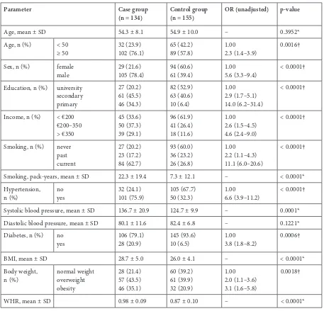
education and income, current smokers and – to a lesser extent – former smokers, patients with di-agnosed hypertension or diabetes, overweight or obese individuals.
There were statistically significant differenc-es in the periodontal status and all periodontal pa-rameters between cases and controls (Table 2). Pa-tients in the study group had worse periodontal indices and a higher number of lost teeth.
In the subsequent analyzes the three chosen periodontal indicators were used in dichotomous forms, dividing patients according to the presence of severe periodontitis, extensive periodontitis and more than 10 lost teeth. A relationship (Table 3) was observed between the severity and the extent of periodontitis and age above 50 years, male gen-der, education, level of income, tobacco
smok-ing and hypertension. However, only the presence of severe periodontitis was associated with body weight.
We observed increased unadjusted ORs of MI for the severity of periodontitis, the extent of periodontitis and the high number of lost teeth (Table 4). After adjusting for non-modifiable CVD risk factors, i.e., age and gender, these val-ues slightly decreased, but a strong and statistically significant relationship between all evaluated pa-rameters and the occurrence of MI still remained. Adjustment for further variables in the multivar-iate models gradually reduced the significance of periodontal parameters, but even after including all the variables (age, sex, smoking, hypertension, diabetes, BMI, education, income), an indepen-dent association between the extent of
periodon-Table 1. Bivariate association between acute myocardial infarction, socio-economic variables and risk factors of acute myocardial infarction
Parameter Case group
(n = 134) Control group (n = 155) OR (unadjusted) p-value
Age, mean ± SD 54.3 ± 8.1 54.9 ± 10.0 – 0.3952* Age, n (%) < 50
≥ 50 32 (23.9)102 (76.1) 65 (42.2)89 (57.8) 1.002.3 (1.4–3.9) 0.0016† Sex, n (%) female
male 29 (21.6)105 (78.4) 94 (60.6)61 (39.4) 1.005.6 (3.3–9.4) < 0.0001† Education, n (%) university
secondary primary
27 (20.2) 61 (45.5) 46 (34.3)
82 (52.9) 63 (40.6) 10 (6.4)
1.00 2.9 (1.7–5.1) 14.0 (6.2–31.4)
< 0.0001†
Income, n (%) < €200 €200–350 > €350
45 (33.6) 50 (37.3) 39 (29.1)
96 (61.9) 41 (26.4) 18 (11.6)
1.00 2.6 (1.5–4.5) 4.6 (2.4–9.0)
< 0.0001†
Smoking, n (%) never past current
27 (20.2) 23 (17.2) 84 (62.7)
93 (60.0) 36 (23.2) 26 (26.8)
1.00 2.2 (1.1–4.3) 11.1 (6.0–20.6)
< 0.0001†
Smoking, pack-years, mean ± SD 22.3 ± 19.4 7.3 ± 12.1 – < 0.0001* Hypertension,
n (%) noyes 32 (24.1)101 (75.9) 105 (67.7)50 (32.3) 1.006.6 (3.9–11.2) < 0.0001† Systolic blood pressure, mean ± SD 136.7 ± 20.9 124.7 ± 9.9 – 0.0001* Diastolic blood pressure, mean ± SD 80.1 ± 11.6 82.4 ± 6.8 – 0.1221* Diabetes, n (%) no
yes 106 (79.1)28 (20.9) 145 (93.6)10 (6.5) 1.003.8 (1.8–8.2) 0.0006†
BMI, mean ± SD 28.7 ± 5.0 26.0 ± 4.1 – < 0.0001* Body weight,
n (%) normal weight overweight obesity
28 (21.4) 57 (43.5) 46 (35.1)
60 (39.2) 61 (39.9) 32 (20.9)
1.00 2.0 (1.1–3.6) 3.1 (1.6–5.8)
0.0018†
WHR, mean ± SD 0.98 ± 0.09 0.87 ± 0.10 – < 0.0001*
Table 2. Bivariate association between acute myocardial infarction and oral health variables
Parameter Case group
(n = 134) Control group (n = 155) OR (unadjusted) p-value
Plaque index (%), mean ± SD 85.7 ± 20.0 65.1 ± 24.8 – < 0.0001* Bleeding on probing index (%),
mean ± SD 49.9 ± 29.9 39.5 ± 26.0 – 0.0033* Pocket depth (mm), mean ± SD 2.9 ± 11 2.2 ± 0.8 – < 0.0001* Clinical attachment loss (mm), mean ± SD 4.2 ± 2.0 2.5 ± 1.5 – < 0.0001* Severity of
peri-odontitis, n (%)‡ no periodontitismild moderate severe
7 (5.2) 7 (5.2) 45 (33.6) 75 (56.0)
38 (24.5) 9 (5.8) 64 (41.3) 44 (28.4)
1.00
4.2 (1.2–15.1) 3.8 (1.6–9.3) 9.3 (3.8–22.5)
< 0.0001†
Extent of
perio-dontitis, n (%)§ > 0 – ≤ 33%> 33 – ≤ 67% > 67%
8 (6.0) 30 (22.4) 96 (71.6)
45 (29.0) 54 (34.8) 56 (36.1)
1.00 3.1 (1.3–7.5) 9.6 (4.2–21.9)
< 0.0001†
Number of teeth lost, median (Q1–Q3) 10 (6–17) 4 (2–8) – < 0.0001*
SD – standard deviation; * Mann-Whitney U test; † χ2 test; ‡ severity of periodontitis defined in accordance with Page and Eke definition [20, 21]; § extent of periodontitis defined in accordance with Arbes Index [10].
titis, expressed as a percentage of sites with CAL ≥ 3 mm and the risk of MI still remained sig-nificant (OR = 2.4; 95% CI 1.1–5.2).
Discussion
The obtained results demonstrate that the se-verity and extent of periodontitis is positively asso-ciated with acute MI, but this relationship is stron-ger with the extent of the disease than its severity. Although many previous studies indicate that peri-odontitis increases the risk of CVD, this associa-tion requires verificaassocia-tion.
Periodontitis and CVD share several com-mon risk factors with well-documented impact, such as age, low socio-economic status and ciga-rette smoking [3, 6, 7]. The most important vari-able in the assessment of a causal relationship be-tween periodontitis and CVD is smoking, because it represents both a confounder, as well as a mod-ifying agent [6]. Statistical analysis adjusted for smoking does not rule out residual confounding, but recent research suggests that the relationship between periodontitis and CVD can be indepen-dent of smoking [14]. In the present study, the im-pact of smoking as a confounder was closely mon-itored. Similarly to other reports, smoking was determined by dividing the subjects into smok-ers at the time of the study, in the past, and never smokers [11, 12].
Among the periodontal parameters related to systemic diseases, CAL seems to be the most im-portant, because it is a measure of cumulative
life-time experience of periodontitis. However, peri-odontitis cannot be verified by measurements of single variable and the evaluation of disease pres-ence requires measurement of PD and BoP. Pre-vious studies assessing the relationship between periodontitis and MI indicate a clear discrepancy of applied periodontal parameters and definitions of periodontitis, which makes comparisons of da-ta obda-tained by different researchers harder. Some studies evaluated the alveolar bone loss on radio-graphs, BoP and root furcation involvement [22], alveolar bone loss on radiographs and PD [14], CAL [10, 15], PD and CAL on selected teeth [12, 16], and PD and CAL for all teeth excluding third mo-lars [11, 13].
OR 2.08 (95% CI = 1.47–2.94) for women after ad-justment for age, BMI, physical activity, diabetes and smoking. Likewise, López et al. [11] received crude OR 3.78 (95% CI = 1.60–8.92) and adjust-ed OR 3.17 (1.31–7.63) after adjusting for diabe-tes, systolic blood pressure and smoking. This can partly be explained by the fact that the study by López et al. [11] included a younger age group of 30–50 years, where lower CAL values can be ex-pected. On the other hand, Cueto et al. [12] ob-served a stronger association between
periodonti-tis and MI with OR = 4.42 (95% CI = 2.22–8.82). After adjusting for other risk factors in a multi-variate analysis (sex, age, smoking, hypertension, diabetes, hypercholesterolemia, regular physi-cal activity), OR reached the value of 3.31 (95% CI = 1.42–7.71). Arbes et al. [10] showed that periodontitis increases the risk of MI on a sam-ple from the NHANES III study. Unadjusted OR for CAL ≥ 3 mm in 0–33% of tested sites was 2.2 (95% CI = 1.3–3.8), and in 33–67% of test-ed sites it equaltest-ed 5.5 (95% CI = 3.4–9.1) and
Table 3. Association between the extent (Arbes Index) and severity (Page and Eke definition) of periodontitis and well-known risk factors of acute myocardial infarction
Parameter Extent of periodontitis Severity of periodontitis
extensive periodon-titis (n = 152)
no extensive periodontitis (n = 137)
p-value severe perio-dontitis (n = 119)
no severe periodonti-tis (n = 170)
p-value
Age, n (%) < 50
≥ 50 32 (21.2)119 (78.8) 65 (47.4)72 (52.5) < 0.0001* 23 (19.3)96 (80.7) 74 (43.8)95 (56.2) < 0.0001*
Sex, n (%) female
male 53 (34.9)99 (65.1) 70 (51.1)67 (48.9) 0.0053* 33 (27.7)86 (72.3) 90 (52.9)80 (47.1) < 0.0001* Education, n (%) university
secondary primary
38 (25.0) 68 (44.7) 46 (30.3)
71 (51.8) 56 (40.9) 10 (7.3)
< 0.0001* 26 (21.8) 56 (47.1) 37 (31.1)
83 (48.8) 68 (40.0) 19 (11.2)
< 0.0001*
Income, n (%) < €200 €200–€350 > €350
56 (36.8) 54 (35.5) 42 (27.6)
85 (62.0) 37 (27.0) 15 (11.0)
< 0.0001* 44 (37.0) 42 (35.3) 33 (27.7)
97 (57.1) 49 (28.8) 24 (14.1)
0.0013*
Smoking habits, n (%) neverpast
current
48 (31.6) 31 (20.4) 73 (48.0)
72 (52.6) 28 (20.4) 37 (27.0)
0.0003* 37 (31.1) 22 (18.5) 60 (50.4)
83 (48.8) 37 (21.8) 50 (29.4)
0.0010*
Smoking, pack-years, mean ± SD 19.3 ± 19.3 8.6 ± 13.4 < 0.0001† 19.3 ± 22.7 10.7 ± 15.7 < 0.0001† Hypertension, n (%) no
yes 61 (40.4)90 (59.6) 76 (55.5)61 (44.5) 0.0105* 43 (36.4)75 (63.6) 94 (55.3)76 (44.7) 0.0016* Systolic blood pressure,
mean ± SD 135.8 ± 19.8 129.4 ± 17.7 0.0125† 137.8 ± 19.1 129.0 ± 18.4 0.0016† Diastolic blood pressure,
mean ± SD 80.5 ± 10.5 81.0 ± 10.7 0.8577† 80.8 ± 10.4 80.6 ± 10.9 0.7265† Diabetes, n (%) no
yes 127 (83.6)25 (16.4) 124 (90.5)13 (9.5) 0.0805* 98 (82.4)21 (17.6) 153 (90.0)17 (10.0) 0.0583* BMI, mean ± SD 27.8 ± 4.9 26.7 ± 4.4 0.1115† 28.0 ± 4.7 26.8 ± 4.6 0.0781† Body weight,
n (%) normal weight overweight obesity
40 (26.7) 68 (45.3) 42 (28.0)
48 (35.8) 50 (37.3) 36 (26.9)
0.2184* 27 (23.1) 57 (48.7) 33 (28.2)
61 (36.5) 61 (36.5) 45 (27.0)
0.0384*
WHR,
in > 67% of tested sites 9.8 (95% CI = 4.5–21.0). After adjusting for age, sex, race, wealth, smok-ing, diabetes, high blood pressure, BMI, and blood cholesterol, the authors obtained OR of 1.4 (95% CI = 0.8–2.5), 2.3 (95% CI = 1.2–4.4) and 3.8 (95% CI = 1.5–9.7), respectively. In a recent study, the extent and severity of periodontitis were significantly associated with acute MI size as measured by troponin I and myoglobin lev-els [15]. However, only the extent index account-ed for serum levels of myoglobin, total leukocytes and neutrophils.
The biological model of the association be-tween periodontitis and MI takes into account several potential mechanisms [2]. They include the direct impact of microorganisms on the vas-cular endothelium, the influence of cytokines and proinflammatory mediators, cross-reactivity lead-ing to autoimmune reactions, and genetically de-termined common susceptibility to periodonti-tis and atherosclerosis. The greater is the extent of periodontitis, the bigger is the chronic dissem-ination of bacteria, toxins and inflammatory me-diators into the bloodstream, which may explain the results of our study. Immunologically compe-tent cells stimulated by bacterial lipopolysaccha-rides secrete excessive amounts of pro-inflamma-tory mediators (IL-1, IL-6, TNF-α, PGE2), which stimulate the liver to produce acute phase proteins (CRP), intensify platelet aggregation processes and increase plasma viscosity [2]. The presence of in-flammation affects the various stages of atheroscle-rotic plaque development – from initiating the for-mation of plaque to its rupture [6, 7].
Recent studies provided new information on the association between periodontitis and MI. Lund
Häheim et al. [23] proved a positive correlation be-tween the high titer of serum antibodies against specific periopathogens and the risk of MI. Anoth-er study confirmed a positive association between the total burden of periodontal pathogens – pri-marily the number of Porphyromonas gingivalis and MI [24]. On the other hand, in the most re-cent research, plasma antibodies against periodon-tal pathogens were inversely associated with coro-nary heart remodeling, but not with the extent of coronary atherosclerosis [25].
However, not all scientific studies confirm a re-lationship between periodontitis and MI [18–20]. Physicians’ Health Study [18] and the National Survey Questionnaire Sweden [19], which includ-ed more than 5,000 people, showinclud-ed no impact of periodontitis on MI. In the two quoted studies though, the results were based solely on self-re-ported periodontal and oral status. This method-ology might be the cause of the erroneous classi-fication, which, in turn, could have translated into the obtained results.
Our research showed a relationship between the number of lost teeth and MI, similarly to the studies by other authors [14, 22, 26, 27]. Most re-cently, a linear relationship between tooth loss and the degree of arterial stiffness that differed depend-ing on sex was suggested [28]. In our study, OR of MI in patients with a large number of extracted teeth (> 10) was calculated at 4.2 (95% CI = 2.5–7.2) and remains significant after adjusting for age, sex, smoking, hypertension and diabetes (OR = 2.7; 95% CI = 1.2–5.9). After completing the model with education and income, an independent cor-relation between the number of lost teeth and the risk of MI was no longer observed. Periodontitis
Table 4. Crude and adjusted OR estimates (with 95% CI) for the association between case status and the periodontal parameters: Severity of periodontitis in accordance with Page and Eke definition, extent of periodontitis in accordance with Arbes Index and number of lost teeth
Unadjusted Model I * Model II † Model III ‡
OR (95%
CI) p-value (χ2 test) OR (95% CI) p-valueWald OR (95% CI) Waldp-value OR (95% CI) Wald p-value
Severity of
perio dontitis § 3.2 (2.0–5.2) < 0.0001 2.0 (1.2–3.5) 0.0109 1.2 (0.6–2.4) 0.6057 0.9 (0.4–1.9) 0.7852 Extent of
periodontitis # 4.5 (2.7–7.4) < 0.0001 3.7 (2.1–6.4) < 0.0001 3.2 (1.6–6.6) 0.0011 2.4 (1.1–5.2) 0.0203 Number of lost
is the main cause of tooth loss in the middle-aged and elderly [29]. However, in this study tooth loss has been registered in a cross-sectional design and thus might have not been entirely due to periodon-tal disease. A relationship between tooth loss and MI can result from many other factors, including dental caries and its complications, bacteremia as-sociated with teeth extraction and the presence of confounders, such as socio-economic status and various behavioral factors. Nevertheless, most re-searchers did not confirm a relationship between the number of teeth and the risk of MI [11, 19, 30]. Still, all the quoted studies have some limitations; namely, the study by Buhlin et al. [19] was a sur-vey which did not include clinical verification, the study by López et al. [11] included individuals aged 30–50 years, and the study by Syrjälä et al. [30] in-volved elderly people above 75 years of age.
This research has several constraints that need attention when interpreting the presented data. The study group included patients during hos-pitalization, and a periodontal examination was performed two or three days after MI episode, thus CVD could have negatively affected the oral health. Nonetheless, due to the chronic nature of periodontitis, the impact of hospitalization on the results seems to be doubtful. Plaque indices were not analyzed, as probably the period of hospital-ization could have negatively impacted the scores in this area. In addition, CAL and PD measure-ments were made at four, rather than six, sites, which could have led to an underestimation of the occurrence of periodontitis. In the authors’ opin-ion, however, a more comprehensive periodontal examination was contraindicated in patients after MI due to their general condition. Patients who died of MI after admission to the hospital were not included in the study; therefore, a cause-effect re-lationship between periodontitis and MI could be underestimated, which is more probable than an overestimation of these associations.
Also, the use of blind experiment methodology on the examiner would be preferable but
imprac-tical due to the hospitalization of all the patients in the study group in the cardiology ward. None-theless, the researcher evaluating the state of peri-odontal tissues was calibrated.
Particular attention should be paid to errors associated with the impact of confounders and modifying factors. There are no studies evaluat-ing the associations between periodontitis and MI, which would also permit us to control all potential confounding variables. In our study potential con-founding variables were assessed and an attempt was made to construct statistical models in logis-tic regression analysis, controlling the most impor-tant of these variables.
To sum up, according to the authors’ knowl-edge, this study is the first one that assessed the effects of both the severity (Page and Eke defini-tion) and the extent of periodontitis (Arbes Index) on the incidence of MI and the largest case-con-trol study of this kind that was conducted in Cen-tral Europe and in Poland [31]. It is also one of the few works that have assessed the relationship be-tween the number of teeth and MI [11, 17, 22, 26, 27, 30]. The authors demonstrated a strong posi-tive association between the extent of periodonti-tis and MI, independent of classical CVD risk fac-tors, including tobacco smoking. Still, further and more methodologically perfected studies on larger samples are needed in order to assess the strength of the relationship between periodontitis and MI. The most important factor seems to be protec-tion against errors associated with the influence of confounders and modifying factors, and the use of uniform diagnostic standards of periodonti-tis. The demonstration of a causal relationship be-tween periodontitis and MI could have significant clinical implications, because it would mean that periodontal treatment may be an element of pri-mary and/or secondary prevention of MI. If such a relationship is not causal, then the fact that both periodontitis and MI are diseases of high morbid-ity and they largely relate to the same population is still important from a public health perspective.
References
[1] Wojtyniak B, Goryński P, Moskalewicz B: The health situation of Poles and it determinants. National Institute of Public Health – National Institute of Hygiene, Warszawa 2012, ISBN 83-89379-63-9, 59–74.
[2] Schenkein HA, Loos BG: Inflammatory mechanisms linking periodontal diseases to cardiovascular diseases. J Periodontol 2013, 84, Suppl 4, 51–69.
[3] Tonetti MS, Van Dyke TE: Periodontitis and atherosclerotic cardiovascular disease: Consensus report of the joint EFP/AAP workshop on periodontitis and systemic diseases. J Periodontol 2013, 84 Suppl 4, 24–29.
[4] Bahekar AA, Singh S, Saha S, Molnar J, Arora R: The prevalence and incidence of coronary heart disease is sig-nificantly increased in periodontitis: A meta-analysis. Am Heart J 2007, 154, 830–837.
[6] Lockhart PB, Bolger AF, Papapapanou PN, Osinbowale O, Trevisan M, Levison ME, Tauber KA, Newburegr JW, Gornik HL, Gewitz MH, Wilson WR, Smith SC, Baddour LM: Periodontal disease and atherosclerotic vascular disease: Does the evidence support an independent association? A scientific statement from the American Heart Association. Circulation 2012, 125, 2520–2544.
[7] Perk J, De Backer G, Gohlke H, Graham I, Reiner Z, Verschuren M, Albus C, Benlian P, Boysen G, Cifkova R, Deaton C, Ebrahim S, Fisher M, Germano G, Hobbs R, Hoes A, Karadeniz S, Mezzani A, Prescott E, Ryden L, Scherer M, Syvänne M, Scholte op Reimer WJ, Vrints C, Wood D, Zamorano JL, Zannad F: European guide-lines on cardiovascular disease prevention in clinical practice (v. 2012). The fifth Joint Task Force of the European Society of Cardiology and Other Societies on Cardiovascular Risk Prevention in Clinical Practice (constituted by representatives of nine societies and by invited experts). Eur Heart J 2012, 33, 1635–1701.
[8] Górska R, Pietruska M, Dembowska E, Wysokińska-Miszczuk J, Włosowicz M, Konopka T: Prevalence of peri-odontal diseases in 35–44 year-olds in the large urban agglomeration. Dent Med Probl 2012, 49, 19–27.
[9] Eke PI, Dye BA, Wei L, Slade GD, Thornton-Evans GO, Borgnakke WS, Taylor GW, Page RC, Beck JD, Genco RJ:
Update on prevalence of periodontitis in the United States: NHANES 2009 to 2012. J Periodontol 2015, 86, 611–622.
[10] Arbes SJ, Slade GD, Beck JD: Association between extent of periodontal attachment loss and self-reported history of heart attack: An analysis of NHANES III data. J Dent Res 1999, 78, 1777–1782.
[11] López R, Oyarzún M, Naranjo C, Cumsille F, Ortiz M, Baelum V: Coronary heart disease and periodontitis – a case control study in Chilean adults. J Clin Periodontol 2002, 29, 468–473.
[12] Cueto A, Mesa F, Bravo M, Ocaňa-Riola R: Periodontitis as risk factor for acute myocardial infarction. A case control study of Spanish adults. J Periodont Res 2005, 40, 36–42.
[13] Andriankaja OM, Genco RJ, Dorn J, Dmochowski J, Hovey K, Falkner KL, Trevisan M: Periodontal disease and risk of myocardial infarction: The role of gender and smoking. Eur J Epidemiol 2007, 22, 699–705.
[14] Dietrich T, Jimenez M, Krall Kaye EA, Vokonas PS, Garcia RI: Age-dependent associations between chronic periodontitis/edentulism and risk of coronary heart disease. Circulation 2008, 117, 1668–1674.
[15] Marfil-Álvarez R, Mesa F, Arrebola-Moreno A, Ramirez-Hernández JA, Magán-Fernández A, O’Valle F, Galindo-Moreno P, Catena A: Acute myocardial infarct size is related to periodontitis extent and severity. J Dent Res 2014, 93, 993–998.
[16] Parkar SM, Modi GN, Jani J: Periodontitis as risk factor for acute myocardial infarction: A case control study. Heart Views 2013, 14, 5–11.
[17] Noguchi S, Toyokawa S, Miyoshi Y, Suyama Y, Inoue K, Kobayashi Y: Five-year follow-up study of the associa-tion between periodontal disease and myocardial infarcassocia-tion among Japanese male workers: MY Health Up Study. J Public Health (Oxf) 2015, 37, 605–611.
[18] Howell TH, Ridker PM, Ajani UA, Hennekens CH, Christen WG: Periodontal disease and risk of subsequent cardiovascular disease in U.S. male physicians. J Am Coll Cardiol 2001, 37, 445–501.
[19] Buhlin K, Gustafsson A, Håkansson J, Klinge B: Oral health and cardiovascular disease in Sweden. Results of a national questionnaires study. J Clin Periodontol 2002, 29, 254–259.
[20] Johansson SC, Ravald N, Pagonis C, Richter A: Periodontitis in patients with coronary artery disease: An 8-year follow-up. J Periodontol 2014, 85, 417–425.
[21] Slots J: Periodontology: Past, present, perspectives. Periodontology 2000 2013, 62, 7–19.
[22] Holmlund A, Holm G, Lind L: Severity of periodontal disease and number of remaining teeth are related to the prevalence of myocardial infarction and hypertension in a study based on 4,254 subjects. J Periodontol 2006, 77, 1173–1178.
[23] Lund Häheim L, Olsen I, Nafstad P, Schwarze P, Rønningen KS: Antibody levels to single bacteria or in combina-tion against myocardial infarccombina-tion. J Clin Periodontol 2008, 35, 473–478.
[24] Paspuleti MK, Nagireddy RR, Dinahalli R, Anumala D, Kishore Kumar A, Chavan V: Microbiological tests to identify a link between periodontitis and acute myocardial infarction – an original research. Iran J Microbiol 2013, 5, 391–395.
[25] de Boer SP, Cheng JM, Rangé H, Garcia-Garcia HM, Heo JH, Akkerhuis KM, Meilhac O, Cosler G, Pussinen PJ, van Geuns RJ, Serruys PW, Boersma E, Kardys I: Antibodies to periodontal pathogens are associated with coro-nary plaque remodeling but not with vulnerability or burden. Atherosclerosis 2014, 237, 84–91.
[26] Spivakovsky S: Myocardial infarction and tooth extraction associated. Evid Based Dent 2012, 13, 110. DOI: 10.1038/ sj.ebd.6400894.
[27] Liljestrand JM, Havulinna AS, Paju S, Männistö S, Salomaa V, Pussinen PJ: Missing teeth predict incident car-diovascular events, diabetes, and death. J Dent Res 2015, May, 19. pii: 0022034515586352. [Epub ahead of print].
[28] Asai K, Yamori M, Yamazaki T, Yamaguchi A, Takahashi K, Sekine A, Kosugi S, Matsuda F, Nakayama T, Bessho K: Tooth loss and atherosclerosis: The Nagahama study. J Dent Res 2015, 94, Suppl 3, 52–58.
[29] Seirafi AH, Ebrahimi R, Golkari A, Khosropanah H, Soolari A: Tooth loss assessment during periodontal main-tenance in erratic vs. complete compliance in a periodontal private practice in Shiraz, Iran: A 10-year retrospective study. J Int Acad Periodontol 2014, 16, 43–49.
[30] Syrjäla AMH, Ylöslo P, Hartikainen S, Sulkava R, Knuuttila ML: Number of teeth and myocardial infarction and stroke among elderly never smokers. J Negat Results Biomed 2009, 8, 6. DOI: 10.1186/1477-5751-8-6.
Address for correspondence:
Bartłomiej Górski
Department of Periodontology and Oral Mucous Membrane Disease Medical University of Warsaw
ul. Miodowa 18 00-246 Warszawa Poland
Tel.: +48 22 831 21 36 E-mail: bartek_g3@tlen.pl Conflict of interest: None declared Received: 10.06.2015