F
RANCISZEKK
OKOT1, I
ZABELAU
LMAN2, M
ASAMITZUN
AKAZATO3, T
OMASZI
RZYNIEC1, 4,
A
NDRZEJW
IĘCEK1Plasma and Urinary Uroguanylin in Preeclamptic
and Healthy Pregnant Women and Their Fetuses
Stężenie uroguaniliny w surowicy krwi i w moczu u zdrowych kobiet
ciężarnych, ciężarnych z preeklampsją oraz u ich płodów
1 Department of Nephrology, Endocrinology, and Metabolic Diseases, Silesian University Medical School,
Katowice, Poland
2 Department of Obstetrics and Gynecology, Silesian University Medical School, Katowice, Poland 3 Department of Internal Medicine, Miyazaki Medical College, Miyazaki, Japan
4 Department of Health Promotion, Silesian University Medical School, Katowice, Poland Adv Clin Exp Med 2006, 15, 5, 789–795
ISSN 1230−025X
ORIGINAL PAPERS
© Copyright by Silesian Piasts University of Medicine in Wrocław
Abstract
Background.Preeclampsia is characterized by elevated blood pressure, proteinuria, and abnormal water−electro− lyte metabolism. Uroguanylin (UG) is a member of a new family of natriuretic, diuretic, and kaliuretic peptides which may indirectly influence blood pressure.
Objectives.This study aimed to establish the pathophysiological role of uroguanylin in preeclamptic and healthy pregnant women and their fetuses.
Material and Methods.Uroguanylin was measured in cubital vein blood obtained from 11 non−pregnant women, 14 preeclamptic, and 13 healthy pregnant women some minutes before delivery and in amniotic fluid and umbili− cal cord blood of the fetuses. In addition, UG was assessed in the urine of pregnant women collected 1–3 days be− fore delivery. Uroguanylin was measured using the RIA method.
Results.Preeclamptic women showed significantly lower UG plasma levels than healthy pregnant women and
non−pregnant women (2.9 ±0.6 vs. 5.6 ±0.5 and 8.2 ±1.0 fmol/ml, respectively). In preeclamptic women the UG level in umbilical cord blood was lower (although not significantly) than in healthy pregnant women (3.4 ±0.6 vs. 5.1 ±0.6 fmol/ml). Urinary UG excretion in non−pregnant women (72.4 ±19.3 pmol/day) was not significantly higher than in healthy pregnant women (51.3 ±11.3 pmol/d), but was significantly higher than in preeclamptic women (24.2 ±4.6 pmol/d). Finally, the UG concentration in the amniotic fluid of the preeclamptic women was significantly reduced compared with that of healthy pregnant women (41.1 ±8.2 vs. 68.6 ±6.0 fmol/ml).
Conclusions.Abnormal UG secretion seems to be involved in the pathogenesis of the abnormal water−electrolyte homeostasis in preeclampsia (Adv Clin Exp Med 2006, 15, 5, 789–795).
Key words:preeclampsia, hypertension, uroguanylin.
Streszczenie
Wprowadzenie.Preeklampsja charakteryzuje się nadciśnieniem tętniczym, białkomoczem i zaburzeniami gospo− darki wodno−elektrolitowej. Uroguanilina jest nowym członkiem rodziny peptydów działających natriuretycznie i kaliuretycznie, a więc związkiem potencjalnie wpływającym na ciśnienie tętnicze krwi.
Cel pracy.Określenie roli patofizjologicznej uroguaniliny u ciężarnych zdrowych i z preeklampsją oraz u ich płodów.
Materiał i metody.Uroguanilinę oznaczono we krwi żylnej pobranej z żyły łokciowej u 11 nieciężarnych kobiet, u 14 ciężarnych z preeklampsją i 13 zdrowych ciężarnych (kilka minut przed rozwiązaniem), w płynie owodnio− wym ciężarnych oraz we krwi pępowinowej ich płodów. Uroguanilinę oznaczono ponadto w moczu kobiet niecię− żarnych i u ciężarnych (zebranym 1–3 dni przed rozwiązaniem).
The recent definition of preeclampsia involves mostly arterial hypertension and proteinuria and ignores severe edemata, although the last should not be omitted for clinical purposes [1]. Although the pathogenesis of preeclampsia seems to be very complex, abnormal interaction between fetal and maternal tissues seems to be the triggering mecha− nism of this syndrome [2]. Recent studies are con− sistent with the hypothesis that there seems to be both a maternally and a paternally transmitted genetic predisposition to preeclampsia [3]. The morphological hallmark of preeclampsia is abnor− mal endovascular invasion of the cytotrophoblast in the maternal spiral arteries with subsequent abnor− mal function of the fetal−uterus unit [4]. Among the many metabolic abnormalities induced by impaired trophoblast invasion, disturbances in the water−elec− trolyte balance are to be mentioned [5, 6]. As is well known, normal pregnancy is characterized by an increase in GFR and renal blood flow and by a sig− nificant expansion of maternal plasma volume, which is accompanied by a decrease in systemic vascular resistance [4, 5]. In preeclampsia, GFR and RBF are reduced. Simultaneously, plasma volume is contracted, although total sodium and water retention are of the some magnitude or even greater than in normal pregnancy. Thus a proportionally greater increase in the interstitial fluid space than in the vascular compartment is observed. The redistri− bution of body fluids seems to be due to complex regulatory factors leading to abnormal endothelial cell function and increasing capillary permeability [4, 5,7]. Excessive sodium and water retention is an important clinical symptom of preeclampsia. In spite of many studies, the pathogenesis of abnormal sodium and water retention in preeclampsia has not been completely clarified [for review, see: 4–6].
In recent years a new family of natriuretic pep− tides has been identified which comprises at least three hormones: guanylin [8], uroguanylin [9–10], and lymphoguanylin [11]. These natriuretic hor− mones seem to be involved in blood pressure reg− ulation by influencing sodium excretion by the kidneys and sodium resorption by the gastroin− testinal tract [12–15]. As uroguanylin shows a natriuretic and diuretic effect, it was of interest to study plasma uroguanylin and urinary excretion of this hormone in preeclamptic and healthy pregnant women, that is in pathophysiological settings char− acterized by excessive sodium and water retention.
Material and Methods
This study comprised 11 non−pregnant women (mean age: 28.9 ±1.9 years), 13 healthy pregnant women (mean age: 25.1±1.0 years), and 14 preeclamptic women (mean age: 25.1 ±1.0 years). The preeclamptic women were characterized by proteinuria (mean: 2.1 ±0.27 g/day) and hyperten− sion (MAP: 124.1 ± 3.3 mmHg). They also showed lower platelet counts in blood (167.4 ± 12.4 T/l) than non−pregnant (212.2 ±2.3 T/l) and healthy pregnant women (236 ± 12.5 T/l). The mean gestosis index was 5.2 ±1.2.
All the pregnant women were admitted to the hospital 1–3 days before the calculated time of delivery. Blood samples for measuring uroguanylin were obtained from the cubital vein in the fasting non−pregnant women, while in the pregnant women blood was taken some minutes before delivery as well as umbilical cord blood and amniotic fluid. The study protocol was accept− ed by the local ethics committee.
Uroguanylin was assessed by the RIA procedure according Kinoshita et al. [16, 17] with some modi− fications. In brief, the blood samples were collected in polypropylene tubes containing sodium EDTA (1 mg/ml of blood) and aprotinin (500 units/ml of blood) and centrifuged within 15 minutes at 3000 rpm at 4C. Two ml of supernatant plasma was dilut-ed by an equal volume of a 0.9% NaCl solution and applied to a Sep Pack C 18 cartridge (Waters Associates, Milford, MA, USA) which was previ-ously equilibrated with 0.9% NaCl solution. Then the columns were washed with 5 ml of 0.9% saline and 5 ml of a 10% acetonitrile solution containing 0.1% trifluoroacetic acid (TFA). Desorption of the adsorbed uroguanylin was performed with 3 ml of 60% acetonitrile containing 0.1% TFA. The eluate was evaporated in a stream of air and the dry residue dissolved in 0.4 ml of 0.05 M phosphate buffer con-taining 0.25% albumin (BSA), 0.08M NaCl, 0.05% sodium azide, and 0.1% triton X-100. This solution was used for the RIA procedure.
A volume of 0.1 ml of the uroguanylin extract was incubated for 48 hrs at 4C with antiuroguanylin antibodies (kindly supplied by Dr. Nakazato) (final dilution: 1 : 9600) and 0.1 ml of 125I-labeled ligand
(12,000–16,000 cpm). Separation of the free and bound ligand was done by adding 0.5 ml of 23% polyethylene glycol to the incubation mixture. The
niż u ciężarnych z preeklampsją (24,2 ±4,6 pmol/d). Stężenie uroguaniliny w płynie owodniowym ciężarnych z preeklampsją było istotnie mniejsze niż u zdrowych ciężarnych (41,1 ±8,2 vs. 68,6 ±6 fmol/ml).
Wnioski.Udział nieprawidłowej sekrecji uroguaniliny w patogenezie zaburzeń gospodarki wodno−elektrolitowej u ciężarnych z preeklampsją jest prawdopodobny (Adv Clin Exp Med 2006, 15, 5, 789–795).
125I-labeled ligand was obtained by iodination of
[Tyro]-uroguanylin by the chloramine T method
[18]. Radio-labeled [Tyro]-uroguanylin was
adsorbed on a Sep Pack cartridge and desorbed by 60% acetonitrile containing 0.1% TFA. A calibration curve was obtained using [Tyro]-uroguanylin as
a standard. The inter- and intra-assay deviations were 10% and 7%, respectively. All samples were processed in duplicate.
Measurement of uroguanylin was done in urine specimens which were centrifuged at 3000 rpm for 15 minutes at 4C. A volume of 0.5 ml of super-natant was diluted with 0.9% NaCl solution and applied to a Sep Pack C-18 cartridge. Then the car-tridge was washed with 0.9% saline and 10% ace-tonitrile. The adsorbed uroguanylin was desorbed by 60% acetonitrile containing 0.1% TFA and the eluate evaporated. The dry residue was dissolved in 1 ml of phosphate buffer. 0.1 ml of this extract was further processed like the plasma extracts.
Data entry and statistical analysis were per− formed with Statistica 6.0 (Stat Soft). Descriptive data were expressed as the means and standard errors of means (SEM). Statistical evaluation of the results was performed using the Mann−Whitney U test for unpaired variables and the Student t test for paired variables. Correlation coefficients were calculated according the tau Kendall correlation test.
Results
The pregnant women of both the examined groups did not differ in gestational age or signifi− cantly from the ages of the non−pregnant women.
The preeclamptic women showed significant− ly elevated MAP (124.1 ±3.3 mm Hg) compared with healthy pregnant women (97.1 ±1.7 mm Hg) and non−pregnant women (96.2 ±2.8 mm Hg). In preeclamptic women the blood pressure measured six months after delivery was systolic 123 ±1.2, diastolic 74.7 ±1.1, and MAP 90.9±1.1 mm Hg. The mean weight of the fetuses of the healthy pregnant women was 3362 ± 132 g and of the preeclamptic women 3469±183 g (difference sta− tistically not significant). The mean weight of the placenta from the healthy pregnant women was higher (542 ±24 g) than that from preeclamptic women (485 ±24 g) (difference statistically not significant).
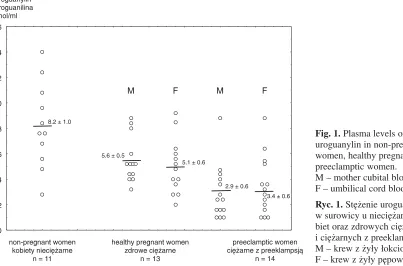
As can be seen in Figure 1, the pregnant women of both groups showed lower uroguanylin plasma levels than the non−pregnant women. This difference was statistically significant between non−pregnant and preeclamptic women (8.2 ± 1.0 vs. 2.9 ± 0.6 fmol/ml, p= 0.0003). The difference between the healthy pregnant and the preeclamptic women was also statistically significant (5.6 ±0.5 vs. 2.9 ±0.6,
p= 0.0013). Uroguanylin plasma levels in umbilical cord blood of the preeclamptic women were lower (not significantly) than those of the healthy pregnant women (3.4 ±0.6 vs. 5.1 ±0.6 fmol/ml).
The uroguanylin concentration in the amniotic fluid of the preeclamptic women was significantly lower (p = 0.01) than in the healthy pregnant women (Fig. 2).
Urinary uroguanylin excretion was signifi− cantly lower in the preeclamptic women than in the healthy pregnant and the non−pregnant women
2 4 6 8 10 12 14 16
8.2 ± 1.0
5.6 ± 0.5
2.9 ± 0.6
3.4 ± 0.6 M F M F
preeclamptic women ciężarne z preeklampsją
n = 14 healthy pregnant women
zdrowe ciężarne n = 13 non−pregnant women
kobiety nieciężarne n = 11 0
5.1 ± 0.6 uroguanylin
uroguanilina fmol/ml
Fig. 1.Plasma levels of uroguanylin in non−pregnant women, healthy pregnant, and preeclamptic women. M – mother cubital blood, F – umbilical cord blood
(24.2 ±4.6 vs. 51.3 ±11.3 vs. 72.4 ±19.3 pmol/d,
p= 0.045 and p= 0.012, respectively). Although urinary uroguanylin in the healthy pregnant women was lower than in the non−pregnant ones (51.3 ±11.3 vs. 72.4 ±19.3), this difference was statistically not significant.
In the non−pregnant healthy women, a signifi− cant positive correlation was found between MAP and plasma uroguanylin level (tau = 0.56, p= 0.017) and between MAP and urinary uroguanylin excre− tion (tau = 0.46 p= 0.05). In the healthy pregnant women, a significant negative correlation was found between MAP and maternal urinary uroguanylin excretion (tau = –0.49, p= 0.02). Such a correlation was absent in the preeclamptic women.
Only in the preeclamptic women a significant positive correlation was found between plasma uroguanylin level in the maternal blood and umbil− ical cord blood (tau = 0.85, p= 0.000021). No sig− nificant correlation was noted between uroguanylin concentration in amniotic fluid and plasma uroguanylin level in maternal or fetal blood in both the examined groups of pregnant women.
Discussion
As shown in this study, pregnant women (both healthy and preeclamptic) have lower plasma lev− els of uroguanylin in maternal peripheral blood and reduced urinary uroguanylin excretion com− pared with healthy non−pregnant women. These differences between pregnant and non−pregnant women became statistically significant in the
preeclamptic women. In addition, the preeclamptic women showed lower (but statistically not signifi− cant) uroguanylin plasma levels in umbilical cord blood, but significantly lower uroguanylin concen− trations in amniotic fluid and uroguanylin excre− tion in urine than healthy pregnant women. Finally, preeclamptic women, in contrast to healthy pregnant ones, did not show any relation− ship between urinary uroguanylin excretions and MAP. In the light of these findings the question arises whether uroguanylin is of pathophysiologi− cal relevance in preeclampsia.
Uroguanylin is a polypeptide containing 16 amino acids [10, 19, 20]. It is a member of a new family of natriuretic peptides which comprises guanylin [8], uroguanylin [9, 10], and lym− phoguanylin [11]. It shows structural homology with the heat−stable enterotoxins (STs) that cause traveler’s diarrhea [11]. All guanylins and STs are ligands for the guanylate cyclase C (GC−C) recep− tor [13]. After activation of this receptor, cyclic GMP (cGMP) is generated, which is the intracel− lular messenger of guanylins actions [for review see: 21]. GC−C belongs to the family of guanylate cyclases which comprises guanylate cyclase A (GC−A) and guanylate cyclase B (GC−B). The ligands for GC−A are, in order of decreasing affin− ity, atrial natriuretic peptide (ANP), brain natri− uretic peptide (BNP), and C−type natriuretic pep− tide (CNP), while for GC−B they are CNP, ANP, and BNP, respectively [13, 22]. Uroguanylin is present not only in the gastrointestinal tract [23–30], but also in the pancreas [28, 31], heart[ 24], kidney [24, 29, 32, 33; for review see: 34, 35], brain [28], and other organs [28].
40 80 120 160 200
72.4 ± 19.3
51.3 ± 11.3
24.2 ± 4.6
preeclamptic women ciężarne z preeklampsją
n = 14 healthy pregnant women
zdrowe ciężarne n = 13 uroguanylin
uroguanilina pmol/d
non−pregnant women kobiety nieciężarne
n = 11
Fig. 2.Uroguanylin concentration in amniotic fluid of healthy preg− nant and preeclamptic women
It is generally presumed that guanylins are involved in the salt and water balance by influenc− ing Cl– and HCO
3– secretion into the intestinal lumen [14, 36, 37] and water sodium, and potassi− um excretion by the kidneys [38]. This effect seems to be the result of guanylins’ actions on membrane GC−C by an autocrine and or paracrine pathway mediated by cGMP [8, 39]. As uroguanylin and guanylin increase water sodium and potassium excretion even in GC−C−null mice, it seems that the mechanism(s) of their action may also be GC−C independent [40]. Quite recently the existence of two signaling pathways for guanylin peptides in principal cells of mouse cortical col− lecting duct were found [41]. One pathway is cGMP and protein kinase G (PKG) dependent but not mediated by guanylate cyclase C, while the second is a cGMP−independent signaling pathway for these peptides which apparently involves phos− pholipase A2 (PLA2) and arachidonic acid [41]. Intestinal guanylin and uroguanylin are secreted not only into the lumen of the gastrointestinal tract, but also into the circulatory blood, from where they are cleared by the kidneys [28, 33] and where they exert a natriuretic, diuretic, and kali− uretic effect [21, 38; for review see: 34, 35]. The guanylins seem to link the gastrointestinal tract and kidneys in a potential endocrine axis which participates in monitoring water−electrolyte home− ostasis [for review see: 12, 34, 35, 42, 43].
Until now, only scarce reports on the clinical relevance of guanylins have been available. The intake of a high−salt diet is accompanied by a sig− nificant increase in urinary excretion of uroguanylin [16]. This increase shows a significant positive cor−
relation with natriuresis, kaliuresis, and urinary cGMP excretion [16]. In patients with chronic renal failure, both plasma guanylin [44] and uroguanylin [16] are elevated and positively correlated with the severity of renal failure. Plasma levels of guanylin [44] and uroguanylin [45] are also significantly ele− vated in dialyzed patients with end−stage renal fail− ure compared with healthy controls. Elevated uroguanylin plasma levels were also found in ede− matous nephritic patients [17] and patients with congestive heart failure [46]. In the latter, urinary uroguanylin was substantially increased [46]. The present authors found higher values of urinary uroguanylin in patients with essential hypertension than in normotensive subjects [47].
As shown in experimental studies in guinea pigs, pregnancy is characterized by a significant increase in myometrical cGMP production [48]. As shown by Buhimshi et al., this increase in cGMP production is due to an increased GC−A activity which is responsive to ANP [48]. These authors suggest that the enhanced production of cGMP in the pregnant myometrium is due to increased particulate GC−A activity induced by a natriuretic peptide in a paracrine manner [48]. As shown by Itoh et al., amniotic fluid obtained from pregnant women contains high concentrations of BNP [49]. This natriuretic peptide, by acting in a paracrine manner on myometrical GC−A, could be responsible for the high myometrical content of cGMP [49]. The relationship between amniotic uroguanylin and cGMP concentrations in pregnant myometrium has not been studied until now. As uroguanylin is present in human amniotic fluid, it seems likely that this hormone, acting on GC−C,
20 40 60 80 100 120 140
68.6 ± 6.0
41.1 ± 8.2
0
preeclamptic women ciężarne z preeklampsją healthy pregnant women
zdrowe ciężarne uroguanylin
uroguanilina fmol/ml
Fig. 3.Urinary uroguanylin excre− tion in non−pregnant, healthy preg− nant, and preeclamptic women
may contribute to increased cGMP concentration in the pregnant myometrium. This speculation is not consistent with studies reported by Buhimschi et al., in which no influence of uroguanylin on par− ticulate GC−C in the myometrium was noted [48]. Taking into account the above−mentioned effects of uroguanylin on water−electrolyte home− ostasis as well as the results presented in this paper (significantly lower uroguanylin levels in
plasma and amniotic fluid, markedly reduction of uroguanylin excretion in urine, and lower uroguanylin plasma levels in umbilical cord blood in preeclamptic women compared with healthy pregnant ones), it seems likely that uroguanylin is involved in the pathogenesis of preeclampsia. Further studies are necessary to elucidate the role of this hormone in the pathogenesis of abnormal water−electrolyte metabolism in preeclampsia.
References
[1] Higgins JR, de Swiet M:Blood pressure measurement and classification in pregnancy. Lancet 2001, 357, 131–135.
[2] Roberts JM, Cooper DW:Pathogenesis and genetics of preeclampsia. Lancet 2001, 357, 53–56.
[3] Esplin MS, Fausett MD, Fraser A, Kerber R, Mineau G, Carrillo J:Paternal and maternal component of the
predisposition to preeclampsia. N Engl J Med 2001, 344, 867–872.
[4] Dekker GA, Sibai BM:Etiology and pathogenesis of preeclampsia: current concepts. Am J Obstet Gynecol 1998, 179, 1359–1375.
[5] Kokot F:Starvation in the midst of plenty – the problem of volaemia in pregnancy. Nephrol Dial Transplant 1997, 12, 388–391.
[6] Lindheimer MD, Akbari A: The kidney and hypertension in pregnancy. In: Hypertension: a Companion to
Brenner and Rector’s The Kidney. Eds.: Oparil S and Weber MA. WB Saunders Comp., St Louis 2000, 688–701.
[7] Brown MA, Zammit VC, Lowe SA:Capillary permeability and extracellular fluid volumes in pregnancy induced hypertension. Clin Sci 1989, 77, 599–604.
[8] Currie MG, Fok KF, Kato J, Moore RJ, Hamra FK, Duffin KL:Guanylin: an endogenous activator of intesti−
nal guanylate cyclase. Proc Natl Acad Sci USA 1992, 89, 947–951.
[9] Hamra FK, Forte LR, Eber SL et al.: Uroguanylin: structure and activity of a second endogenous peptide that stimulates intestinal guanylate cyclase. Proc Natl Acad Sci USA 1993, 90, 10464–10468.
[10] Kita T, Smith CE, Fok KF et al.:Characterization of human uroguanylin: a member of the guanylin peptide fam− ily. Am J Physiol 1994, 266, F342–F348.
[11] Forte LR, Eber SL, Fan X et al.:Lymphoguanylin: cloning and characterization of a unique member of the
guanylin peptide family. Endocrinology 1999, 140, 1800–1806.
[12] Forte LR, Fan X, Hamra FK:Salt and water homeostasis: uroguanylin is a circulating peptide hormone with natriuretic activity. Am J Kidney Dis 1996, 28, 296–304.
[13] Krause WJ, London RM, Freeman RH, Forte LR:The guanylin and uroguanylin peptide hormones and their
receptors. Acta Anat Basel 1997, 160, 213–231.
[14] Forte LR, Curie MG:Guanylin: a peptide regulator of epithelial transport. FASEB J 1995, 9, 643–650.
[15] Forte LR, London RM, Freeman RH, Krause WJ:Guanylin peptides: renal actions mediated by cyclic GMP.
Am J Renal Physiol 2000, 278, F180–F191.
[16] Kinoshita H, Fujimoto S, Nakazato M et al.:Urine and plasma levels of uroguanylin and its molecular forms in renal diseases. Kidney Int 1997, 52, 1028–1034.
[17] Kinoshita H, Fujimoto S, Fukae A, Yokota N, Hisanaga S, Nakazato M: Plasma and urine levels of
uroguanylin, a new natriuretic peptide, in nephrotic syndrome. Nephron 1999, 81, 160–164.
[18] Kokot F, Stupnicki R:Radioimmunological and radiocompetitive methods used in clinics. 2nded. PZWL Warsaw
1986, 22–33.
[19] Hill O, Cetin Y, Cieslak A, Magert HJ, Forssman WG:A new human guanylate cyclase−activating peptide
(GCAP−II, uroguanylin): Precursor cDNA and colonic expression. Biochim Biophys Acta 1995, 1253, 146–149.
[20] Cetin Y, Forssman WG:GCAP−II: Isolation and characterization of the circulating form of human uroguanylin. FEBS Lett 1995, 374, 34–38.
[21] Fonteles MC, Greenberg RN, Monteiro HS, Currie MG, Forte LR: Natriuretic and kaliuretic activities of
guanylin and uroguanylin in the isolated perfused rat kidney. Am J Physiol 1998, 275, F191–F197.
[22] Leitman DC, Waldman SA, Murad F:Regulation of particulate guanylate cyclase by natriuretic peptides and
Escherichia coliheart−stable enterotoxin. Adv Pharmacol 1994, 26, 67–86.
[23] Nakazato M, Yamaguchi H, Date Y et al.:Tissue distribution, cellular source, and structural analysis of rat immunoreactive uroguanylin. Endocrinology 1998, 139, 5247–5254.
[24] Fan X, Hamra FK, Freeman RH et al.:Uroguanylin: Cloning of preprouroguanylin cDNA, mRNA expression
in the intestine and heart, and isolation of uroguanylin and prouroguanylin from plasma. Biochem Biophys Res Commun 1996, 219, 457–462.
[25] Li Z, Perkins AG, Peters MF, Campa MJ, Goy MF:Purification, cDNA sequence, and tissue distribution of rat uroguanylin. Regul Pept 1997, 68, 45–56.
[26] Fan X, Hamra FK, London RM et al.:Structure and activity of uroguanylin and guanylin from the intestine and urine of rats. Am J Physiol 1997, 273, E957–E964.
[28] Fan X, Wang Y, London RM et al.:Signaling pathways for guanylin and uroguanylin in the digestive, renal, cen− tral nervous, reproductive, and lymphoid systems. Endocrinology 1997, 138, 4636–4648.
[29] Miyazato M, Nakazato M, Matsukura S et al.:Uroguanylin gene expression in the alimentary tract and extra− gastrointestinal tissue. FEBS Lett 1996, 398, 170–174.
[30] Date Y, Nakazato M, Yamaguchi H et al.:Enterochromaffin−like cells: A cellular source of uroguanylin in rat stomach. Endocrinology 1999, 140, 2398–2404.
[31] Kulaksiz H, Cetin Y:Uroguanylin and guanylate cyclase C in the human pancreas: expression and mutuality of lig− and/receptor localization as indicators of intracellular paracrine signaling pathways. J Endocrinol 2001, 170, 267–275.
[32] Cui L, Blanchard RK, Cousins RJ:Dietary zinc deficiency increases uroguanylin accumulation in rat kidney. Kidney Intern 2001, 59, 1424–1431.
[33] London RM, Eber SL, Visweswariah SS et al.:Structure and activity of OK GC: a kidney receptor guanylate cyclase activated by guanylin peptides. Am J Physiol 1999, 276, F882–F891.
[34] Kokot F, Ficek R:Guanylins – are they of nephrological relevance? Nephron 2000, 84, 201–205.
[35] Forte LR, London RM, Freeman RH, Krause WJ:Guanylin peptide: renal actions mediated by cyclic GMP.
Am J Renal Physiol 2000, 278, F180–F191.
[36] Hamra FK, Eber SL, Chin DT et al.:Regulation of intestinal uroguanylin/guanylin receptor mediated respons− es by mucosal acidity. Proc Natl Acad Sci USA 1997, 94, 2705–2710.
[37] Joo NS, London RM, Kim HD et al.:Regulation of intestinal Cl– and HCO
3–secretion by uroguanylin. Am
J Physiol 1998, 274, G633–G644.
[38] Greenberg RN, Hill M, Crytzer J et al.:Comparison of effects of uroguanylin, guanylin, and Escherichia coli heat stable enterotoxin STa in mouse intestine and kidney: evidence that uroguanylin is an intestinal natriuretic hormone. J Invest Med 1997, 45, 276–282.
[39] Hamra FK, Fan X, Krause WJ et al.:Prouroguanylin and proguanylin: Purification from colon, structure and modulation of bioactivity by proteases. Endocrinology 1996, 137, 257–265.
[40] Carrithers SL, Hill MJ, Johnson BR et al.:Renal effects of uroguanylin and guanylin in vivo. Braz J Med Biol Res. 1999, 32, 1337–1344.
[41] Sindić A, Velic A, Basoglu C et al.:Uroguanylin and guanylin regulate transport of mouse cortical collecting duct independent of guanylate cyclase C. Kidney Intern. 2005, 68, 1008–1017.
[42] Semrad CE:Guanylin: where it’s at! Why’s it there? Gastroenterology 1997, 113, 1036–1038.
[43] Giannella RA:Escherichia coli heat−stable enterotoxins, guanylins, and their receptors: what are they and what do they do? J Lab Clin Med 1995, 125, 173–183.
[44] Kinoshita H, Nakazato M, Yamaguchi H et al.:Increased plasma guanylin levels in patients with impaired renal function. Clin Nephrol 1997, 47, 28–32.
[45] Fukae H, Kimoshita H, Fujimoto S et al.:Plasma concentration of uroguanylin in patients on maintenance dial− ysis therapy. Nephron 2000, 84, 206–210.
[46] Carrithers SL, Eber SL, Forte LR, Greenberg RN:Increased urinary excretion of uroguanylin in patients with congestive heart failure. Am J. Physiol Heart Circ Physiol 2000, 278, 538–547.
[47] Kokot F, Nakazato M, Adamczak M et al.:Plasma and urinary uroguanylin in patients with essential hyperten− sion – relationship to plasma renin activity. Proceedings in Nephrology Publisher House “Russian Physicians”, Moscow 2001, 55–65 (in Russian).
[48] Buhimshi JA, San Martin−Clark O, Aguan K, Thompson LP, Weiner CP:Differential alterations in responsiveness in particulate and soluble guanylate cyclases in guinea pig myometrium. Am J Obstet Gynecol 2000, 183, 1512–1519.
[49] Itoh H, Sagawa N, Hasegawa M et al.:Expression of biologically active receptors of natriuretic peptides in the human uterus during pregnancy. Biochem Biophys Res Commun 1994, 203, 602–607.
Address for correspondence:
Franciszek Kokot
Department of Nephrology, Endocrinology, and Metabolic Diseases Silesian University Medical School,
ul. Francuska 20 40−027 Katowice Poland
tel.: +48 32 255 26 95 fax: +48 32 255 37 26
e−mail: [email protected]
Conflict of interest: None declared
Received: 11.05.2006 Revised: 7.07.2006 Accepted: 21.09.2006
Praca wpłynęła do Redakcji: 11.05.2006 r. Po recenzji: 7.07.2006 r.