Introduction
The palms of the hands and the soles of the feet are covered with two totally distinct classes of marks. The most conspicuous are the creases or folds of skin which interested the people in palmistry [1]. Scientifically, now, palmistry is dermatoglyphics.
Dermatoglyphics is the science and art of surface markings of skin, especially hands and feet [2]. It is the dermal ridge configuration of palms, digits, and soles. Toward the end of the 19th century,
Galton put forth a rule called “proof of no change,” which states that an individual dermatoglyphics pattern remains unchanged throughout his/her lifetime.
The ridge formation occurs in the 13th week of prenatal life and pattern
formation is completed in the 19th week. The ridges are influenced
ABSTRACT
Dermatoglyphics is focused on studying the fine patterned dermal ridges on volar surfaces of soles, palms, and ridges. The volar pads are mound-shaped elevations on each finger above the proximal end of the distal metacarpal bone. Most dermatoglyphics which are correlated with genetic abnormalities, are used in biomedical studies. This study aims to systematically review and to assess the correlation between dermatoglyphics and orofacial disease in human population. An electronic search was initiated in the PubMed, Embase, and Google Scholar databases about the articles discussing the relationship between dermatoglyphics and orofacial diseases using specific keyword search terms. A PIO analysis was done to identify the articles. The search methodology yielded and was excluded and included. 32 articles were generated among which 9 articles were excluded and 23 articles were included finally to discuss the relationship between orofacial disorders and dermatoglyphics. Out of the 23 articles, 5 were research articles and 18 review articles. Extraction of data revealed a positive correlation between dermatoglyphics and orofacial disorders. Dermatoglyphics can be used as a predictive model to diagnose orofacial disease conditions.
Keywords: Dermatoglyphics, genetic, investigations, patterned ridges
Dermatoglyphics: A tool in dentistry
Shruthi Chandrasekaran
1, Ramya Chellammal
1, Dhanraj M. Ganapathy
21Department of Prosthodontics, Saveetha Dental College, Saveetha University, Chennai, Tamil Nadu, India, 2Department of Prosthodontics, Saveetha Dental College, Saveetha University, Chennai, Tamil Nadu, India
Correspondence: Dhanraj M. Ganapathy, Department of Prosthodontics, Saveetha Dental College, Saveetha University, 162, Poonammale High Road, Chennai -
600 077, Tamil Nadu, India.
by blood vessel-nerve pairs at the border between the dermis and epidermis during this prenatal development and factors such as inadequate oxygen supply, unusual distribution of sweat glands, and alterations in epithelial growth could alter the ridge patterns. The ridged skin is a sensitive indicator of intrauterine dental anomalies because it originates from the same ectodermal layer in the 6th–7th
week of embryonic life. In a similar way, development of dermal ridges and congenital deafness seems to be interlinked as they develop around the same time [3].
Fingerprints are unique to individuals and will remain unique throughout their lifetime. Multiple genes determine fingerprint configurations and the study of fingerprints reveal vital genetic and medical information about an individual. Since it is not even found similar even in monozygotic twins, studying them can determine a number of parameters which could be helpful in diagnosing and treating individuals. Thus, it is considered an important tool in assessing the genetic trait, evaluation of children with suspected genetic disorders, and also in forensics.
Dermatoglyphics has drawn attention in the field of dentistry. It has been to unveil various pathological conditions and genetic abnormalities such as dental caries, periodontal diseases, oral submucous fibrosis, cleft lip and palate, malocclusions, Down’s syndrome, and Klinefelter’s syndrome. Taking an instance of dental
How to cite this article: Chandrasekaran S, Chellammal R, Ganapathy DM.
Dermatoglyphics: A tool in dentistry. J Adv Pharm Edu Res 2017;7(3):248-252.
Source of Support: Nil, Conflict of Interest: None declared.
Access this article online
Website: www.japer.in E-ISSN: 2249-3379
caries, it is found that individuals with patterns such as plain loop, double loop, arch with whorl, tented arch, and central pocket loop have a high susceptibility to dental caries. Furthermore, since dermatoglyphics are genetically controlled characteristics, any deviation from the normal features indicates a genetic abnormality.
Dermatoglyphics possess qualities of an adjunctive diagnostic tool in identification of various orofacial dental disorders. The advantages of dermatoglyphics as a diagnostic aid are the relative simplicity, versatility, and faster interpretation of the findings. However, its usage in the field of dentistry and stomatology needs to be explored further.
Aim
This study aims to systematically review and to assess the correlation between dermatoglyphics and orofacial disease in human population.
Materials and Methods
An electronic search was initiated in the PubMed, Embase, and Google Scholar databases about the articles discussing the relationship between dermatoglyphics and orofacial diseases using specific keyword search terms involving dermatoglyphics, dermatography, fingerprints, palmar creases, orofacial diseases, orofacial disorders, dental diseases, dental caries, periodontitis, syndromes, malocclusion, bruxism, oral submucous fibrosis, and squamous cell carcinoma were used. A PIO analysis was done using these keywords to identify the articles. The search methodology yielded 32 articles and 9 were excluded and 23 included.
Data extraction
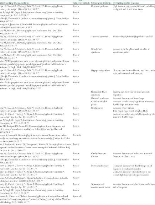
The data from the selected articles were retrieved [Table 1] and analyzed by two independent investigators and the findings were pooled and tabulated.
Results
Out of the 23 articles included for the study, 5 were research articles and 18 were review articles. The 5 research articles included 2 articles on malocclusion, 1 on dental caries, 1 on periodontitis, and 1 on precancerous lesions of oral cavity. Summation of the research findings suggested a weak positive correlation between dental caries, malocclusion, certain precancerous lesions, squamous cell carcinoma, cleft lip and palate, and dermatoglyphics.
Discussion
Dermatoglyphics has played a pivotal role in personal identification and forensic investigations. In the upcoming period, dermatoglyphics seems to have drawn attention to the field of dentistry, to identify simple oral disease such as dental caries, periodontitis to even syndromic manifestations such as Down’s syndrome and Klinefelter’s syndrome.
Priya et al. have studied extensively into dermatoglyphics and orofacial disease correlation. The study has reported varied patterns being
consistent with various orofacial diseases. For instance, high frequency of creases, bilateral, radial loops on digits 4 and 5, and ulnar loops seen in patients with Down’s syndrome[4] which are consistent with
other studies conducted by Soni et al.,[5] Lakshmi et al.,[6] and
Denny et al.[7] who have also reported increases bilateral radial
loops. In Turner’s syndrome, a characteristic short 5th finger,
bilateral hypothenar pattern is reported by Lakshmi et al.[6] which
again is confluent with the study conducted by Preus et al. showing similar results.[8] In respect to Klinefelter’s syndrome, a similar
inference has been drawn by Priya et al.,[4] Preus et al.,[8] and
Forbes et al.[9] which emphasizes on an increase in height of axial
triradius in hypothenar areas. Priya et al.[4] have also reported
findings in cases of Rubinstein-Taybi syndrome and Kanner syndrome which show four or more arches bilaterally in fingertips and increased frequency of lower loops, respectively.
When dental diseases are taken into consideration, numerous studies have been reported by others in regards to dental caries, periodontitis, squamous cell carcinoma, oral submucous fibrosis, etc.
A study was carried out by Sharma et al., to determine if there was any significant correlation between salivary bacterial interactions, dermatoglyphics, and dental caries.[10] The results revealed that the
control group had increases frequency of whorl pattern in all palmar digits and they were highly susceptible to dental caries. Similarly, the study conducted by Bhat et al. also showed the increases whorl frequency.[11] One more significant finding was reported by Priya et
al., which was an higher frequency of arches and radial loops in subjects susceptible to dental caries.[4]
Following dental caries, periodontal disease is the commonly reported dental disease. An extensive study was carried out by Atasu et al., to study each variant of periodontitis. The fingertip palm and sole prints of 36 patients with juvenile periodontitis (JP), 45 patients with rapidly progressive periodontitis and 38 patients with adult periodontitis (AP) were compared with 39 periodontally healthy (PH) individuals to study patterns occurring in individuals and to correlate them with their periodontal conditions. When the fingertip patterns of the patients were compared with those of PH individuals, the decreased frequencies of twinned and transversal ulnar loops on all fingers of the patients with JP, a decreased frequency of double loops on all fingers and an increased frequency of radial loops on the right second digits of the patients with RPP and the increased frequencies of concentric whorls and transversal ulnar loops on all fingers of the patients with AP, an increased frequency of t″ triradii on the palms of the patients with JP, the increased frequencies of IV and H loops and tb triradii on the palms of the patients with RPP, and an increased frequency of e triradii on the soles of the patients with JP were found.[12]
Similarly, Issrani et al. have also shown an decreased frequency of radial loops in the second digit in progressive periodontitis.[13]
Oral submucous fibrosis which is reported to be a potentially malignant disorder has also some characteristic dermatoglyphic patterns. The studies conducted by Priya et al,[4] and Lakshmi et al. [6] have shown an increased frequency of arches in the thenar area.
Squamous cell carcinoma seems to manifest an increased frequency of whorls in lower half of the palm as reported by Denny et al.,[7]
Articles citing the condition Nature of article Clinical conditions Dermatoglyphic features Review
Review
Review
Review
Review
Downs’s syndrome High frequency of creases, bilateral, radial loops on digits 4 and 5, and ulnar loops
Review
Review
Turner’s syndrome Short 5th finger, bilateral hypothenar pattern
Review
Review
Review
Klinefelter’s
syndrome Increase in the height of axial triradius in hypothenar pattern Priya NS, Sharada P, Chaitanya Babu N, Girish HC. Dermatoglyphics in
dentistry: An insight. J Dent 2013;4:144-7.[4]
Soni A, Singh SK, Gupta A. Implications of dermatoglyphics in dentistry. J Dentofacial Sci 2013;2:27-30.[5]
Prabha JL, Thenmozhi R. A short review on dermatoglyphics. J Pharm Sci Res 2014;6:200-2.[6]
Rajangam S, Janakiram S, Thomas IM. Dermatoglyphics in Down’s syndrome. J Indian Med Assoc 1995;93:10-3.[14]
Preus M, Fraser FC. Dermatoglyphics and syndromes. Am J Dis Child 1972;124:933-43.[8]
Priya NS, Sharada P, Chaitanya Babu N, Girish HC. Dermatoglyphics in dentistry: An insight. J Dent 2013;4:144-7.[4]
Preus M, Fraser FC. Dermatoglyphics and syndromes. Am J Dis Child 1972;124:933-43.[8]
Priya NS, Sharada P, Chaitanya Babu N, Girish HC. Dermatoglyphics in dentistry: An insight. J Dent 2013;4:144-7.[4]
Preus M, Fraser FC. Dermatoglyphics and syndromes. Am J Dis Child 1972;124:933-43.[8]
Forbes AP. Fingerprints and palm prints (dermatoglyphics) and palmar-flexion creases in gonadal dysgenesis, pseudohypoparathyroidism and Klinefelter’s syndrome. N Engl J Med 1964;270:1268-77.[9]
Priya NS, Sharada P, Chaitanya Babu N, Girish HC. Dermatoglyphics in dentistry: An insight. J Dent 2013;4:144-7.[4]
Prabha JL, Thenmozhi R. A short review on dermatoglyphics. J Pharm Sci Res 2014;6:200-2.[6]
Forbes AP. Fingerprints and palm prints (dermatoglyphics) and palmar-flexion creases in gonadal dysgenesis, pseudohypoparathyroidism and Klinefelter’s syndrome. N Engl J Med 1964;270:1268-77.[9]
Review
Review
Review
Hypoparathyroidism Characterized by broad brands and short, with wide and increased arch patterns
Rubinstein-Taybi
syndrome Bilateral and show four or more arches in fingertips Kanner’s syndrome Increased frequency of lower loops Cleft lip and cleft
palate Increased triradii count, significant increase in double loops and ulnar loops Review
Review
Review
Review
Research
Review
Dental caries Increased whorl pattern
Total finger ridge count is higher. High frequency of arches and radial loops, along with ulnar and double loops
Review
Review
Oral submucous
fibrosis Increased frequency of arches and increased frequency in thenar area
Review
Research
Review
Periodontal disease Decreased frequency of double loops on all fingers in juvenile periodontitis
Decreased frequency of radial loops in the second digit in progressive periodontitis Priya NS, Sharada P, Chaitanya Babu N, Girish HC. Dermatoglyphics in
dentistry: An insight. J Dent 2013;4:144-7.[4]
Denny C, Ahmed J, Shenoy N, Binnal A. Dermatoglyphics in Dentistry-A Review. Int J Cur Res Rev 2013;5:30-3.[7]
Soni A, Singh SK, Gupta A. Implications of dermatoglyphics in dentistry. J Dentofacial Sci 2013;2:27-30.[5]
Bhat PK, Badiyani BK, Aruna CN. Dermatoglyphics-A new diagnostic tool in detection of dental caries in children. Indian J Forensic Med Toxicol 2012;6:24-8.[15]
Sharma A, Somani R. Dermatoglyphic interpretation of dental caries and its correlation to salivary bacteria interactions: An in vivo study. J Indian Soc Pedod Prev Dent 2009;27:17.[16]
Bhat P, Badiyani B, Aruna CN, Chengappa S, Bhaskar N. Dermatoglyphics-A new diagnostic tool in detection of dental caries among deaf and mute children. Int J Clin Dent Sci 2012;2:80-4.[11]
Priya NS, Sharada P, Chaitanya Babu N, Girish HC. Dermatoglyphics in dentistry: An insight. J Dent 2013;4:144-7.[4]
Prabha JL, Thenmozhi R. A short review on dermatoglyphics. J Pharm Sci Res 2014;6:200-2.[6]
Denny C, Ahmed J, Shenoy N, Binnal A. Dermatoglyphics in Dentistry-A Review. Int J Cur Res Rev 2013;5:30-3.[7]
Denny C, Ahmed J, Shenoy N, Binnal A. Dermatoglyphics in Dentistry-A Review. Int J Cur Res Rev 2013;5:30-3.[12]
Issrani R, Prabhu N, Mathur S, Mishra G, Sinha S. Dermatoglyphics in health and diseases-a review. JSM Dent 2013;2:1044.[13]
Denny C, Ahmed J, Shenoy N, Binnal A. Dermatoglyphics in Dentistry-A Review. Int J Cur Res Rev 2013;5:30-3.[7]
Soni A, Singh SK, Gupta A. Implications of dermatoglyphics in dentistry. J Dentofacial Sci 2013;2:27-30.[5]
Venkatesh, Elluru, et al. “Palmar dermatoglyphics in oral leukoplakia and oral squamous cell carcinoma patients.” Journal of Indian Academy of Oral Medicine and Radiology 20.3 (2008): 94.[17]
Review
Review
Researsch
Squamous cell
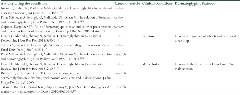
carcinoma and tumor Increased frequency of whorls seen in the lower half of the palm Table 1: Data extraction for dermatoglyphics and orofacial conditions
Articles citing the condition Nature of article Clinical conditions Dermatoglyphic features Review
Review
Review Issrani R, Prabhu N, Mathur S, Mishra G, Sinha S. Dermatoglyphics in health and diseases-a review. JSM Dent 2013;2:1044.[13]
Polat MH, Azak A, Evlioglu G, Malkondu OK, Atasu M. The relation of bruxism and dermatoglyphics. J Clin Pediatr Dent 1999;24:191-4.[18]
Gupta A, Karjodkar FR. Role of dermatoglyphics as an indicator of precancerous and cancerous lesions of the oral cavity. Contemp Clin Dent 2013;4:448.[19] Denny C, Ahmed J, Shenoy N, Binnal A. Dermatoglyphics in Dentistry-A Review. Int J Cur Res Rev 2013;5:30-3.[7]
Sharma A, Kapoor D. Dermatoglyphics, dentistry and diagnosis-a review. Baba Farid Univ Dent J 2010;1:45-8.[10]
Polat MH, Azak A, Evlioglu G, Malkondu OK, Atasu M. The relation sof bruxism and dermatoglyphics. J Clin Pediatr Dent 1999;24:191-4.[20]
Review
Review
Research
Bruxism Increased frequency of whorls and decreased ulnar loops
Denny C, Ahmed J, Shenoy N, Binnal A. Dermatoglyphics in Dentistry-A Review. Int J Cur Res Rev 2013;5:30-3.[7]
Reddy BR, Sankar SG, Roy ET, Govulla S. A comparative study of
dermatoglyphics in individuals with normal occlusions and malocclusions. J Clin Diagn Res 2013;7:3060.[21]
Tikare S, Rajesh G, Prasad KW, Thippeswamy V, Javali SB. Dermatoglyphics-A marker for malocclusion? Int Dent J 2010;60:300-4.[22]
Review
Research
Research
Malocclusion Increased whorl pattern in Class I and Class II malocclusion
Table 1: Data extraction for dermatoglyphics and orofacial conditions
Venkatesh et al., to determine whether any specific dermatoglyphic pattern exists which can help in predicting the occurrence of oral squamous cell carcinoma. 30 subjects were studied in comparison with 30 controls and the results which were conflicting to the previous studies as there were increased arches and loops seen in the study group.[17] A similar
cross-sectional study was conducted by Gupta et al., with 120 subjects and showed consistent results with the study conducted by Venkatesh et al. of increased arches and loops.[10]
Increases whorl frequency decreased ulnar loops are the characteristics seen in bruxism as studied by Denny et al.[7] and Sharma et al.[16]
Malocclusion too has been extensively studied to predict any specific dermatoglyphic pattern associated with it. A study was conducted by Reddy et al. using dermatoglyphics to predict and compare Class I, Class II, division 1, division 2, and Class III malocclusions. A total of 96 subjects were divided into three malocclusion groups, i.e., Class I (control group), Class II, division 1, division 2, and Class III (experimental group) in the ages of 12–14 years. The dermatoglyphic findings revealed that the craniofacial Class II, division 1, division 2 pattern was associated with increased frequency of arches and ulnar loops and decreased frequency of whorls, whereas in Class III, there was an increased frequency of arches and radial loops with decreased frequency of ulnar loops.[21] However, a study conducted by Tikare et al.
showed conflicting results. The study aimed at assessing the relationship between fingerprints and malocclusion among a group of high schoolchildren aged 12–16 years in Dharwad, Karnataka, India, in 696 high schoolchildren and results showed no correlation between dermatoglyphics and malocclusion.[22]
This review observed only a few studies with proper research designs. Further studies with meticulous research designs could contribute a lot of credible evidence regarding the association between dermatoglyphics and various orofacial disorders.
Conclusion
Dermatoglyphics is an upcoming integral part of medicine and science these days. Fingerprints known to be unique and unalterable hence act as excellent tools for personal identification, morphological, and genetic research as well as for population studies. The correlation of dental problems is still in its nascent stages, and presently, it is safe to say that the various fingerprint patterns can be considered as an indicator for the occurrence of congenital orofacial abnormalities and dental diseases. Dermatoglyphics has moved from obscurity to acceptability as a diagnostic tool. Extensive study and research in this field are required to determine its validity. In the future, it may serve as an important tool that can predict the future health of a person.
References
4. Priya NS, Sharada P, Chaitanya Babu N, Girish HC. Dermatoglyphics in dentistry: An insight. J Dent 2013;4:144-7.
7. Denny C, Ahmed J, Shenoy N, Binnal A. Dermatoglyphics in Dentistry-A Review. Int J Cur Res Rev 2013;5:30-3.
5. Soni A, Singh SK, Gupta A. Implications of dermatoglyphics in dentistry. J Dentofacial Sci 2013;2:27-30.
Prabha JL, Thenmozhi R. A short review on dermatoglyphics. J Pharm Sci Res 2014;6:200-2.
3.
Dermatoglyphics: A new diagnostic tool in detection of dental caries among deaf and mute children. Int J Clin Dent Sci 2011;2:80-4
Bhat PK, Badiyani BK, Aruna CN, Chengappa S, Bhaskar NN. NS Priya, P Sharada, N Chaitanya Babu, HC Girish. Dermatoglyphics in Dentistry: An Insight, 10.5005/jp-journals-10015-1221: 144-47. 2.
Watson J. Reading for the body. 2012, University of Georgia Press.
1.
6.
Preus M, Fraser FC. Dermatoglyphics and syndromes. Am J Dis Child 1972;124:933-43.
17. Venkatesh E, Bagewadi A, Keluskar V, Shetti A. Palmar dermatoglyphics in oral leukoplakia and oral squamous cell carcinoma patients. J Indian Acad Oral Med Radiol 2008;20:94.
13. Issrani R, Prabhu N, Mathur S, Mishra G, Sinha S. Dermatoglyphics in health and diseases-a review. JSM Dent 2013;2:1044.
18. Polat MH, Azak A, Evlioglu G, Malkondu OK, Atasu M. The relation of bruxism and dermatoglyphics. J Clin Pediatr Dent 1999;24:191-4.
4 Rajangam S, Janakiram S, Thomas IM. Dermatoglyphics in down’s syndrome. J Indian Med Assoc 1995;93:10-3.
9. Forbes AP. Fingerprints and palm prints (dermatoglyphics) and palmar-flexion creases in gonadal dysgenesis, pseudohypoparathyroidism and Klinefelter’s syndrome. N Engl J Med 1964;270:1268-77.
11. Bhat P, Badiyani B, Aruna CN, Chengappa S, Bhaskar N. Dermatoglyphics-A new diagnostic tool in detection of dental caries among deaf and mute children. Int J Clin Dent Sci 2012;2:80-4.
19. Gupta A, Karjodkar FR. Role of dermatoglyphics as an indicator of precancerous and cancerous lesions of the oral cavity. Contemp Clin Dent 2013;4:448.
10.
George SM, Philip B, Madathody D, Mathew M, Paul J, Dlima JP. An assessment of correlation between dermatoglyphic patterns and sagittal skeletal discrepancies. J Clin Diagn Res 2017;11:ZC35.
Reddy BR, Sankar SG, Roy ET, Govulla S. A comparative study of dermatoglyphics in individuals with normal occlusions and malocclusions. J Clin Diagn Res 2013;7:3060.
21.
Tikare S, Rajesh G, Prasad KW, Thippeswamy V, Javali SB. Dermatoglyphics-A marker for malocclusion? Int Dent J 2010;60:300-4.
22.
Sharma A, Somani R. Dermatoglyphic interpretation of dental caries and its correlation to salivary bacteria interactions: An in vivo study. J Indian Soc Pedod Prev Dent 2009;27:17.
16.
Bhat PK, Badiyani BK, Aruna CN. Dermatoglyphics-A new diagnostic tool in detection of dental caries in children. Indian J Forensic Med Toxicol 2012;6:24-8.
15.
Atasu M, Kuru B, Firatli E, Meriç H. Dermatoglyphic findings in periodontal diseases. Int JAnthropol 2005;20:63-75.
12.
Sharma A, Kapoor D. Dermatoglyphics, dentistry and diagnosis-A review. Baba Farid Univ Dent J 2010;1:45-8.