_____________________________________________________________________________________________________ *Corresponding author: E-mail: yjeffagboola@yahoo.com;
The Effect of Aerosols on the Air Microflora of the
Indoor Air
E. O. Jeff-Agboola
1, O. E. Dada
2and Y. A. Jeff-Agboola
3*1
Department of Biological Sciences, Joseph Ayo Babalola University, Ikeji Arakeji, Osun State, Nigeria.
2
Department of Biological Sciences, Elizade University, Ilara-Mokin, Ondo State, Nigeria.
3
Department of Biological Sciences, University of Medical Sciences, Ondo City, Nigeria.
Authors’ contributions
This work was carried out in collaboration among all authors. Authors OED and EOJA designed the study, performed the statistical analysis. Author YAJA wrote the protocol and authors EOJA and YAJA wrote the first draft of the manuscript, managed the analyses of the study. Author OED and YAJA managed the literature searches. All authors read and approved the final manuscript.
Article Information
DOI: 10.9734/JAMB/2019/v16i130116 Editor(s): (1)Dr. P. Rama Bhat,PG Biotechnology, Alva’s College, Mood bidri, Karnataka, India. Reviewers: (1)Hideharu Shintani, University of Chuo Tokyo, Japan. (2)K. Robinson, Victor, Rivers State University, Nigeria. (3)Bharat Raj Singh, Dr. APJ Abdul Kalam Technical University, India. Complete Peer review History:http://www.sdiarticle3.com/review-history/48118
Received 08 February 2019 Accepted 15 April 2019 Published 25 April 2019
ABSTRACT
This research work assessed the microflora of rooms sprayed with different insecticides and air freshners with the aim of investigating the effect of the aerosols on the types of microflora in the room environment. Eight (8) different samples of chemical aerosols were used they are: Mobile insecticide(Imidacloprid), Raid multipurpose insect killer(1R-trans Phenothrin), Morten Insecticide(pyrethroids), Rambo Insecticide(pyrethroid compound). as categorized as Insecticides, while Febreze(hydroxypropyl beta-cyclodextrin), Air wick(Dipropylene glycol monomethyl ether (aka dipropylene glycol methyl ether), Glade(allyl 3-cyclohexylpropionate, allyl caproate, benzyl alcohol, butylated hydroxytoluene (BHT) and Top breeze(Cyclodextrin) were purchased as air fresheners/fragrance and eight (8) different rooms were used. Microorganisms isolated from the rooms before and after spraying with aerosols were: Staphylococus aureus, Lactobacillus jensenii, Bacillus coagulans, Aspergillus flavus, Aspergillus niger, Micrococcus spp., Aerococcus viridans,
Pediococcus cerevisiae, Streptococcus spp., Aspergillus fumigatus and Aspergillus niger. The result of eight different rooms sprayed with different aerosol as Insecticide and air fresheners showed that, some aerosols were able to inhibit some organisms that were initially present in some rooms while there were introduction of another organisms from some aerosols into some rooms. The occurrence of Staphylococus aureus (100%) was the highest in all the rooms followed by Aspergillus niger
(87.5) and A. flavus (75%). Lactobacillus jensenii, Bacillus coagulans and Micrococcus spp. had the lowest frequency of occurrence (12.5%).
Keywords: Air environment; aerosols; microflora; Indoor; microbial load.
1. INTRODUCTION
1.1 Background to the Study
Each day people are exposed to millions of bio aerosols, including whole microorganisms, which can have both beneficial and detrimental effects. Assessment of the indoor of the built environment, the aerobiomes is important and they are bacteria, viruses, fungi and their spores are examples of bio aerosols present in the air, inhaled by human beings. According to Smith [1] major sources of these bioaerosols are: humans, pets, plants, plumbing systems, heating, ventilation, and air-conditioning systems, dust, suspension; aesthetic pollutant and the outdoor environment. Recent advances in molecular sequencing have generated a rush to characterize the microbiome of various environments including indoor and outdoor air [2]. This is because humans spend over 90% of their time indoors [3], Researchers have observed that there are diverse microbial communities in indoor environments such as schools, houses, and hospital [4,5] rooms within the same building. For instance, Dunn [6] revealed that microbial isolates in the bedroom differs from that of the bathroom within the same building.
Despite rapid advances in the characterization of airborne microbial communities through rRNA surveys, metagenomics, proteomics, and metabolomics, limited information is available about actual concentrations of airborne microorganisms in built environments. In one of the few studies of concentrations of total bacteria and viruses in indoor air by air sampler, a researcher [7] found virus-like and bacteria-like particle concentrations of approximately 105 and 106 particles m3 in various indoor and outdoor air environment, respectively [8]. More over an average viable airborne fungi concentration of 80 CFU/m3 were reported in samples collected from schools, hospitals, residences, and industrial buildings; However, in some instances
concentrations were as high as 104 CFU m3. Such information should be forthcoming as methods for quantitative metagenomics analyses air samplers become more powerful [9,10].
in many cases differs from that observed in outdoor environments. Although less frequent than the possible dangers caused by exposure to pollen and acari, fungal exposure causes hypersensitive reactions which characterize allergic respiratory pathologies like bronchial asthma and rhinitis [18]. Fungi may elicit allergic symptoms similar to those caused by pollen.
With an ever-increasing population utilizing different types of aerosols as insecticides and air fresheners, in order to improve and sustain health and vitality; and consuming products in which these supplements are used as room flavors, it is essential that these products are safe for human use. A very critical indicator of safety is the microbiological quality of these products. To improve the prediction of dispersion models and the environmental health assessment on the one hand and to get an insight on the airborne micro-organisms in other relevant environments, e. g. living spaces. However, these studies give insight in the internal structure of bio-aerosols and the distribution of micro-organisms on airborne particles themselves for developing guidelines in order to achieve and maintain safe microbial levels in these products.
Therefore, the aim of the study are to, isolate microorganism in air environment of
rooms sprayed with selected chemical aerosols and investigate the effect of the aerosols on the load of microflora in the room environment.
2. MATERIALS AND METHODOLOGY
2.1 Study Area
The sampling area was an inbuilt living rooms in a house at Akure and the aerosols were purchased from Shoprite shopping mall located at alagbaka, Akure, Ondo State, Nigeria.
2.2 Collection of the Samples
Eight (8) different samples of chemical aerosols were purchased from shoprite shopping mall, alagbaka, Akure, Ondo State, Nigeria. The selected aerosols were; insecticideImidacloprid, 1R-trans Phenothrin, pyrethroids, pyrethroid compound. as categorized as Insecticides, while hydroxypropyl beta-cyclodextrin,dipropylene
glycol methyl ether,allyl 3-cyclohexylpropionate, allyl caproate, benzyl alcohol, butylated hydroxytoluene (BHT) and Cyclodextrin were purchased as air fresheners/fragrance.
2.3 Experimental Design
The experimental design is 8x3; eight (8) rooms were sprayed with each of the eight selected chemical aerosols, Petri-dishes were prepared aseptically in triplicates and exposed to each room 10 minutes after spraying with insecticides and air fresheners.
2.4 Microbial Isolation and Determination of Total Viable Counts
The method used for isolation and identification of microorganisms was as described [19]. Twenty (20 ml) of nutrient agar and acidified potato dextrose agar cooled to 45˚C was poured separately onto each of the plates in triplicate and the plates were gently swirled and allowed to solidify. The plates were exposed to air in the room before and after spraying with aerosols for 10 minutes. Thereafter, the nutrient agar plates were incubated in an inverted position at 37±2˚C for 24 hours for isolation of mesophilic bacteria while Potato Dextrose Agar plates were incubated at 28±2˚C for 72 hours. Anaerobic plates were inverted in the anaerobic jar at 37±2˚C for 24 hours for isolation of anaerobic organisms present in the samples. After incubation, colonies on the plates were counted using colony counter and the number of viable cells obtained to be the total viable counts of the isolates. The viable colonies were sub cultured from mixed culture plate to obtain a pure culture. The colonies were then identified directly by their size, shape, colour of the pigment (chromo-genesis), opacity, elevation, surface, edge and consistency and stored on agar slants for further biochemical tests.
2.5 Determination of Microbiology of the Air Samples
reduction test, sugar fermentation test, oxidative fermentation (O/F) test, methyl red Voges-proskaur test, citrate test, oxidase test and motility test.
2.6 Identification of Fungal Isolates
Moulds were identified based on cultural and morphological features using light microscope also number of colony isolated was recorded [21,22]. Cultural characterization was based on the rate of growth, presence of aerial mycelium, colour of aerial mycelium as well as colour on the obverse and reverse of the plates. Microscopic identification was based on spore and conidiophore morphology.
2.7 Calculation of Percentage Frequency of the Isolates
The isolation frequency (Fq) of each isolate from the eight rooms was calculated according to the formula by Gonzalez [23]. This was used to determine the distribution of the isolates in the eight sample rooms.
2.8 Data Analysis
The experiment was conducted using a completely randomized design. Means of three replicates were computed using computer software Microsoft Excel.
3. RESULTS AND DISCUSSION
This present study was conducted to isolate and identify airborne microbes in some rooms sprayed with insecticides and air fresheners with a view to identify the microflora of the rooms and determine their sensitivity to the aerosols. A total of ten organisms were isolated from eight rooms during the course of this study. Seven bacterial genera were identified from the sampling sites as shown in Table 2 comprising Staphylococcus aureus, Lactobacillus jensenii, Bacillus coagulans, micrococcus spp., Aerococcus viridans, Pediococcus cerevisiae and
Streptococcus spp. while Aspergillus was the only mould generally identified Aspergillus niger, A. flavus and A. fumigatus are the specific species of Aspergillus reported. The result of eight different rooms sprayed with different aerosol as Insecticide and air fresheners are as follows:
Table 1 revealed the bacteria Isolated before and after spraying all the rooms with different aerosols are: Staphylococus aureus, Lactobacillus jensenii, Bacillus coagulans, Micrococcus spp, Aerococcus viridans, Pediococcus cerevisiae, Streptococcus spp.
Table 2 shows the fungi isolated before and after spraying; Aspergillus flavus, Aspergillus niger, Aspergillus fumigatus and Aspergillus niger. Before spraying the room with Mobil Insecticides, the microorganisms isolated were:
Staphylococcus aureus, Lactobacillus jensenii, Bacillus coagulans, Aspergillus flavus and
Aspergillus niger, after spaying the room with Imidacloprid, the Insecticide was able to inhibit the growth of Lactobacillus jensenii, Bacillus coagulans, however, there was an introduction of a new organisms (Micrococcus spp.) which was not present initially. The microorganisms isolated were able to inhibit the growth of Lactobacillus jensenii, Bacillus coagulans and Aspergillus flavus that were present in the room after spraying. However, there was an introduction of new organisms (Micrococcus spp.) which was not present initially before spraying the room with 1R-trans Phenothrin, microbes reported were:
Staphylococcus aureus, Aerococcus viridans,
and Pediococcus cerevisiae. Streptococcus spp., Aspergillus fumigatus, Aspergillus flavus, after spraying there was inhibition of Streptococcus
spp. only by pyrethroids thereafter before spraying pyrethroid into the rooms, microorganism isolated were: Staphylococcus aureus, Aerococcus viridans, Pediococcus cerevisiae. Streptococcus spp., Aspergillus fumigatus, A. flavus, A. niger after spraying it was discovered that pyrethroid was able to inhibit all the organisms present initially except
Staphylococus aureus and Aspergillus flavus.
Similarly, before spraying hydroxypropyl beta-cyclodextrin air fresheners, the microorganisms reported were: Staphylococcus aureus, Streptococcus spp., Aspergillus fumigatus and A. niger. Then after spraying, it was discovered that hydroxypropyl beta-cyclodextrinwas not able to inhibit all the initial organisms present. There was an introduction of three new organisms which are: Lactobacillus jensenii, Bacillus coagulans, Aspergillus flavus, likewise before spraying with Air wick, microorganism present were:
spraying both BHT and Cyclodextrin into the rooms, the following microorganisms were isolated: Staphylococcus aureus, Streptococcus
spp., Pediococcus cerevisiae. Aspergillus flavus
and Aspergillus niger and for Cyclodextrin spray, the isolates were: Staphylococcus aureus, Pediococcus cerevisiae. Aspergillus fumigatus,
and A. niger. However, after spraying the room, it was discovered that there was no difference between the type of organism present before and after spraying the room with BHT. Similarly, there was no difference between the type of organism
present before and after spraying the room with Cyclodextrin. However, there was an introduction of A. flavus. The occurrence of Staphylococcus aureus (100%) was highest in all the rooms followed by Aspergillus niger (87.5) and A. flavus
(75%). Lactobacillus jensenii, Bacillus coagulans
and Micrococcus spp. had the lowest frequency of occurrence (12.5%) as shown on Table 3 and Figs.1-8. The result of the morphological, microscopic and biochemical characterization of all the organisms isolated before and after spraying are shown in Tables 4-7.
Fig. 1. The mean values of colony counted from each room before and after spraying with imidacloprid aerosol
S. aureus and A. flavus were recorded as ≥300 Cfu/m3/sec
Fig. 2. The mean valuesof colony counted from each room before and after spraying with 1R-trans phenothrin aerosol
0 0.5 1 1.5 2 2.5 3 3.5
C
fu
/m
3
/s
ec
No of colony counted before spraying
0 0.5 1 1.5 2 2.5 3
C
fu
/m
3/s
ec
No of colony counted before spraying
Fig. 3. The mean values of colony counted from each room before and after spraying with pyrethroids aerosol
Fig. 4. The mean valuesof colony counted from each room before and after spraying with pyrethroid aerosol
The highest percentage occurrence (100%) was
Staphylococus aureus followed by Aspergillus niger (87.5) and A. flavus (75%). While
Lactobacillus jensenii, Bacillus coagulans and Micrococcus spp. had the lowest frequency of
occurrence (12.5%). These pathogens could be linked with several infectious
organisms responsible for gastroenteritis, respiratory tract infections, urinary tract infections and skin disorders. As Staphylococcus aureus
belong to the normal flora of the human skin and nose, revealed that this organism may be originated from the nose and skin flora of the occupant of the rooms.
However, this higher incidence of
Staphylococcus aureus obtained from this study correlates with several and similar findings of the studies conducted by several researchers. A study conducted by a researcher [24] who found that Staphylococcus aureus was the predominant bacteria isolated from an hospital enviroment. This study also supported the finding of [25], in which the occurrence was reported to be 38% in a research conducted to detect the airborne microorganism from a college. In a review of indoor bioaerosols, [26] suggested that the penetration efficiency of bioaerosols is close to 100% in a naturally ventilated building,
0 0.5 1 1.5 2 2.5
C
fu
/m
3
/s
ec
No of colony counted before sprayingNo of colony counted after spraying (CFU/m3/sec)
0 0.5 1 1.5 2 2.5 3 3.5
C
fu
/m
3
/s
ec
No of colony counted before spraying
meaning that all bioaerosols following through leaks and openings in the building environment arrive indoors. In fact, researcher [27] showed that concentrations of bacteria-like and virus-like particles were approximately two times higher in outdoor air than in indoor air, suggesting that human occupant might not be the only component shaping the microbial structure of indoor air environment.
The microbial community structure of indoor air varies geographically, depending on the external factors such as temperature, humidity, oxygen etc. However, some specific chemical air
pollutants insecticides and fresheners like the samples used in the experiment, affected the distribution of some microorganisms because microorganisms were discovered before spaying and some of the microbes found before spraying might not be seen after spraying due to the fact that the chemical aerosols inhibited the growth of some of these microbes, this shows that these microbes are very sensitive to the aerosols. For those microbes that were seen after spraying, they were not inhibited by the chemical aerosols, this means they adapt or tolerate the condition, so the spray do not have effect on the microbes.
Fig. 5. The mean values of colony counted from each room before and after spraying with hydroxypropyl beta-cyclodextrin aerosol
Fig. 6. The mean values of colony counted from each room before and after spraying with dipropylene glycol methyl etheraerosol
0 0.2 0.4 0.6 0.8 1 1.2 1.4 1.6 1.8 2
C
fu
/m
3
/s
ec
No of colony counted before spraying
No of colony counted after spraying (CFU/m3/sec)
0 0.5 1 1.5 2 2.5 3
C
fu
/m
3
/s
ec
No of colony counted before spraying
Fig. 7. The mean values of colony counted from each room before and after spraying with BHT aerosol
Fig. 8. The mean values of colony counted from each room before and after spraying with cyclodextrinaerosol
S. aureus were recorded as ≥300 Cfu/m3/sec
From midacloprid Insecticides the microorganisms reported were: Staphylococcus aureus, Lactobacillus jensenii, Bacillus coagulans, Aspergillus flavus and A. niger.
However, after spaying the room with the same insecticides, the Insecticide was able to inhibit the growth of Lactobacillus jensenii, Bacillus coagulans, from the report, there was an introduction of a new organisms (Micrococcus
spp.) which was not present initially. Furthermore the insecticide was able to inhibit the growth of Lactobacillus jensenii, Bacillus
coagulans and Aspergillus flavus that were present in the room after spraying. However,
there was an introduction of a new organism (Micrococcus spp.) which was not present initially.
Before spraying the room with 1R-trans Phenothrin, the microbes isolated were:
Staphylococus aureus, Aerococcus viridans, Pediococcus cerevisiae. Streptococcus spp., Aspergillus fumigatus, A. flavus, A. niger and after spraying there was inhibition of
0 0.5 1 1.5 2 2.5 3 3.5
C
fu
/m
3
/s
e
c
No of colony counted before spraying
No of colony counted after spraying (CFU/m3/sec)
0 0.5 1 1.5 2 2.5 3 3.5
C
fu
/m
3/s
e
c
No of colony counted before spraying
Streptococcus spp. only by pyrethroids Insecticide. Before spraying pyrethroid into the rooms, microorganism identified were:
Staphylococus aureus, Aerococcus viridans, Pediococcus cerevisiae. Streptococcus spp., Aspergillus fumigatus, A. flavus, A. niger after spraying it was discovered that the Insecticide was able to inhibit all the organisms present initially except Staphylococus aureus and As pergillus flavus.
Similarly, before spraying hydroxypropyl beta-cyclodextrin, the initial microorganisms identified were: Staphylococus aureus, Streptococcus
spp., Aspergillus fumigatus and Aspergillus niger
but after spraying it was discovered that the chemical was not able to inhibit all the initial organisms present. There was an introduction of three new organisms which are: Lactobacillus jensenii, Bacillus coagulans, Aspergillus flavus,
and also before spraying with Air wick microorganism present are: Staphylococus aureus, Streptococcus spp., Aspergillus flavus
and A. niger, and after spraying the it was discovered that there was no difference between the type of organism present before and after spraying the room with dipropylene glycol methyl ether. Similarly before spraying both BHT and Cyclodextrin into the rooms the microorganism that were isolated were: Staphylococus aureus, Streptococcus spp., Pediococcus cerevisiae. Aspergillus flavus and A. niger and for Cyclodextrin, the isolates were; Staphylococus aureus, Pediococcus cerevisiae. Aspergillus fumigatus, and A. niger after spraying it was discovered that there was no difference between the type of organism present before and after spraying the room with BHT and there was no difference between the type of organism present before and after spraying the room with Cyclodextrin. However, there was an introduction of A. flavus, so a single community profile cannot be applied to all indoor settings to account for the influence of outdoor air.
Adams [28] sought to determine how outdoor air and human occupancy affected bacterial microbial communities in a mechanically ventilated, office-like building. Although the authors found that human occupancy was associated with increased levels of bioaerosols associated with the human body, occupancy did not have the most profound effect on the microbiome. Rather, microbial communities observed in indoor air were closely related with those in outdoor air, and changes in microbial communities in outdoor air were mirrored by
changes in indoor air. The observation recorded in this study showed an overlap in the microbial taxa in aerosol samples collected in indoor air. The observation indicated high abundances of
Staphylococus aureus, Lactobacillus jensenii, Bacillus coagulans, Micrococcus spp., Aerococcus viridans, Pediococcus cerevisiae
and Streptococcus spp., which are typically classified as outdoor-associated microorganism. This study led to the conclusion that outdoor air might exert a stronger influence on microbial communities than does human occupancy in the built environment that is well ventilated and has moderate occupancy.
Compared to airborne bacteria, fungi are even more strongly correlated between indoor and outdoor air [28]. Typically most airborne fungi found indoors are presumed to originate from outdoors, except in water-damaged buildings. In residential homes, Adams showed that indoor and outdoor air were dominated by Cryptococcus victoriae, Cladosporium spp., Epicoccum
spp.,and Penicillium spp. and that the fungal community structure varied seasonally contrary to this finding. Lee [29] found an indoor/outdoor (I/O) ratio of 0.345 for total fungal spores and 0.025 for pollen grains. Additionally, indoor fungal and pollen concentrations followed trends in outdoor air concentrations. The low I/O ratio
for pollen grains reflected the low penetration efficiency of large particles into the
built environment compared to smaller spores.
This result is also inconformity with the result obtained by Barberan et al. [30], who reported
Staphylococcus aureus as the highest bacteria isolated from their study.
In the present study Staphylococcus aureus was the dominant isolated organism and this bacterium is a common causative agent of various human diseases, it is responsible for many gastrointestinal tract infections, respiratory tract infections and skin disorders [31]. The reasons for high percentage frequency of occurrence of bacteria in this study could be due to low minimal usage of disinfection procedures against airborne pathogens,
10
Table 1. Morphology and microscopic characteristic of the bacterial isolates
C o d e S h a p e o n P la te s C h ro m o g e n e s is O p a c it y E le v a ti o n S u rf a c e E d g e C o n s is te n c y G ra m r e a c ti o n s h a p e s A rr a n g e m e n t o f c e ll s S p o re S p o re p o s it io n M o ti li ty
1 Circular Insoluble Opaque Low Convex
Smooth/ glistering
Entire Smooth tve rod Chains -ve -ve -ve
2 Circular Insoluble Opaque Raised Dull Tentate friamble tve rod singly Oval Spore
Central tve
3 filamentous Insoluble Opaque Effuse Smooth Rhizoid Friamble tve cocci Pairs/ cluster -ve -ve -ve 4 filamentous Slightly soluble translucent raised Dull Rhizoid friamble tve cocci Pair/tetrad -ve -ve -ve 5 Circular Slightly soluble Opaque Raised Smooth/ glistering Entire Smooth tve cocci cluster -ve -ve -ve 6 Circular Slightly soluble Opaque Raised Smooth/ glistering Entire smooth tve cocci tetrad -ve -ve -ve 7 Circular Insoluble Opaque Raised Smooth Entire smooth tve cocci chains -ve -ve -ve
Key: 1= Lactobacillus jensenii, 2= Bacillus coagulans, 3= Aerococcusviridans, 4= Pediococcuscerevisiae, 5=Staphylococcus aureus, 6= Micrococcus spp., 7=Streptococcus spp; +ve positive; -ve negative
Table 2. Morphological identification of the fungi isolates
Isolate Morphological
characteristics
Microscopic identification
Aspergillus flavus Obverse: yellow-green becoming green with age. Reverse: creamish-yellow
Conidial head showing verrucose stipe, domed-shaped vesicle and philades borne directly on vesicle
Aspergillus fumigatus Obverse: bluish-green Reverse: creamish-green.
Conidia head with philiades, metulae is absent.
Aspergillus niger Obverse: blackish-brown often with yellow mycelium Reverse: creamish-yellow to yellow.
Table 3. Biochemical characteristic of the bacterial isolates
ASP GA GL MN SC LA MA AR XY RA SO LM GH SH CA CO UR IN CI Probable ORG
-ve -ve +ve -ve +ve -ve +ve -ve -ve -ve -ve -ve -ve +ve -ve -ve -ve -Ve -Ve Lactobacillus Jensen
-ve -ve +ve -ve -ve -ve -ve -ve +ve -ve -ve -ve -ve +ve +ve -ve -ve -ve -ve Bacillus coagulans
-ve -ve +ve +ve +ve +ve +ve -ve -ve -ve -ve -ve -ve +ve +ve -ve -ve -ve -ve Aerococcus viridans
-ve -ve +ve -ve +ve +ve -ve +ve -ve +ve +ve +ve -ve +ve +ve -ve -ve -ve -ve Pediococcuscerevisiae
-ve +ve A +ve A +ve A +ve A +ve A +ve A +ve A +ve A +ve A +ve A +ve +ve +ve +ve +ve +ve -ve +ve Staphylococusaureus
Tve -ve -ve -ve -ve -ve -ve -ve -ve -ve -ve ND ND ND ND ND ND ND ND Streptococcus spp.
-ve -ve -ve -ve -ve -ve -ve -ve -ve -ve -ve -ve +ve -ve +ve +ve -ve -ve -ve Micrococcus spp.
Keys: ND- not determined, +ve - positive, -ve -negative, ASP- ascospore, GA-galactose; GL- Glucose, MN-manitol, SC-Sucrose, LA- Lactose, MA -Maltose , AR- Arabinose , XY- Xylose , RA- Raffinose, SO- Sorbitol , LM- Litmus Milk, GH-Gelatin, SH -Starch Hydrolysis, CA- Catalase, CO-Coagulase, UR -Urease, IN -Indole, CI- Citrate
Table 4. List of bacteria isolates from rooms before and after spraying with aerosol
Room code
Type of aerosol used Type of microorganisms isolated from the room before spraying with aerosol (control rooms)
Type of microorganisms isolated from the room after spraying with aerosol for 10 minutes
Remarks
A Imidacloprid Staphylococus aureus, Lactobacillus jensenii,
Bacillus coagulans
Staphylococus aureus, and Micrococcus spp..
The Insecticide was able to inhibit the growth of Lactobacillus jensenii, Bacillus coagulans,However, there was an introduction of a new organisms (Micrococcus spp.) which was not present initially B 1R-trans Phenothrin Staphylococusaureus, Aerococcusviridans,
Pediococcuscerevisiae. Streptococcusspp.
Staphylococus aureus 1R-trans Phenothrin was able to inhibit all organisms presents initially
except Staphylococcus aureus
C pyrethroids Staphylococusaureus, Aerococcusviridans, Streptococcus spp.
Staphylococus aureus, Aerococcus viridans
There was inhibition of Streptococcus spp. only by pyrethroids Insecticide
D permethrin Staphylococusaureus, Aerococcusviridans, Pediococcuscerevisiae. Streptococcus spp.
Staphylococus aureus permethrin Insecticide was able to inhibit all the organisms present
initially except Staphylococcus aureus
E hydroxypropyl beta-cyclodextrin
Staphylococus aureus, Streptococcus spp. Staphylococus aureus,
Streptococcus spp., Lactobacillus jensenii, Bacillus coagulans,
hydroxypropyl beta-cyclodextrin air freshener was not able to inhibit all the initial organisms present. There was an introduction of three new organisms which are: Lactobacillus jensenii, Bacillus coagulans
F dipropylene glycol methyl ether
Staphylococus aureus, Streptococcus spp. Staphylococcus aureus, Streptococcus
spp.
There was no difference between the type of organism present before and after spraying the room with dipropylene glycol methyl ether
G BHT Staphylococcus aureus, Streptococcus
spp., Pediococcus cerevisiae.
Staphylococus aureus, Streptococcus
spp., Pediococcus cerevisiae.
There was no difference between the type of organism present before and after spraying the room with BHT
H Cyclodextrin Staphylococus aureus, Pediococcus cerevisiae.
Staphylococus aureus, Pediococcus cerevisiae.
Table 5. Fungi isolates from rooms before and after spraying with aerosol
Room code
Type of aerosol used Type of microorganisms isolated from the room before spraying with aerosol
Type of microorganisms isolated from the room after spraying with aerosol for 10 minutes
Remarks
A Imidacloprid Aspergillus flavus, Aspergillus niger Aspergillus niger The Insecticide was able to inhibit the growth of Aspergillus flavus.
B 1R-trans Phenothrin Aspergillus fumigatus, Aspergillus flavus, Aspergillus niger
The Insecticide was able to inhibit all organisms presents
C pyrethroids Aerococcus viridan, Aspergillus fumigatus, Aspergillus flavus
Aerococcus viridans Aspergillus fumigatus
and Aspergillus flavus
There was no inhibition of any microorganism
D permethrin Aspergillus fumigatus, Aspergillus flavus, Aspergillus niger
Aspergillus flavus The Insecticide was able to inhibit all the organisms present
initially except Aspergillus flavus
E hydroxypropyl beta-cyclodextrin
Aspergillus fumigatus and Aspergillus niger
Aspergillus fumigatus, Aspergillus flavus, Aspergillus niger
The freshener was not able to inhibit all the initial organisms present. There was an introduction of a new organisms as
Aspergillus flavus,
F dipropylene glycol methyl ether
Aspergillus flavus and Aspergillus niger Aspergillus flavus and Aspergillus niger There was no difference between the type of organism present before and after spraying the room
G BHT Aspergillus flavus and Aspergillus niger Aspergillus flavus and Aspergillus niger There was no difference between the type of organism present
before and after spraying the room H Cyclodextrin Aspergillus fumigatus, and Aspergillus
niger
Aspergillus fumigatus, A. flavus and
Aspergillus niger
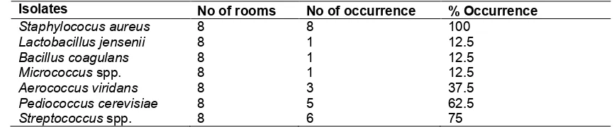
Table 6. Percentage (%) occurrence of bacteria isolates
Isolates No of rooms No of occurrence % Occurrence
Staphylococus aureus 8 8 100
Lactobacillus jensenii 8 1 12.5
Bacillus coagulans 8 1 12.5
Micrococcus spp. 8 1 12.5
Aerococcus viridans 8 3 37.5
Pediococcus cerevisiae 8 5 62.5
Streptococcus spp. 8 6 75
Table 7. Percentage occurrence (%) of fungi isolates
Isolates No of rooms No of occurrence % Occurrence
Aspergillus flavus 8 6 75
Aspergillus niger 8 7 87.5
Aspergillus fumigatus 8 5 62.5
4. CONCLUSION
Conclusively, it was important to determine the type of microflora present in the built environment. The outcome of this research revealed that some aerosols were able to inhibit some organisms that were initially present in experimental rooms while there were introduction of another organisms from some aerosols into some rooms. This shows that, airborne microbiome can be emitted into any environment through the use of aerosols.
COMPETING INTERESTS
Authors have declared that no competing interests exist.
REFERENCES
1. Smith DJ, Timonen HJ, Jaffe DA, Griffin DW, Birmele MN, Perry KD. Intercontinental dispersal of bacteria and archaea by transpacific winds. Appl Environ Microbiol. 2013;79:1134–1139. 2. Smith DJ, Jaffe DA, Birmele MN, Griffin
DW, Schuerger AC, Hee J. Free tropospheric transport of microorganisms from Asia to North America. Microbial Ecol. 2012;64:973–985.
3. DeLeon-Rodriguez N, Lathem TL, Rodriguez-RLM, Barazesh JM, Anderson BE, Beyersdorf AJ. Microbiome of the upper troposphere: Species composition and prevalence, effects of tropical storms, and atmospheric implications. Proc Natl Acad Sci. 2013;110:2575–2580.
4. Kelley ST, Gilbert JA. Studying the microbiology of the indoor environment. Genome Biol. 2002;14:2-202.
5. Rintala H, Pitkäranta M, Toivola M, Paulin L, Nevalainen A. Diversity and seasonal dynamics of bacterial community in indoor environment. BMC Microbiol. 2008;8:56. 6. Tringe SG, Zhang T, Liu X, Yu Y, Lee
WH, Yap J. The airborne metagenome in an indoor urban environment. J. Pone. 2008;10:1371.
7. Kembel SW, Jones E, Kline J, Northcutt D, Stenson J, Womack AM. Architectural design influences the diversity and structure of the built environment microbiome. ISMEJ. 2012;6: 1469–1479. 8. Dunn RR, Fierer N, Henley JB, Leff JW,
Menninger HL. Home life: Factors structuring the bacterial diversity found within and between homes. J Pone. 2013;10:13-71.
9. Adams RI, Miletto M, Lindow SE, Taylor JW, Bruns TD. Airborne bacterial communities in residences: Similarities and differences with fungi. J Pone. 2014;10:13-71.
10. Prussin AJ, Garcia II, Marr EB. Total concentrations of virus and bacteria in indoor and outdoor air. Environ Sci Technol Lett. 2015;2:84–88.
11. Shelton BG, Kirkland KH, Flanders WD, Morris GK. Profiles of airborne fungi in buildings and outdoor environments in the United States. Appl Environ Microbiol. 2002;68:1743–1753.
12. Frank JA, Sørensen SJ. Quantitative metagenomic analyses based on average genome size normalization. Appl Environ Microb. 2011;77:2513–2521.
14. Duhaime MB, Deng L, Poulos BT, Sullivan MB. Towards quantitative metagenomics of wild viruses and other ultra-low concentration DNA samples: A rigorous assessment and optimization of the linker amplification method. Environ Microbiol. 2012;14:2526–2537.
15. Klepeis NE, Nelson WC, Ott WR, Robinson JP, Tsang AM, Switzer P. The national human activity pattern survey (NHAPS): A resource for assessing exposure to environmental pollutants. J Expo Anal Env Epid. 2001;11:231–252. 16. Macher JM, Chatigny MA, Burge HA.
Sampling airborne microorganisms and aeroallergens. In Air Sampling Instruments for evaluation of atmospheric contaminants, cap. 23. 8th Edition ACGIH, Cincinnati, Ohio; 1995.
17. Grinshpun SA, Chang CW, Nevalainen A, Willeke K. Inlet Characteristic of Bioaerosol Samplers. J. Aerosol Sci. 1994;25(8):1503-1522.
18. Abt E, Suh HH, Allen G, Koutrakis P. Characterization of indoor particle sources: A study conducted in the metropolitan Boston area. Environ Health Persp. 2000;108:35–44.
19. Knights D, Kuczynski J, Charlson ES, Zaneveld J, Mozer MC, Collman RG. Bayesian community-wide culture-independent microbial source tracking. Nat Meth. 2011;8:761–763.
20. Stetzenbach LD, Harry R. Microorganisms and indoor air quality. Clinical Microbiology Newsletter. 1998;20:19. 21. Sudharsanam S, Swaminathan S,
Ramalingam A, Thangavel G, Annamalai R, Steinberg R. Characterization of indoor bioaerosols from a hospital ward in a tropical setting Afr Health Sci. 2012;12(2):217-225.
22. Sautour M, Sixt N, Dalle F, L'Ollivier C, Fourquenet V, Calinon C, Profiles and seasonal distribution of airborne fungi in indoor and outdoor environments at a French hospital Sci Total Environ. 2009;407:3766-3771.
23. Tormo-Molina R, Gonzalo-Garijo MA, Fernández-Rodríguez S, Silva-Palacios Monitoring the occurrence of indoor fungi in a hospital. Rev Iberoam Micol. 2012;29(4):227-234.
24. Yagoub SO, Elagbashi A. Isolation of potential pathogenic bacteria from the air of hospital Delivery and nursing rooms. Int J Appl Sci. 2010;10(11):1011-1014. 25. Sheik GB, Abd Al Rheam AI, Al Shehri
ZS, Al Otaibi OM. Assessment of Bacteria and fungi in air from college of applied medical sciences (Male) at AD-Dawadmi, Saudi Arabia. Int Res J Biol Sci. 2015;4(9):48-53.
26. Naruka K, Gaur J. Distribution pattern of airborne bacteria and fungi at market area. American Eurasian J Sci Res. 2014;9(6):186-192.
27. Bond TC. Bounding the role of black carbon in the climate system: A scientific assessment. J.Geophys. Res. – Atmospheres. 2013;11:118.
28. Adams RI, Bateman AC, Bik HM, Meadow JF. Microbiota of the indoor environment: A meta- analysis. Microbiome. 2015;3:1– 18.
29. Lee LD, Hachem RY, Berkheiser M, Hackett B, Jiang Y. Hospital environment and invasive aspergillosis in patients with hematologic malignancy. Am J Infect Control. 2012;40:247–249.
30. Barberan A, Ladau J, Leff JW, Pollard KS, Menninger HL, Dunn RR. Continental-scale distributions of dust-associated bacteria and fungi. Proc Natl Acad Sci U. 2015;112:5756–5761.
31. Chen C, Zhao B. Review of relationship between indoor and outdoor particles: I/O ratio, infiltration factor and penetration factor. Atmos Environ. 2011;45:275–288. 32. Cyrys J, Pitz M, Bischof W, Wichmann H,
Heinrich J. Relationship between indoor and outdoor levels of fine particle mass, particle number concentrations and black smoke under different ventilation conditions. J Expo Sci Env Epid. 2004;14:275–283.
_________________________________________________________________________________ © 2019 Jeff-Agboola et al.; This is an Open Access article distributed under the terms of the Creative Commons Attribution License (http://creativecommons.org/licenses/by/4.0), which permits unrestricted use, distribution, and reproduction in any medium, provided the original work is properly cited.
Peer-review history: