CrossMark
Published by DiscoverSys
INTRODUCTION
Oral cavity cancer (OCC) is a malignancy which arises from the mucosal epithelium lining the oral cavity, the organs of the oral cavity and sali-vary glands (especially the minor salisali-vary glands) which were located on the wall of the oral cavity. The scope of the organs in the oral cavity were the upper and lower lips, tongue anterior two-thirds, buccal mucosa, floor of mouth, gingiva, maxilla and mandible, trigonum retromolare, palatum durum and molle triangule.1,2
Oral cancer was the sixth most found cancer in the world with an estimated 500,000 new cases each year. Frequency and risk factors differ in different geographic regions. OCC contributed to 3-5% of all human malignancy.3
OCC was a major health problem and was associated severe morbidity. The mortality rate was 120,000 deaths annualy.4,5 The incident is estimated at 270,000 cases in men (5% of all cancers) and 119,000 (2.5%) in women. Two-thirds of the cases (67%) were reported from developing countries with a ratio of male and female 3-4:1.3 Locally, the incidence of Oral Cavity Squamous Cell Carcinoma (OCSCC) based of epidemiological data in Sanglah Hospital
Denpasar was approximately 71.7% (2002 - 2011) and there has been significant increase annualy.6
More than 90% of malignancy of the oral cavity was squamous cell carcinoma. Despite many prog-ress in diagnostic and therapy for OCSCC, current modality still unable to detect precancerous lesions (leukoplakia and erythroplakia) which is the reason why majority of patients come with advanced disease (stages III and IV). Furthermore, approxi-mately 20-50% cases had unknown clinical lymph node metastases to the neck and primary tumor recurrence within one year after treatment with poor prognosis.
Current consideration in determining oral cancer treatment decisions (the tumor node metastasis, histopathological type and grade) were considered unsatisfactory. Therefore, biomolecular markers were considered important for diagnostic and prognostic indicators.2
P16 is a tumor suppressor gene which produced p16 protein that binds to CDK4/CDK6, causing cell cycle arrest. Normal p16 activity is required for cell cycle regulation from the G1 phase to the S phase.8 Mutations in p16 gene produce mutant p16 protein ABSTRACT
Background: Oral cavity cancer is considered as a major health problem worldwide and has been associated with high recurrence rate and poor prognosis. Oral cavity squamous cell carcinoma (OCSCC) in Sanglah General Hospital Denpasar contributed to 71.7% of all head and neck cancer from 2002-2011 with an increasing trend annually. Advances in our understanding of OCSCC have not improved the outcome in OCSCC management significantly. Therefore, many studies have focused on the roles of biomolecular markers in OCSCC. One of biomolecular marker that has been the focus of many OCSCC studies is p16.
Method: This was an analytic cross sectional study with 31 samples to determine the association of p16 expression with age group, tumor location, stage, and grade in OCSCC. Data was analyzed descriptively
and the association between variables were evaluated with Chi-Square or Fisher’s Exact Test with a p value <0.05 was considered significant.
Results: From 31 samples collected, the mean age was 56.19±15.197 years old. 17 samples (54.8%) were female and 17 samples (54.8%) were from the ≤60 years old age group. Majority of the tumor was located on the tongue (14 samples; 45.2%) with postero-inferior group location (19 samples; 61.3%). Almost half of the samples were in Stage IVA-B (15 samples; 48.4%) and within high grade group (24 samples; 77.4%) meanwhile 21 samples (67.7%) were in low p16 expression group. There was a significant association between p16 expression with tumor grade group (p=0,044).
Conclusion: p16 expression was significantly associated with tumor grade in OCSCC patients.
Keywords: Oral Cavity Squamous Cell Carcinoma, p16, clinicopathological features.
Cite This Article: Ampur, O., Sudarsa, I., Suryawisesa, I. 2016. Association of p16 expression with clinicopathological features of oral cavity squamous cell carcinoma patients in Bali. Bali Medical Journal 5(3): 362-366. DOI:10.15562/bmj.v5i3.205
*Correspondence to: Ampur, O. Yansen, Surgical Oncology Sub-Division, Department of General Surgery, Udayana Faculty of Medicine/Sanglah General Hospital Denpasar.
supad9@yahoo.co.id
1Surgical Oncology Sub-Division, Department of General Surgery, Udayana Faculty of Medicine/ Sanglah General Hospital Denpasar
Association of p16 expression with
clinicopathological features of oral cavity
squamous cell carcinoma patients in Bali
Ampur, O.Y, Sudarsa, I.W,1* Suryawisesa,I.B.1
Volume No.: 5
Issue: 3
First page No.: 362
P-ISSN.2089-1180
E-ISSN.2302-2914
which is unable to bind CDK4/CDK6, therefore causing uncontrolled cell cycle and results in cellu-lar transformation.9
Several studies have shown reduced or absence of p161NKA expression in premalignant lesions of the oral cavity and OCSCC.10-14 A study in Japan in 1999 showed changes in α transcripts of p16 gene in all OCSCC examined and obtained multiple patterns of gene inactivation.15 Naggar et.al found reduced or absence of p16 expression in 89% of OCSCC cases. Nakahara et.al reported that 67.9% of p16 is under expressed in OCSCC. Meanwhile, a study with adequate tools to assess oral epithelial dyspla-sia and reduced expression of p16 was conducted by Greer et.al. He found reduced expression of p16 in three early stages of OCSCC. The same study also reported Smokeless tobacco keratosis (STK) and alveolar ridge keratosis (ARK) was very weak but relatively strong in all SCC.16
Yuen et.al had 229 head and neck squamous cell carcinomas patients in which 48% had low expres-sion of p16 and a higher incidence of p16 under expression was found in the larynx and pharynx compared to the pharynx and oral cavity.17 Yuen et al found that p16 under expression was signifi-cantly associated with the tumor location, size, and history of radiotherapy. They found a weak correlation between p16 expression with younger age, higher tumor grade, as well as a more poste-rior location. Nevertheless, there has been no established consensus about p16 expression with clinicopathological features of oral cavity squamous cell carcinoma with majority of researches showed mixed results for p16 correlation with clinicopath-oliogical features of OCSCC.10,14,18
Given the high incidence of OCSCC in Indonesia (especially Bali), generally unsatisfactory clinical outcome, and variations of results between litera-tures, we interested to investigate the association of p16 expression with clinicopathological features (age, tumor location, tumor stage, and tumor grade) of OCSCC in Bali.
MATERIAL AND METHOD
A cross-sectional analytical study was conducted to determine the correlation of p16 protein expres-sion with clinicopathological features of OCSCC patients in Bali. The sampling method was retro-spective which includes OCSCC patients treated at the Department of Surgery Udayana University / Sanglah General Hospital in Denpasar Bali, from January 1st 2010 until March 31st 2013. The data of tumor location, histopathologic type, tumor stage and tumor grade were collected from OCSCC patient’s medical records in the Department of Pathology Anatomy Udayana University/Sanglah
General Hospital. Paraffin block were cut into 5μm thickness, de-waxed for 15 minutes in xylene and rehydrated with ethanol. Slides were treated with 3% hydrogen peroxidase solution for 30 minutes at room temperature. After being washed three times with phosphate buffered saline, microwave antigen retrieval was performed in citrate buffer (pH 6) for 5 minutes. We used a 1/250 dilution, followed by immunohistochemical staining of p16 (Avidin biotin complex immunoperoxidase and Anti-CDKN2A/p16INK4a antibody [EPR1473] ab108349 (Abcam).
Sample age was determined by age listed in medical records at the time of diagnosis. In this study, samples were grouped into two groups: ≤60 years and >60 years of age. Tumor location was determined from intraoperative tumor location. Oral cavity organ locations were the lips, tongue, buccal, gingival maxilla and mandible, hard and soft palate. Tumor locations were grouped into: antero-superior location (lip, palate and maxillary gingiva) and postero-inferior location (tongue, buccal, and mandibular gingiva). Tumor stage was determined by AJCC tumor staging system (2002). Stage I, II, III were categorized as low stage and stage IV A-B and IV C were categorized as advanced stage. Well differentiated tumors were categorized as low grade, while moderate and poorly differentiated tumors were categorized as high grade.
Descriptive analysis was conducted to deter-mine samples baseline characteristics as well as proportion of variables. The association between p16 and other variables was evaluated by chi-square or Fisher’s exact test. P-value of <0.05 was consid-ered significant.
RESULTS
Of the 36 samples collected, five samples did not meet the inclusion criteria. Two with no paraffin blocks, two samples showed normal histopatho-logic features, and one sample with damaged paraf-fin block. Of the 31 patients who were the subjects of research, the youngest was 19 years old and the oldest was 80 years old with a mean age of 56.19 ± 15.197 years. In this study there were 14 samples (54.8%) within ≤60 years age group and 14 samples (45.2%) within >60 years age group. A total of 23 samples (74.2%) were from Bali and 8 samples (25.8%) were Non Balinese.
grade. A total of 21 samples (67.7%) showed weak p16 expression and 10 samples (32.3%) showed strong p16 expression. Overall, the characteristics of the sample are presented in Table 1.
Association between p16 Expression with Oral Cavity Cancer Age Group
There were 17 samples in the ≤60 years age group with 12 samples (70.6%) showed weak expres-sion of p16 and 5 samples (29.4%) showed strong expression of p16. From 14 samples in >60 years age group there were 9 samples (64.3%) with weak expression of p16 and 5 samples (35.7%) showed strong expression. Fisher’s exact test showed no significant association between p16 expression with age in this study group (Table 2).
Relationship between p16 Expression with Oral Cavity Cancer Location
There were 12 samples with tumor located in the antero-superior region, as many as 7 samples (58.3%) showed weak expression of p16 and 5 samples (41.7%) showed strong expression of p16. The postero-inferior tumor group contained 19 samples of which 14 samples (73.7%) showed weak expression of p16 and 5 samples (26.3%) showed strong expression of p16 (Table 3).
Fisher’s exact test results demonstrate no signif-icant association between p16 expression with tumor location in this study (p = 0.308).
Association of p16 Expression with Oral Cavity Cancer Stage
From 15 samples with low stage, as many as 10 samples (66.7%) showed weak expression of p16 and 5 samples (33.3%) showed strong expression of p16.
Within the group of advanced stage tumors, there were 16 samples of which 11 samples (68.8%) showed weak expression of p16 and 5 samples (31.3%) showed strong expression of p16 (Table 4). Fisher’s exact test results demonstrate no significant association between p16 expression with tumor stage in this study (p=0.602).
The study also found no significant association between p16 expression with variables T, N, and M staging system (Table 5).
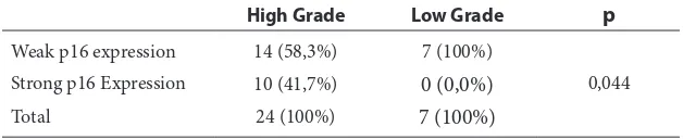
Association between p16 Expression with Oral Cavity Cancer Grade
From 7 samples with low-grade tumors, a total of 7 samples (100.0%) showed weak expression of p16 and no sample showed strong expression of p16. In the group of high-grade tumors, there were 24 samples of which 14 samples (58.3%) showed weak expression of p16 and 10 samples (41.7%) showed strong expression of p16 (Table 6). Fisher’s exact test Table 1 OCSCC Sample Baseline Characteristics
Variable n
Age
≤60 years 17 (54,8%)
>60 years 14 (45,2%)
Sex
Male 14 (45,2%)
Female 17 (54,8%)
Tumor Location
Tongue 14 (45,2%)
Gingiva 6 (19,4%)
Palatum 4 (12,9%)
Buccal 5 (16,1%)
Lip 2 (6,5%)
Tumor Location Group
Antero-superior 12 (38,7%)
Postero-inferior 24 (61,3%)
Stadium
Stadium II 1 (3,2%)
Stadium III 14 (45,2%)
Stadium IV A-B 15 (48,4%)
Stadium IV C 1 (3,2%)
Tumor Grade
Well Differentiated 7 (22,6%)
Moderately Differentiated 22 (71,0%)
Poorly Differentiated 2 (6,5%)
P16 expression
+1 15 (48,4%)
+2 6 (19,4%)
+3 7 (22,6%)
+4 3 (9,6%)
Table 2 p16 Expression Correlation with Age Group
Age ≤60 years Age >60 years p
Weak p16 expression 12 (70,6%) 9 (64,3%)
0,503 Strong p16 Expression 5 (29,4%) 5 (35,7%)
Total 17 (100%) 14 (100%)
Table 3 Association of p16 Expression with Tumor Location
Antero-Superior Postero-Inferior p
Weak p16
expression 7 (58,3%) 14 (73,7%)
0,308 Strong p16
Expression 5 (41,7%) 5 (26,3%)
results demonstrate the value of p = 0.044 (p <0.05), so that there was a significant association between p16 expression with tumor grade in this study.
DISCUSSION
In this study, 31 samples were selected from 36 patients included. A total of 23 samples (74.2%) were Balinese and 8 samples (25.8%) were Non Balinese. The youngest was 19 years of age and the oldest was 80 years of age. Fourteen samples (45.2%) were male and 17 samples (54.8%) were female. The mean age of the sample was 56.19 ± 15.197 years with 17 samples ≤60 years (54.8%) and 14 samples >60 years (45.2%). Most tumors were located on the tongue (14 samples, 45.2%) with most at postero-inferior region (19 samples, 61.3%). Most samples were in Stage IVA-B (15 samples, 48.4%), the high-stage group (16 samples, 51.6%), and had a moderately differentiated grade (22 samples, 71.0%) within the high grade group (16 samples, 51.6%). A total of 21 samples (67.7%) showed a weak p16 expression.
In terms of age, we found the tendency for OCSCC to be younger (≤ 60 years, 17 samples, 54.8%) in this study, which was in accordance with data obtained by Westra and Curado.19,20 There was a difference in terms of male to female ratio in this study compared to others (14 male samples (45.2%) and 17 female samples (54.8%); 8,2:10). We suspect
that other factors such as smoking, alcohol, tobacco chewing, oral sex, and education, which were not analyzed in this study contributed to this difference in result. We found that the OCSCC patients in Bali were relatively young, but the majority were women. OCSCC found in this study was mostly on the tongue (In accordance with the findings of Westra 2009) with the majority of the group located on the postero-inferior region. Therefore, further research on this matter is still needed.
The role of p16 in the carcinogenesis and clin-icopathology of OCSCC has become a topic of research for the last two decades.21 Pande et al in 1998 found a significant correlation between tumor progression with tumor stage. However, there are still many differences between the results of studies on the role of p16 in OCSCC.14,15,22 Nakahara et al (2001) found that weak expression of p16 was found in 67.9% of squamous cell carcinoma samples, with similar results to that obtained in our study where weak p16 expression were found in 21 samples (67.7%). However, Yuen et al (2002) in his study on the expression of p16 in OCSCC found weak p16 expression by 48%. Differences between this study and Yuen et al (2002) is in terms of the number of samples, Yuen et al used 225 samples that may be more representative of the population there. During sample calculations, we used the proportional value of p16 under expression of 0.6, where the figure was obtained from the research Nakahara et al (2001). This value indicates that the difference in the proportion of p16 under expression on OCSCC likely differs between each population and requires further research with a larger sample size.
Significant correlation between p16 expression with OCSCC was not found in this study (p = 0.602), as well as between p16 expression with age group (p = 0.503). Vairaktaris et al (2007) found that p16 under expression was found at the beginning of the carcinogenesis process of OCSCC, but they did not find a significant association between p16 under expression with dysplasia phase in OCSCC carcinogenesis. In this study, we did not obtain any significant correlation between p16 protein expres-sion with tumor location (p = 0.595). Yuen et al (2002) found that more frequent p16 under expres-sion were found in laryngeal carcinoma compared to carcinoma of the oral cavity and pharynx. One weakness of this study is limited diagnostic facil-ities for laryngeal and pharyngeal carcinomas. Therefore, we limited the study only on oral cavity carcinoma. What was interesting in this study is that although there was no significant correlation between p16 expression with T-N-M status (p = 0.597, p = 0.483, p = 548 respectively), we found a significant correlation between p16 expression with OCSCC grade (p = 0.044). However, we could Table 4 Association of p16 Expression Correlation with Tumor
Stage Group
Low Stadium Advanced Stadium p
Weak p16 expression 10 (66,7%) 11 (68,8%)
0,602 Strong p16 Expression 5 (33,3%) 5 (31,3%)
Total 15 (100%) 16 (100%)
Table 5 Association of p16 Expression with T-N-M Status in OCSCC
Variable Weak p16 expression P
T T0-3
T4 12 (57,1%)9 (42,9%) (Fisher’s exact test0,597 )
N N0
N1-2 15 (71,4%)6 (28,6%) (Fisher’s exact test0,483 )
M M0
M1 20 (95,2%)1 (4,8%) (Fisher’s exact test0,548 )
Table 6 Association of p16 Expression Correlation with Tumor Grade Group
High Grade Low Grade p
Weak p16 expression 14 (58,3%) 7 (100%)
0,044 Strong p16 Expression 10 (41,7%) 0 (0,0%)
not determine the value of the odds ratio because we did not find any samples which showed strong expression of p16 tumor with low grade.
CONCLUSION
A significant correlation was found between p16 expression with OCSCC grade. This study was a preliminary study; a more robust research has to be designed to better understand the role of p16 in OCSCC.
REFERENCES
1. Sidransky, D. Cancer of the head and neck. In: DeVita VT, Lawrence TS, SA. R, editors. Devita, Hellman & Rosenberg’s cancer: principles & practice of oncology. 8th Edition ed. Philadelphia Lippincott Williams & Wilkins. 2008. 2. Azamris, Burmansyah, Tjindarbumi, D., Achmad, D.,
Dlidir, D. Panduan penatalaksanaan kanker rongga mulu. In: Manuaba TW, editor. Panduan penatalaksanaan kanker solid PERABOI. 2010. Jakarta: Sagung seto,:98-130. 2010. 3. Ferlay, J., Parkin, D.M., Pisani, P. editors. GLOBOCAN
2002 : Cancer incidence, mortality and prevalence world-wide. Lyon IARC Press. 2004
4. Das, B.R., Nagpal, J.K.. Understanding the biology of oral cancer.Med Sci Monit ,8:258–67. 2002
5. Sudbo, J., Reith, A. 2005. The evolution of predictive oncol-ogy and molecular based therapy for oral cancer preven-tion. Int J Cancer;115:339–45.
6. Rekam medis RSUP Sanglah, SMF Ilmu Bedah Fakultas Kedokteran Universitas Udayana/RSUP Sanglah,Denpasar Bali.
7. Alex, J.C., Klenoff, J. Head and neck cancers. In: Krag, D.N., editor. Surgical oncology. Landes Bioscience, hal 66-80. 2000.
8. Kumar, V., Cotran, S.R., and Robbins, L.S. Robbins Basic Pathology. 7th ed. Saunders Philadelphia London Toronoto Montreal Sydney Tokyo. 174-185. 2003.
9. Lang, J.C., Borchers, J., Danahey, D, et al. Mutation Status of Overexpressed p16 in Head and Neck Cancer: Evidence for Germline Mutation of p16/p14-ARF. J Oncol 21 : 401-408. 2002.
10. Naggar, A.K., Lai, S., Clayman, G.L., Zhou, J.H., Tucker, S.A., Myers, J., et al, Expression of p16, Rb, and cyclin D1 gene product in oral and laryngeal squamous carci-noma biological and clinical implications. Human patol-ogy,30:1013-18. 1999.
11. Shintani, S., Nakahara, Y., Mihara, N., Oeyama, Y., Matsumura, T. 2001. Inactivation of the p14, p15 and p16
genes is a frequent event in human oral squamous cell car-cinomas. Oral oncology, 37:498-504.
12. Nakahara, Y., Shintani, S., Mihara, M., Kiyota, A., Ueyama, Y., Matsumura, T. Alterationof Rb, p16 and cyclin D1 in the tumorgenesis of oral squamous cell carcinomas.
Cancer letters,160:3-8. 2001.
13. Ruesga, M.T., Sagredo, A.A., Rodriquez, M.J., Aguirregaviria, J.I., Videgain, J., Rodriquez, C. et al. p16 promoter hypermethylation in oral scrappings of oral squmous cell carcinomas risk patients Cancer let-ters,250:140-5. 2007.
14. Cao, D., Begum, S., Ali, S.Z., Westra, W.H. Expression of p16 in benign and malignant cystic squamous lesions of the neck. Human patology,41:535-9. 2010.
15. Akanuma D., Uzawa N., Yoshida M.A., Negishi A., Amagasa T., Ikeuchi T. Inactivation pattern of the p16 (INK4a) gene in oral squamous cell carcinoma cell lines.
Oral oncology, 35:476-83. 1999.
16. Greer, R.O., Meyers, A., Said, S.M., Shroyer, K.R. Is p16 INK4a protein expresion in oral ST lesions reliable precan-cerrous marker. Int .J.Oral.Maxilofac.surg, 37;840-6. 2008. 17. Yuen, W.M., Man, K.Y. Clinicopathological significance
of p16 gene expression in the surgical treatment of head and neck squamous cell carcinoma. J clinical pathology 2002;55;58-60. 2002.
18. Muirhead, D.M., Hoffman, H.T., Robinson, R.A. Correlation of clinicopathological features with immuno-histochemical expression of cell cycle regulatory proteins p16 and retinoblastoma: distinct association with kerati-nisation and differentiation in oral cavity squamous cell carcinoma. J Clin Pathol,59:711–5. 2006.
19. Westra, H.W. The Changing Face of Head and Neck Cancer in the 21st Century: The Impact of HPV on the
Epidemiology and Pathology of Oral Cancer. Head and Neck Pathol.; 3:78–81. 2009.
20. Curado, M.P. dan Hashibe, M. Recent changes in the epi-demiology of head and neck cancer. Curr.Opin.Oncol., 21, (3) 194-200. [cited 21 Maret 2013]. Available from: http://
sfx.ub.rug.nl.proxy-ub.rug.nl/sfx_local?sid=Entrez:Pub-Med&id=pmid:19363341. 2009.
21. Pande, P., Mathur, M., Shukla, N.K., Ralhan, R. pRb and p16 protein alterations in human oral tumorgenesis. Oral oncology,34:396-403. 1998.
22. Vairaktaris, E., Yapijakis, C., Psyrri, A., Spyridonidou, S., Yannopoulo, A., Lazaris, A., et al. Loss of tumour suppres-sor p16 expression in initial stages of oral oncogenesis. Anticancer Research, 27:979. 2007.