CrossMark
Published by DiscoverSys
ABSTRACT
Background: Neonatal sepsis is a common occurrence in our part of the world, characterized by signs and symptoms of bacterial infection during first 28 days of life. Diagnosis of neonatal sepsis may be difficult as the early signs of sepsis may be subtle and not specific.
Objective: This study was to determine the accuracy of a simple and cost effective hematological scoring system (HSS) in the early diagnosis of neonatal sepsis using blood culture as a gold standard.
Methods: The cross-sectional study enrolled 62 neonates who were clinically suspected of sepsis. The neonatal hematological parameter
was measured in all cases. All subjects were analyzed according to HSS. Blood culture was taken for the gold standard of proven sepsis.
Results: Proven sepsis confirmed by blood culture was found in 34% of neonates. The HSS was found to have a sensitivity of 80.9%, specificity of 92.7%, positive predictive value of 85%, negative predictive value of 90.5%, positive likelihood ratio of 11.1, negative likelihood ratio of 0.2, and accuracy of 88.7%.
Conclusion: The HSS is a simple cost effective tool that can be used to early diagnose of neonatal sepsis.
Keywords: hematological, scoring, neonatal, sepsis
Cite This Article: Pramana, K., Kardana, I., Nilawati, G. 2016. Diagnosis accuracy of hematological scoring system in early identification of neonatal sepsis. Bali Medical Journal 5(3): 488-492. DOI:10.15562/bmj.v5i3.310
INTRODUCTION
Sepsis is the most common cause of neonatal mortality and morbidity. Neonatal sepsis is a clini-cal syndrome characterized by signs and symptoms of infection with accompanying bacteremia in the first month of life. The early signs of neonatal sepsis may be subtle.1 In developing countries, the incidence of neonatal sepsis varies from 1.8-18 per 1000 live birth and mortality rate is 12-68 per 1000 live birth.2 Despite continuing advances in diagno-sis and treatment, it remains one of the important causes of higher mortality and morbidity. The inci-dence of neonatal sepsis is 15.5% with mortality rate is 13.68% in Jakarta.3 In Sanglah hospital, a study from January until December 2010 reported the incidence of neonatal sepsis to be 5% with a mortality rate of 30.4%.4
Neonatal sepsis is often undetected and causes death within a short time. Clinical signs and symptoms of sepsis in children are rare in neonatal sepsis, making it difficult to diagnose. A definite diagnosis is made by blood culture. However, the procedure is time-consuming (takes more than 48-72 hours), yields positive result in 30-40%, and the facilities for the test might not be avail-able in many laboratories.5 Investigations such as C-reactive protein (CRP), immature to total neutrophil (IT) ratio, total number of leukocytes and platelets, if not combined with other investi-gations become non-specific and can’t be used to diagnose neonatal sepsis.6
Timely diagnosis of neonatal sepsis is critical because in neonates the illness can progress more rapidly than an adult. Early diagnosis of neonatal sepsis is still a great challenge. So, the significance of various screening tests, either singly or in combi-nation is still observed. The need is for an infallible test for bacteremia that is easily performed, quick, simple, and cost-effective.7 A hematological scoring system (HSS) of Rodwell is preferable for early diag-nosis of neonatal sepsis because it includes some parameters. The HSS is formulated that assigned a score of one for each of seven findings, namely abnormal leukocyte count, abnormal total neutro-phil count, elevated band cell, elevated IT ratio, elevated immature to mature neutrophil (IM) ratio, decreased platelet count, and degenerative changes in neutrophils.8 The present study was undertaken to evaluate and highlight the accuracy of HSS in the early detection of neonatal sepsis.
METHODS
This study is a hospital-based cross-sectional study of all the neonates during the first week after birth in Sanglah Hospital from March to May 2015. Neonates who were clinically suspected to have a bacterial infection within the first week of life, based on perinatal risk factors and clinical features were taken as a subject. Some of these neonates were asymptomatic but were evaluated for sepsis because of maternal intrapartum sepsis risk factors
Department of Child Health, Udayana University School of Medicine, Sanglah General Hospital, Denpasar, Bali-Indonesia
*Correspondence to: Kissinger
Puguh Pramana, MD, Pediatrician, Department of Child Health, Udayana University School of Medicine, Sanglah General Hospital, Jl. Pulau Nias, Denpasar, Bali, Indonesia, Telephone/Fax: (+62) 361 244038.
kissingerpp@gmail.com
Volume No.: 5
Issue: 3
First page No.: 488
P-ISSN.2089-1180
E-ISSN.2302-2914
Doi: http://dx.doi.org/10.15562/bmj.v5i3.310
ORIGINAL ARTICLE
Diagnosis accuracy of hematological scoring system
in early identification of neonatal sepsis
like premature rupture of membranes, maternal urinary tract infection, maternal intrapartum fever, chorioamnionitis, and excessive vaginal discharge. The other sepsis risk factors were fetal distress, foul-smelling amniotic fluid, birth asphyxia, very low birth weight, and preterm infants. Neonates with incomplete data and laboratory result obtained from other hospital were excluded.
Blood samples of all subjects were taken for septic work up. The septic works up included complete blood counts measurements, IT ratio, peripheral blood smear, and blood culture with antibiotic sensitivity. The analysis of the peripheral blood smear findings was done by the pathologist blinded to the infection status of the neonates. Blood culture was taken for the gold standard of proven sepsis. Sensitivity, specificity, positive predictive value, negative predictive value, positive and negative likelihood ratio, and accuracy were calculated for the HSS. Data was compiled and statistically analyzed by using computer programs. This study was approved by Medical Research Ethics of Faculty and Hospital Ethical Committee. The variables in HSS with the score are as seen in
Table 1.
There were 7 variables for the HSS which each variable has score 1 if meet the criteria. The inter-pretation of the HSS was total score ≥ 5 considered as neonatal sepsis. Findings of HSS were recorded and later compared with results of blood cultures to assess the diagnosis accuracy of HSS.
RESULTS
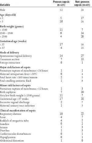
During the study period, there were 62 neonates with clinically suspected of sepsis and no subjects were excluded. Characteristics of the study subjects are presented in Table 2. Male (58%) was more common than female and most of the newborns (65%) were present within more than 3 days of life. Preterm (56%) and very low birth weight (53%) of newborns were more prone to develop sepsis.
Mode of delivery predominated by sponta-neous vaginal delivery in this study. Foul-smelling amniotic fluid and the preterm infants were found 15% and 56%, the commonest major and minor risk factor of sepsis, respectively. On analysis of collected data, we found that respiratory distress was the most common presentation (52%) followed by a history of neonatal fever (18%), and reddish of the orogastric tube (10%).
Proven sepsis is confirmed by blood culture in 34% of the neonates. Klebsiella pneumoniae and Acinetobacter baumanii are the most common organism isolated followed by Enterobacter cloa-cae, coagulase-negative Staphylococci, and Serratia marcescens.
Table 1 Hematological scoring system
Variables Score
Increased of IT ratio > 0.2 1
Increased (> 5,400/mm3) or decreased (< 1,800/mm3) of
polymorphonuclear (PMN) count 1
Increased of immature to mature neutrophil (IM) ratio ≥ 0.3 1
Increased of immature PMN count > 500/mm3 1
Decreased or increased of leukocyte count (≤ 5,000/mm3 or ≥ 30,000/mm3) 1
Degenerative changes in PMN (vacuolization, toxic granule, and
Dohle bodies) 1
Decreased of thrombocyte count ≤ 150,000/mm3 1
Source: Rodwell et al.8
Table 2 Characteristics of the subjects
Variable Proven sepsis (n=21) sepsis (n=41)Not-proven
Male 12 24
Age (days old)
≤ 3
> 3 165 1724
Birth weight (grams)
< 1500 1500 – 2500 > 2500
11 6 4
7 14 20
Gestational age (weeks)
< 37
≥ 37 174 1823
Mode of delivery
Spontaneous vaginal delivery Caesarean section
Forceps extraction
14 7 0
27 13 1
Major risk factors of sepsis
Premature rupture of membranes >24 hours Maternal intrapartum fever >38°C
Fetal heart rate >160 times/minute Foul-smelling amniotic fluid
1 0 2 1
1
4 5 8
Minor risk factors of sepsis
Premature rupture of membranes >12 hours Birth asphyxia
Very low birth weight (<1500 grams) Gestational age <37 weeks
Excessive vaginal discharge Maternal urinary tract infection
1 7 11 17 2 1
3 10
7 18
7 5
Clinical manifestation of sepsis
Respiratory distress Fever
Reddish of orogastric tube Jaundice
Seizure Diarrhea
Cardiovascular disturbances Hypoglycemia
Abdominal distention
10 4 4 0 1 1 0 0 1
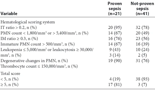
Table 3 shows the characteristic of the HSS. Out of 21 subjects with culture proven sepsis, 17 (81%) neonates had a score ≥ 5, and 4 (19%) had a score < 5. Three (7%) subjects of the not-proven sepsis group had a score ≥ 5 suggesting the presence of sepsis and 38 (93%) had a score < 5 which implies that sepsis was unlikely in these cases. Most of the proven sepsis group had IT ratio > 0.2 and throm-bocyte count ≤ 150,000/mm3. Degenerative changes
of PMN was not frequently found in proven sepsis group.
The hematological scoring system had a sensi-tivity of 80.9%, specificity 92.7%, positive predictive value 85%, negative predictive value 90.5%, positive likelihood ratio of 11.1, negative likelihood ratio 0.2, and accuracy of 88.7%.
DISCUSSION
The major problem in neonatal infections is the identification of the infected infant and the equally important task is of identifying the non-infected infant because of its nonspecific clinical symptoms. It is the current practice to start empirical antibi-otic therapy in all neonates showing infection-like symptoms.9 However, this may expose newborns to a risk of adverse drug effects on one hand and lead to nosocomial complications and the emergence of resistant strains on the other. Accurate and quick diagnosis is, therefore, essential so that timely treat-ment of a potentially fatal disease can be provided and at the same time damages deriving from the unnecessary use of antibiotics be prevented.10
The definitive diagnosis of sepsis is made by a positive blood culture, which requires a minimum of 48-72 hours, yields a positive result in 30-40% of cases. Blood culture remains the gold standard for diagnosis of neonatal sepsis, yet it is import-ant to develop effective screening tools which can
presumably diagnose or exclude neonatal sepsis at the time of presentation.11 Early diagnosis of neona-tal sepsis is still a great challenge. For early diagno-sis of neonatal sepdiagno-sis, the HSS was introduced in the past. It included hematological parameters and showed that such score could accurately predict the presence or absence of infection and be reliable. The HSS assigns a score of one for each of seven hematologic findings and shown to be significantly (P<0.005) associated with sepsis.12 The present study evaluates the usefulness of the HSS as an early indicator of neonatal sepsis. This study was undertaken because it is a simple bedside and cost effective test which can be done even if the neonate has had antibiotics.
In this study, 34% neonates were considered as proven sepsis by blood culture. However, not-proven sepsis group (66%) comprises a difficult diagnostic group and could not be ignored because the fatal infection had been reported in another study in the presence of negative blood culture.8 Among the infected neonates, the predominance of the male was due to the factors regulating the synthesis of immunoglobulin G is situated on the × chromosome. A male has only one × chromo-some, so he is less immunologically protected than the females. Preterm and very low birth weight neonates are more susceptible to infection due to low level of immunoglobulin G and lower defense mechanism.12
Although various tests are used, as a diagnostic tool for neonatal sepsis, the complete blood count with differential is widely used, either singly or in conjunction with other test or clinical findings. As no single individual hematological parameter is superior in comparison to another in predicting neonatal sepsis, a combination of these parame-ters in the form of HSS has been recommended. Hematologic scoring system should improve the efficiency of the complete blood count as a screen-ing test for sepsis until a reliable diagnostic test is available. The HSS has practical advantages; it is applicable to all infants, including those who have received antibiotic therapy prior to evaluation and simplifies the interpretation of hematologic profile. The HSS is the most reliable of the published criteria evaluated which identifies almost all neonates with sepsis or with probable sepsis.13 In this study with a cut-off score ≥ 5, we observed a sensitivity of 80.9%, specificity 92.7%, positive predictive value 85%, negative predictive value 90.5%, positive likelihood ratio of 11.1, negative likelihood ratio 0.2 These results were consistent with other studies.10,14 This scoring system is significant in many other ways, regarding its easy availability, accessibility, low cost, less time consuming and practically possible in all the laboratories, which makes it convenient for any Table 3 Characteristics of the hematological scoring system
Variable
Proven sepsis (n=21)
Not-proven sepsis (n=41) Hematological scoring system
IT ratio > 0.2, n (%)
PMN count < 1,800/mm3 or > 5,400/mm3, n (%)
IM ratio ≥ 0.3, n (%)
Immature PMN count > 500/mm3, n (%)
Leukopenia ≤ 5,000/mm3 or leukocytosis ≥ 30,000/
mm3, n (%)
Degenerative changes in PMN, n (%) Thrombocyte count ≤ 150,000/mm3, n (%)
20 (95) 14 (67) 16 (76) 14 (67) 9 (43) 3 (14) 19 (90)
32 (78) 20 (49) 23 (56) 16 (39) 10 (24) 2 (5) 31 (76)
Total score < 5, n (%)
common man to get a high-risk neonate tested and diagnosed on time.15
Elevated IT ratio was found to be the most reli-able indicator of sepsis in this study, and also in vari-ous other studies like those by Narasimha et al14 and Zulfikri.16 Abnormal leukocyte count is found to be less specific and sensitive than IT ratio. This is in accordance with a study in Pune, India.17 Our study showed that total leukocyte count was not increased in most cases. The result of the present study was in contrast to a study by Anwer and Mustafa which showed that abnormal leukocyte count is the most specific test with better positive and negative predic-tive value than IT ratio. 18 Total leukocyte count is of little clinical use in the diagnosis of neonatal infec-tion because of the wide variainfec-tion in values.
The total PMN count has a limited role in sepsis screening. Neutropenia has been more common in association with sepsis, compared with neutro-philia, probably because of increased adherence to altered endothelial cells and utilization at the site of infection. The elevation of PMN is often very late and inconsistent. A shift to the left in differential leukocyte count with a raised immature neutro-phil count (band form) has been documented in patients with bacterial infection.19 In the present study, total PMN count did not contribute to the incidence of neonatal sepsis. The variation in the results of these parameters in different studies might be due to difference in the blood sampling time, exact method of test employed, the severity of infection, and the age of the neonates.20
Degenerative changes in PMN can help diagnos-ing neonatal sepsis. Moreover, the presence of toxic granules indicates the production of unusual PMN during infection and stress induced leukopoiesis. Activated PMN by bacterial infections can also give a microscopic overview as vacuoles or a large mass of blue at the edges of the cytoplasm called Dohle bodies. Their presence invariably indicates sepsis, but their count is not always increased.21 Degenerative changes in PMN made no significant contribution to the diagnosis in this study.
Neonates with sepsis develop thrombocytope-nia, possibly because of disseminated intravascular coagulation (DIC) and the damaging effects of endo-toxin on platelets. Thrombocytopenia is frequently associated with sepsis and indicated a poor progno-sis. This is thought to be due to increased platelet destruction, sequestration secondary to infections, failure in platelet production due to reduced mega-karyocytes.22 In this study most cases had thrombo-cytopenia. This correlated well with various other studies done in Vermont and India.23,24
The higher the score on HSS, the higher is the chance of sepsis. The simplification and stan-dardization of the interpretation of this global
test are still required. A variety of other rapid detection methods of microorganisms, like DNA probes, automated blood culture system, and fluorometric detection systems are also available globally, but HSS can still be used as a screening test for early diagnosing sepsis and to differentiate infected neonates from the non-infected ones.25 Furthermore, the sensitivity and the specificity of the test are also high, with the certainty of sepsis increasing along with the score.
CONCLUSION
The HSS had a sensitivity of 80.9%, specificity 92.7%, and accuracy of 88.7%. It is a simple, quick, cost effective and readily available tool with high specificity and affordable sensitivity that can be used to early diagnose of neonatal sepsis.
ACKNOWLEDGEMENTS
We thank the colleagues of Neonatal subdivision of Child Health Department, Clinical Pathology Laboratory, and Clinical Microbiology Installation of Sanglah Hospital for technical assistance and collaboration.
REFERENCES
1. Stoll BJ. Infections of the neonatal infant. In: Kleigman RM, Behrman RE, Jenson HB, Stanton BF, editors. Nelson textbook of pediatrics. 18th ed. United States of America: Saunders; 2007. p. 794-809.
2. Watson RS, Carcillo JA, Linde-Zwirble WT, Clermont G, Lidicker J. The epidemiology of severe sepsis in chil-dren in the United States. Am J Respir Care Med. 2003;167:695-701.
3. Juniatiningsih A, Aminullah A, Firmansyah A. Profil mikroorganisme penyebab sepsis neonatorum di Departemen Ilmu Kesehatan Anak Rumah Sakit Cipto Mangunkusumo Jakarta. Sari Pediatri. 2008;10:60-5. 4. Kardana IM. Incidence and factors associated with
mortal-ity of neonatal sepsis. Paediatr Indones. 2011;51:144-8. 5. Movahedian AH, Moniri R, Mosayebi Z. Bacterial culture
of neonatal sepsis. Iranina J Publ Health. 2006;35:84-9. 6. Zeeshan A, Tariq G, Talal W, Shahid M. Diagnostic value
of CRP and hematological parameters in neonatal sepsis. J Coll Physicians Surg Pak. 2005;15:152-6.
7. Arif S, Ehsan A, Arif M, Hussain J, Bano R. Early diagnosis of neonatal sepsis through haematological and biochemi-cal markers. Gomal J Med Sci. 2012;11:178-82.
8. Rodwell RL, Leslie AL, Tudehope DI. Early diagnosis of neonatal sepsis using a hematologic scoring system. J Pediatr. 1998;112:761-7.
9. Aminullah A. Sepsis pada bayi baru lahir. In: Kosim MS, Yunanto A, Dewi R, Sarosa GI, Usman A, editors. Buku ajar neonatologi. 1st ed. Jakarta: BP IDAI; 2010. p. 170-85. 10. Khair KB, Rahman MA, Sultana T, Roy CK, Rahman MQ,
Shahidullah M, et al. Role of hematologic scoring sys-tem in early diagnosis of neonatal septicemia. BSMMU J. 2010;3:62-7.
11. Polin RA. Management of neonates with suspected or proven early-onset bacterial sepsis. Pediatrics. 2012;129:1006-15.
13. Makkar M, Gupta C, Pathak R, Garg S, Mahajan NC. Performance evaluation of hematologic scoring system in early diagnosis of neonatal sepsis. J Clin Neonatol. 2013;2:25-9.
14. Narasimha A, Kumar MLH. Significance of hematological scoring system (HSS) in early diagnosis of neonatal sepsis. Indian J Hematol Blood Transfus. 2011;27:14-7.
15. Ghosh S, Mittal M, Jaganathan G. Early diagnosis of neo-natal sepsis using a hematological scoring system. Indian J Med Sci. 2001;55:495-500.
16. Zulfikri. Diagnosis sepsis neonatal. Sari Pediatri. 2004;6:81-4.
17. Buch AC, Srivastava V, Kumar H, Jadhav PS. Evaluation of haematological profile in early diagnosis of clinically sus-pected cases of neonatal sepsis. Int J Basic Appl Med Sci. 2011;1:1-6.
18. Anwer SK, Mustafa S. Rapid identification of neonatal sep-sis. J Pak Med Assoc. 2000;50:94-8.
19. Vinay BS, Girish GN, Adhikari S, Hugara S. Evaluation of septic screen as a diagnostic tool for neonatal sep-sis in a tertiary hospital at Mysore. Sch J App Med Sci. 2015;3:1005-10.
20. Al-Gwaiz LA, Babay HH. The diagnostic value of absolute neutrophil count, band count and morphologic changes of
neutrophils in predicting bacterial infections. Med Princ Pract. 2007;16:344-7.
21. Hutchison RE, Abraham NZ. Leukocytic disorders. In: McPherson RA, Pincus MR, editors. Henry’s clinical diag-nosis and management by laboratory methods. 21st ed. Philadelphia: Saunders; 2007. p. 545-98.
22. Arif SH, Ahmad I, Ali SM, Khan HM. Thrombocytopenia and bacterial sepsis in neonates. Indian J Hematol Blood Transfus. 2012;28:147-51.
23. Philip AG, Hewitt JR. Early diagnosis of neonatal sepsis. Pediatrics. 1980;65:1036-41.
24. Basu S, Guruprasad, Narang A, Garewal G. Diagnosis of sepsis in the high-risk neonate using a hematologic scoring system. Indian J Hematol Blood Transf. 1999;17:32-4. 25. Supreetha MS, Sathyavathi RA, Shivendra VS, Kariappa TM.
Evaluation of neonatal septicaemia using hematological parameters. Int J Recent Sci Res. 2015;6:2775-8.