Review Article
Iran J Neurol 2015; 14(1): 1-7
Epileptic syndromes: From clinic to
genetic
Abbas Tafakhori1, Vajiheh Aghamollaii2, Sara Faghihi-Kashani3, Payam Sarraf1, Laleh Habibi4
1Department of Neurology, School of Medicine, Imam Khomeini Hospital AND Iranian Center of Neurological Research, Tehran
University of Medical Sciences, Tehran, Iran
2
Department of Neurology, School of Medicine, Roozbeh Hospital AND Iranian Center of Neurological Research, Tehran University of Medical Sciences, Tehran, Iran
3
Department of Neurology, School of Medicine, Tehran University of Medical Sciences, Tehran, Iran
4
Department of Medical Genetics, School of Medicine, Tehran University of Medical Sciences, Tehran, Iran
Keywords
Epilepsy, Genetic, Inheritance, Chromosomal Abnormalities, Mutation
Abstract
Epilepsy is one of the most common neurological disorders. Studies have demonstrated that genetic factors have a strong role in etiology of epilepsy. Mutations in genes encoding ion channels, neurotransmitters and other proteins involved in the neuronal biology have been recognized in different types of this disease. Moreover, some chromosomal aberration including ring chromosomes will result in epilepsy. In this review, we intend to highlight the role of molecular genetic in etiology of epilepsy syndromes, inspect the most recent classification of International League against Epilepsy and discuss the role of genetic counseling and genetic testing in management of epilepsy syndromes. Furthermore, we emphasize on collaboration of neurologists and geneticists to improve diagnosis and management.
Introduction
Epilepsy Definition
“Epilepsy” is derived from the Latin term meaning, “to be attacked.” In medicine, epilepsy is defined as recurring episodes of seizures due to excessive and abnormal synchronous neural activity of the cerebral cortex, which could be induced by cellular or
molecular defects in cerebral tissues.1 In cases of an
altered endocrine or metabolic state, it would be
categorized as structural/metabolic epilepsy.
However, on occasions the underlying disorder could not be recognized, and it would be classified under unknown category. Epilepsies attributed to known genetic disorders are classified as genetic epilepsies2
(refer to the next section).
Annual incidence of seizures in the general population is estimated to be 61/100,000 persons3
with higher occurrence in both extremes of life.4
Clinical diagnosis of epilepsy is carried out mainly by evaluation of patient’s detailed history. Positive electroencephalogram (EEG) results are supportive for confirmation of epilepsy. Nevertheless, negative findings might not exclude the diagnosis of epilepsy.5,6 Clinical presentation of epilepsy may
easily be mistaken with conditions mimicking seizure’s features, including hypoglycemia, sleep disorders, migraines, transient ischemic attacks and transient global amnesia.7,8
Classification
Based on the 2010 report of International League Against Epilepsy (ILAE),9 the etiology of epileptic
seizures is divided into three major classes as discussed below.
Genetic epilepsy: this category (previously known as idiopathic) implies epileptic disorders that are a direct consequence of either known single gene defects or complex inheritances in which the epilepsy
is the essential symptom. Nonetheless, the
Iranian Journal
of Neurology
Iran J Neurol 2015; 14(1) Tafakhori et al. contribution of environmental factors in disease
expression cannot be disregarded.2,9 The recent
alternation of “idiopathic” to “Genetic” has the advantage of highlighting the genetic predisposition, and it no longer conflate other concepts (e.g. prognosis). Most cases display clinical features during childhood or adolescence. Although some suffers from a variety of subtle cognitive and behavioral challenges, the affected patients may have normal intelligence, and EEG might also show generalized discharges.9,10 Genetic epilepsy is further
divided into generalized and partial epilepsy. Childhood absence epilepsy, juvenile myoclonic epilepsy and epilepsy with grand-mal seizures on awakening are examples of genetic generalized and benign focal epilepsy of children is an instance of partial genetic epilepsy.2,9
Structural/metabolic epilepsy: Epilepsies classified
under this category (previously known as
symptomatic) require specific structural or metabolic defects that have been demonstrated to be associated with considerably higher risk of epilepsy. Genetic abnormalities, including mutations and chromosomal abnormalities (e.g. tuberous sclerosis) might be the origin of this category of epilepsy with a particular metabolic or structural disorder inserted between genetic defect and occurrence of epilepsy.9
Unknown epilepsy: this category has replaced the previous classification known as cryptogenic. It should be noted that this category contains epileptic disorders, which the underlying cause is not yet determined and could be a consequence of a genetic or separate defect.9,11
Considering the enhancement of genetic methods and improved neuroimaging techniques, the prevalence of unknown epilepsy is decreasing.11
Role of genetic in etiology of epilepsy
Since establishment of Mendelian Inheritance laws in
1865, modern science launched numerous
investigations to discover the role of genetics in pathology of human diseases. The recognized scholarly debate of nature versus nurture, a popular concept in epilepsy disorders, have influenced research agendas for a century and many pioneers tried to unveil this mystery by studying monozygote (MZ) vs. dizygote twins (DZ).12-15 These approaches
provide an opportunity to decompose the variables into genetic and environmental factors. Identical twins share about 100% of their genes, while fraternal twins share nearly 50%, and both share many aspects of the environment by virtue of being born in the same place and time. Detection of a particular trait to be substantially more common in MZ twins implicates the importance and strength of genetic determinants in expression of the specific trait.16
Concordance of epilepsy has been estimated 62% in MZ pairs compared with 18% in DZ twins.13 Large
twin population studies suggest a higher rate of epilepsy syndromes in MZ pairs,13-15 specifically
generalized epilepsy.13 These findings propose the
involvement of syndrome-specific genetic
determinants in pathology of this group of disorders. It has been estimated that genetic epilepsy affects 0.3-0.5% of general population.1,17 Children of one
parent with genetic epilepsy have a 4-6% risk, while children of both parents with genetic epilepsy have a 12-20% risk.1
Recent reports have highlighted the importance of genetic predisposition in epilepsy syndromes, as ILAE has altered the previous “idiopathic” category to “genetic” and has approved of genetic testing for patients and families affected by epileptic syndromes including X-linked infantile spasm, Dravet syndrome,
Ohtahara syndrome, and early-onset absence
epilepsy.18 Furthermore, the new approaches to
sequence DNA is revealing specific gene defects and linking them to distinct clinical features of genetic epilepsies.8
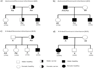
Genetic epilepsies could further divide into four subgroups according to the mechanism of inheritance: (1) genetic epilepsy with Mendelian inheritance, (2) epilepsies with complex inheritance, (3) genetic epilepsies associated with cytogenetic abnormalities and (4) Mendelian disorders in which epilepsy is one of the manifestations. The former class is thought to account for a small number of epilepsies, and the disease occurrence could be tracked through generation. A proper pedigree analysis will affirm whether the phenotype is dominant or recessive, autosomal or X-linked (Figure 1).11 Epilepsies with complex inheritance are
believed to be involved in 50% of epilepsies.11,19
Although familial aggregation is seen through generations, the mode of inheritance cannot easily be identified.
Detection of a specific chromosomal abnormality (either structural or numerical) would be categorized
under genetic epilepsies with cytogenetic
abnormalities.11 This subgroup is mostly associated with
other neurological disorders and facial anomalies. Mendelian disorders in which epilepsy is one of the manifestations indicate multisystem disorders with epilepsy as one of the characteristics. These
syndromes include neurocutaneous and
neurodegenerative disorders and a cluster of metabolic diseases.11 Thus, the genetic counselor
Figure 1. Mendelian modes of Inheritance (a) autosomal recessive inheritance. In this case “a” is the mutated allele of the gene and “A” is non-mutated. Individual who receives mutated allele from both parents (aa) would be affected with disease. Another persons “AA” and “Aa” do not show phenotypes of disease. (b) Autosomal dominant inheritance. In this model “A” (the dominant allele) is mutated allele and can cause disease, so any individual who receives just one mutated allele (AA, Aa) would be affected. (c) X-linked recessive inheritance. This mode has sex-based transmission because the gene is located on X chromosome, therefore females have two alleles of the gene and males have just one allele. If the mother is carrier, 50% of her boys will be affected and none of the girls in such pedigrees would show the phenotype of disease. (d) X-linked Dominant inheritance. In this example, the disease is caused by dominant mutated allele located in chromosome X. So if the father is affected, all the girls would be affected and no boys would show the disease phenotype. If the mother was affected too (Aa or AA) so the boys would have shown the phenotype of disease with different percentage
The following section is dedicated to reviewing the role of chromosomal abnormalities and gene mutations in etiology of epileptic syndromes with some examples. We are referring interested readers for a complete list of genes mutated in epilepsies to two reviews written by Garofalo et al.20 and
Kaneko et al.21
Chromosomal aberrations
Chromosomal aberration is characterized by atypical number or structural abnormality of at least one chromosome that usually leads to genetic disorders. In numerical group, aneuploidy is usually due to abnormal gametogenesis in parents.22 Considering
aneuploidy is accompanied with gaining or losing considerable amount of genetic materials, apart from sex chromosome disorders it is a fatal.23 However,
there are few cases of live birth. These patients usually suffer from facial dysmorphisms and mental
retardation as well as seizure.24,25 Conventional
karyotyping could easily identify numerical
chromosomal aberrations.26
There are several forms of structural chromosomal abnormalities including deletion, duplication and translocation of portion of a chromosome. These defects are not generally fatal, and newborns with structural abnormalities may have developmental delay and facial dysmorphism.27 Epilepsy is one of the
widespread features in this group of anomalies.
Ring chromosomes detection might help
discovering genetic epilepsy.28-30 A ring chromosome
is usually formed through breakage of both ends of the chromosome and fusion of arms. Back et al. have reported a phenotypically normal woman with ring chromosome 20 who had two children suffering from
mental retardation, behavioral disorder, and
Iran J Neurol 2015; 14(1) Tafakhori et al. ring chromosome 20 has a distinct feature of
prolonged high-voltage slow waves and seizures are resistant to medications.28 Ring chromosome 14 has
also been reported to be resistant to antiepileptic therapy.32 The onset of epilepsy in this chromosomal
disorder is often during the first year, mental retardation would be a constant character and the majority of cases have dysmorphic facial features. EEG frequently reveals focal abnormality.32
Chromosome 6q deletion (Long arm of
chromosome 6) and chromosome 22q duplication have been shown to be associated with dysmorphic
facial abnormalities, mental retardation and
epilepsy.33,34
As a result of high-resolution karyotyping, many epileptic seizures have been linked to chromosomal abnormalities.35-37 Aberrations such as microdeletions
and microduplications (microchromosomal defects) that could not be detected by conventional karyology might be identified by molecular cytogenetic
approaches including comparative genomic
hybridization (CGH) array and multiplex ligation-dependent probe amplification (MLPA).38-41 Exploring
the nature of the human genome with high repetitive DNA sequences lead to discovering recurrent rearrangements of regions in some chromosomes such as 15q and 16p that are involved in epilepsy could result in recurrent heritable microdeletions and microduplications.42
Gene mutations
In addition to chromosomal abnormalities, gene mutations also could be associated with epilepsy syndromes. A good example would be genes encoding ion channel subunits.43,44 Excitatory or
inhibitory neurotransmitters in central nervous system45 have also been recognized in Mendelian
forms of epilepsy11,46,47 and thus, following simple
Mendelian mode of inheritance.46,47 Genetic
counseling could help identifying these disorders through a prodigy and risk of disease could be estimated for the next generation.
CHRNA4 gene encodes neural acetylcholine receptor subunit α4.48 It was the first gene to be
associated with epilepsy syndromes. Mutation in this gene has been linked to Autosomal dominant nocturnal frontal lobe epilepsy.49,50 KCNQ2 and
KCNQ3 genes that encode voltage-gated potassium channels were identified in families affected with benign familial neonatal seizures.51,52
At least 37 genes for generalized myoclonic epilepsy and febrile seizures, 47 genes for symptomatic (structural/metabolic) epilepsy and 30 genes for epileptic encephalopathies have been recognized.20 In a recent study of pediatric patients
affected with infantile spasms and Lennox-Gastaut
syndrome, two forms of epileptic encephalopathies, and their parents, researchers found 329 de novo mutations.53 These mutations are significantly more
prominent in genes sets regulated by fragile X protein. Mutation of fragile X protein has been extensively discussed in autism spectrum disorders as it is the most widespread single-gene cause of autism 54. Further
genetic defects involved in epileptic encephalopathies
include MTOR, GABRA1 and FLNA.53
Mutation in SCN1A, a gene encoding voltage-gated sodium channel, has been demonstrated to be involved in Dravet syndrome. The affected patients suffer from severe myoclonic epilepsy during infancy with poor prognosis, as seizures are frequent, prolonged and resistant to treatment. Developmental delay will appear and some would have cognitive impairment. There are reports of mutation in PCDH19, a gene that encodes a calcium-dependent cell-adhesion protein and is located on chromosome X, in female patients with clinical symptoms related to Dravet syndrome.55,56 Interestingly, 11-12% of affected
patients, who did not show any mutation in the mentioned genes, had pathogenic copy number variations (CNVs) in SCN1A gene. These CNVs might be detected by array CGH and MLPA assay.42,57
Marini et al. showed that deletion of 9.3 Mb (49 genes) of chromosome 2q without harming SCN1A gene could also result in Dravert phenotype.57
The prevalence of Unvericht-lunderborg disease or Baltic myoclonic epilepsy, a rare inherited form of epilepsy with progressive myoclonus, is higher in some regions (e.g. Sweden). This disorder has been associated with mutation of CSTB, a gene encoding cystatin B protein responsible for reducing the activity of cathepsins enzymes (protease)58 and is inherited in
an autosomal recessive (AR) pattern.59 Furthermore,
different type of gene mutations including CHRNA4 gene (frontal lobe epilepsy) have been reported in different populations.49,50,60 It seems necessary to
identify specific mutations in distinct population to provide better genetic counseling for epilepsy.21
There are disorders that although epilepsy is one of the symptoms, it is not the core sign. Some examples are discussed below.
Lafora body disease, a neurodegenerative disorder, is a fatal glycogen metabolism disorder with AR inheritance61,62 and has been linked to EMP2A gene
mutation (Lafarin protein).63
Neuronal ceroid lipofuscinoses, a cluster of at least 8 neurodegenerative disorders, a result of lysosomal storage defects and excessive accumulation of lipopigments in brain and other tissues. It has an AR pattern of inheritance.21 CLN1 (PPT1) and CLN3 gene
Myoclonus epilepsy and ragged-red fibers are a rare mitochondrial disorder involving usually mutation of MT-TK gene located on mitochondrial DNA. It would lead to progressive neurological symptoms, including blindness and myoclonic epilepsy.65 Mitochondrial pattern of inheritance is
relatively complex (Figure 2) as maternal mutated mitochondria affects zygote formation.66
Figure 2. Transmission of mitochondrial DNA mutation in a hypothetical pedigree. This mode of inheritance is categorized as non-mendelian transmission because the mutated gene is not located in nuclear DNA. Mitochondria and its DNA (mtDNA) will transmit to next generation through oocyte cytoplasm so just mutated mtDNA from mother could cause the disease. Since, we have too many mitochondria and copies of mtDNA in a cell, the presence of disease and severity of its phenotype will be depended on the amount of mutated mtDNA inside individual’s cells. Heteroplasmy means both mutated and non-mutated mtDNA is present in a cell. Homoplasmy means the whole mtDNAs in a cell are mutated or non-mutated
Malformation of cortical development disorders represents a major spectrum of mental disabilities with severe epilepsies caused by defective neuronal
migration. Mutation of LIS1 gene encoding
microtubule-associated protein is one of the several
genetic defects linked to these disorders.
Lissencepahly with X-link gene mutation (xLIS)67 is
another defective neuronal migration disorder that result in the lack of cerebral folds. Both genetic and non-genetic factors (e.g. viral infections of the fetus) are involved in etiology of these disorders.68
The introduction of new techniques of DNA sequencing has helped identifying point mutations, small insertions, and deletions.38
Genetic counseling and genetic testing in epilepsy management
Epilepsy is a multifactorial disease, and both genetic and environmental components are involved in etiology (Table 1). Various investigations, particularly twin studies, have contributed to detection the role of genetic elements in epilepsy syndromes. These
findings will help predicting the clinical symptoms of the affected individual through genotype-phenotype correlation69 and conduct follow-up of high-risk
pregnancies or an infant born in a family with increased rate of epilepsy.70 It will also aid the
clinician in anticipating the clinical features in advance and manage in accordance.
Detection of specific genetic disorders will improve our understanding of the inheritance pattern. Thus, genetic counseling could better help families by estimating the risk of disease in next generation and family members of epileptic probands, who might be at greater risk for epilepsy syndromes.69 Interestingly,
the same phenotype of epilepsy in different members of a pedigree could be due to different genetic defects.67 Consanguineous marriage will increase the
risk of epilepsy syndrome, especially childhood onset of epilepsy71-73 and is a remarkable challenge for
clinicians and geneticists in societies where it is a common tradition.
Conclusion
We emphasize on cooperation of clinicians (particularly neurologist) and medical genetic experts in eastern societies like Iran, where consanguineous marriage is a common practice. This assistance is highlighted in high-risk families. It should be noted that prior to any genetic testing, patient and family members should be pre-tested in genetic counseling sessions.18
The genetic testing now commercially available for epilepsy includes analysis of 70 genes for detection of point mutations and deletion/duplications using DNA sequencing, CGH array, and MLPA techniques. The specimen used for genetic testing could be whole blood or any other body tissue appropriate for DNA extraction, for example, amniotic fluid, and chorionic villi samples are required for prenatal diagnosis.
Hence, collaboration of neurologist with geneticist in the case of genetic epilepsy will help the diagnosis and in some cases will improve management20.
Conflict of Interests
The authors declare no conflict of interest in this study.
Acknowledgments
We appreciate the help of our department’s staff and supports of the Tehran University of Medical Sciences, Iran.
How to cite this article: Tafakhori A, Aghamollaii V,
Iran J Neurol 2015; 14(1) Tafakhori et al.
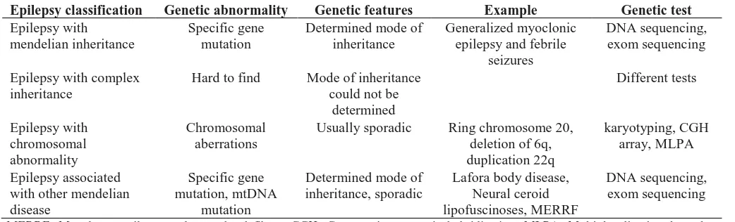
Table 1. Summary of genetic abnormalities in different forms of epilepsies
Epilepsy classification Genetic abnormality Genetic features Example Genetic test
Epilepsy with mendelian inheritance
Specific gene mutation
Determined mode of inheritance
Generalized myoclonic epilepsy and febrile
seizures
DNA sequencing, exom sequencing
Epilepsy with complex inheritance
Hard to find Mode of inheritance could not be
determined Different tests Epilepsy with chromosomal abnormality Chromosomal aberrations
Usually sporadic Ring chromosome 20, deletion of 6q, duplication 22q
karyotyping, CGH array, MLPA
Epilepsy associated with other mendelian disease
Specific gene mutation, mtDNA
mutation
Determined mode of inheritance, sporadic
Lafora body disease, Neural ceroid lipofuscinoses, MERRF
DNA sequencing, exom sequencing
MERRF: Myoclonus epilepsy and ragged-red fibers; CGH: Comparative genomic hybridization; MLPA: Multiplex ligation-dependent probe amplification
References
1. Dekker PA. Epilepsy: A Manual for Medical and Clinical Officers in Africa. Geneva, Switzerland: World Health Organization; 2002.
2. Berg AT, Millichap JJ. The 2010 revised classification of seizures and epilepsy.
Continuum (Minneap Minn) 2013;
19(3 Epilepsy): 571-97.
3. Hauser WA, Annegers JF, Kurland LT. Incidence of epilepsy and unprovoked seizures in Rochester, Minnesota: 1935-1984. Epilepsia 1993; 34(3): 453-68. 4. Beletsky V, Mirsattari SM. Epilepsy, mental
health disorder, or both? Epilepsy Res Treat 2012; 2012: 163731.
5. Engel J. The epilepsies. In: Wyngoorden J, Smith L, Bennet C, Editors. Cecil's
Textbook of Medicine. 19th ed.
Philadelphia, PA: WB Saunders; 1992. p. 2202-13.
6. Noachtar S, Remi J. The role of EEG in epilepsy: a critical review. Epilepsy Behav 2009; 15(1): 22-33.
7. Benbadis S. The differential diagnosis of epilepsy: a critical review. Epilepsy Behav 2009; 15(1): 15-21.
8. Panayiotopoulos CP. A Clinical Guide to Epileptic Syndromes And Their Treatment: Based on the New Ilae Diagnostic Scheme. Oxfordshire, UK: Bladon Medical Pub p. 278; 2002.
9. Berg AT, Berkovic SF, Brodie MJ, Buchhalter J, Cross JH, van Emde BW, et al. Revised terminology and concepts for organization of seizures and epilepsies: report of the ILAE Commission on Classification and Terminology, 2005-2009. Epilepsia 2010; 51(4): 676-85.
10. Tafakhori A, Aghamollaii V, Modabbernia
AH, Ghaffarpour M, Omrani HA,
Harirchian MH, et al. Evaluation of partial epilepsy in Iran: role of video-EEG, EEG, and MRI with epilepsy protocol. Iran J Neurol 2011; 10(1-2): 9-15.
11. Johnson MR, Sander JW. The clinical impact of epilepsy genetics. J Neurol Neurosurg Psychiatry 2001; 70(4): 428-30. 12. Lennox WG. The heredity of epilepsy as
told by relatives and twins. J Am Med
Assoc 1951; 146(6): 529-36.
13. Berkovic SF, Howell RA, Hay DA, Hopper JL. Epilepsies in twins: genetics of the major epilepsy syndromes. Ann Neurol 1998; 43(4): 435-45.
14. Harvald B, Hauge M. Hereditary factors elucidated by twin studies. In: Van Gundia Neel J, Editor. Genetics and the epidemiology of chronic diseases. Washington, DC: U.S. Dept. of Health, Education, and Welfare, Public Health Service, Division of Chronic Diseases; 1965. p. 61-76.
15. Sillanpaa M, Koskenvuo M, Romanov K, Kaprio J. Genetic factors in epileptic seizures: evidence from a large twin population. Acta Neurol Scand 1991; 84(6): 523-6.
16. Bouchard TJ, Propping P. Twins as a tool of behavioral genetics: report of the Dahlem Workshop on What Are the Mechanisms Mediating the Genetic and Environmental Determinants of Behavior? Twins as a Tool of Behavioral. New Jersey, NJ: J. Wiley; 1993. p. 326.
17. Baraitser M. The genetics of neurological disorders. 2nd ed. Oxford, UK: Oxford University Press; 1990. p. 113.
18. Ottman R, Hirose S, Jain S, Lerche H, Lopes-Cendes I, Noebels JL, et al. Genetic testing in the epilepsies--report of the ILAE Genetics Commission. Epilepsia 2010; 51(4): 655-70.
19. Kaneko S, Wada K. Molecular genetic studies of epilepsies. No To Shinkei 1998; 50(12): 1071-7.
20. Garofalo S, Cornacchione M, Di CA. From genetics to genomics of epilepsy. Neurol Res Int 2012; 2012: 876234.
21. Kaneko S, Okada M, Iwasa H, Yamakawa K, Hirose S. Genetics of epilepsy: current status and perspectives. Neurosci Res 2002; 44(1): 11-30.
22. Luthardt FW, Keitges E. Chromosomal Syndromes and Genetic Disease [Online]. [cited 2001]; Available from: URL: http://www.els.net/WileyCDA/ElsArticle/re fId-a0001446.html
23. Langer JC. Prenatal diagnosis of congenital
anomalies. What can and should be done? Can Fam Physician 1993; 39: 595-602. 24. Kumada T, Ito M, Miyajima T, Fujii T,
Okuno T, Go T, et al. Multi-institutional study on the correlation between chromosomal abnormalities and epilepsy. Brain Dev 2005; 27(2): 127-34.
25. Sorge G, Sorge A. Epilepsy and chromosomal abnormalities. Ital J Pediatr 2010; 36: 36.
26. O'Connor C. Karyotyping for chromosomal abnormalities. Nature Education 2008; 1(1): 27.
27. Marshall CR, Noor A, Vincent JB, Lionel AC, Feuk L, Skaug J, et al. Structural variation of chromosomes in autism spectrum disorder. Am J Hum Genet 2008; 82(2): 477-88.
28. Inoue Y, Fujiwara T, Matsuda K, Kubota H, Tanaka M, Yagi K, et al. Ring chromosome 20 and nonconvulsive status epilepticus. A new epileptic syndrome. Brain 1997; 120 (Pt 6): 939-53.
29. Atkins L, Miller WL, Salam M. A ring-20 chromosome. J Med Genet 1972; 9(3): 377-80. 30. Faed M, Morton HG, Robertson J. Ring F
chromosome mosaicism
(46,XY,20r-46,XY) in an epileptic child without apparent haematological disease. J Med Genet 1972; 9(4): 470-3.
31. Back E, Voiculescu I, Brunger M, Wolff G. Familial ring (20) chromosomal mosaicism. Hum Genet 1989; 83(2): 148-54.
32. Specchio N, Trivisano M, Serino D, Cappelletti S, Carotenuto A, Claps D, et al. Epilepsy in ring 14 chromosome syndrome. Epilepsy Behav 2012; 25(4): 585-92. 33. Vignoli A, Scornavacca GF, Peron A, La
BF, Canevini MP. Interstitial 6q microdeletion syndrome and epilepsy: a new patient and review of the literature. Am J Med Genet A 2013; 161A(8): 2009-15. 34. Han K, Holder JL, Schaaf CP, Lu H, Chen
H, Kang H, et al. SHANK3 overexpression causes manic-like behaviour with unique pharmacogenetic properties. Nature 2013; 503(7474): 72-7.
Disord 2005; 7(3): 181-92.
36. Singh R, Gardner RJ, Crossland KM, Scheffer IE, Berkovic SF. Chromosomal abnormalities and epilepsy: a review for clinicians and gene hunters. Epilepsia 2002; 43(2): 127-40.
37. Zuberi SM. Chromosome disorders associated with epilepsy. Handb Clin Neurol 2013; 111: 543-8.
38. Mulley JC, Mefford HC. Epilepsy and the new cytogenetics. Epilepsia 2011; 52(3): 423-32.
39. Mefford HC, Muhle H, Ostertag P, von SS, Buysse K, Baker C, et al. Genome-wide copy number variation in epilepsy: novel susceptibility loci in idiopathic generalized and focal epilepsies. PLoS Genet 2010; 6(5): e1000962.
40. Bedoyan JK, Kumar RA, Sudi J, Silverstein F, Ackley T, Iyer RK, et al. Duplication 16p11.2 in a child with infantile seizure disorder. Am J Med Genet A 2010; 152A(6): 1567-74.
41. Cardoso C, Boys A, Parrini E, Mignon-Ravix C, McMahon JM, Khantane S, et al. Periventricular heterotopia, mental retardation, and epilepsy associated with 5q14.3-q15 deletion. Neurology 2009; 72(9): 784-92.
42. Mulley JC, Nelson P, Guerrero S, Dibbens L, Iona X, McMahon JM, et al. A new molecular mechanism for severe myoclonic epilepsy of infancy: exonic deletions in SCN1A. Neurology 2006; 67(6): 1094-5. 43. Cossette P. Channelopathies and juvenile
myoclonic epilepsy. Epilepsia 2010; 51(Suppl 1): 30-2.
44. Jurkat-Rott K, Lerche H, Weber Y,
Lehmann-Horn F. Hereditary
channelopathies in neurology. Adv Exp Med Biol 2010; 686: 305-34.
45. Werner FM, Covenas R. Classical neurotransmitters and neuropeptides involved in generalized epilepsy: a focus on antiepileptic drugs. Curr Med Chem 2011; 18(32): 4933-48.
46. Jefferys JG. Advances in understanding basic mechanisms of epilepsy and seizures. Seizure 2010; 19(10): 638-46.
47. Scharfman HE. The neurobiology of epilepsy. Curr Neurol Neurosci Rep 2007; 7(4): 348-54.
48. Anand R, Lindstrom J. Chromosomal localization of seven neuronal nicotinic acetylcholine receptor subunit genes in humans. Genomics 1992; 13(4): 962-7. 49. Steinlein OK, Mulley JC, Propping P,
Wallace RH, Phillips HA, Sutherland GR, et al. A missense mutation in the neuronal nicotinic acetylcholine receptor alpha 4 subunit is associated with autosomal dominant nocturnal frontal lobe epilepsy. Nat Genet 1995; 11(2): 201-3.
50. Hirose S, Iwata H, Akiyoshi H, Kobayashi K, Ito M, Wada K, et al. A novel mutation of CHRNA4 responsible for autosomal dominant nocturnal frontal lobe epilepsy. Neurology 1999; 53(8): 1749-53.
51. Biervert C, Schroeder BC, Kubisch C, Berkovic SF, Propping P, Jentsch TJ, et al. A potassium channel mutation in neonatal human epilepsy. Science 1998; 279(5349): 403-6. 52. Singh NA, Charlier C, Stauffer D, DuPont
BR, Leach RJ, Melis R, et al. A novel potassium channel gene, KCNQ2, is mutated in an inherited epilepsy of newborns. Nat Genet 1998; 18(1): 25-9. 53. Allen AS, Berkovic SF, Cossette P, Delanty
N, Dlugos D, Eichler EE, et al. De novo mutations in epileptic encephalopathies. Nature 2013; 501(7466): 217-21.
54. Goodlin-Jones BL, Tassone F, Gane LW, Hagerman RJ. Autistic spectrum disorder and the fragile X premutation. J Dev Behav Pediatr 2004; 25(6): 392-8.
55. Akiyama M, Kobayashi K, Ohtsuka Y. Dravet syndrome: a genetic epileptic disorder. Acta Med Okayama 2012; 66(5): 369-76.
56. Dibbens LM, Tarpey PS, Hynes K, Bayly MA, Scheffer IE, Smith R, et al. X-linked protocadherin 19 mutations cause female-limited epilepsy and cognitive impairment. Nat Genet 2008; 40(6): 776-81.
57. Marini C, Mei D, Temudo T, Ferrari AR, Buti D, Dravet C, et al. Idiopathic epilepsies with seizures precipitated by fever and SCN1A abnormalities. Epilepsia 2007; 48(9): 1678-85.
58. Pennacchio LA, Lehesjoki AE, Stone NE, Willour VL, Virtaneva K, Miao J, et al. Mutations in the gene encoding cystatin B in progressive myoclonus epilepsy (EPM1). Science 1996; 271(5256): 1731-4.
59. Norio R, Koskiniemi M. Progressive
myoclonus epilepsy: genetic and
nosological aspects with special reference to 107 Finnish patients. Clin Genet 1979; 15(5): 382-98.
60. Phillips HA, Marini C, Scheffer IE, Sutherland GR, Mulley JC, Berkovic SF. A de novo mutation in sporadic nocturnal frontal lobe epilepsy. Ann Neurol 2000; 48(2): 264-7.
61. Lehesjoki AE, Koskiniemi M, Pandolfo M,
Antonelli A, Kyllerman M, Wahlstrom J, et al. Linkage studies in progressive myoclonus epilepsy: Unverricht-Lundborg and Lafora's diseases. Neurology 1992; 42(8): 1545-50.
62. Serratosa JM, Delgado-Escueta AV, Posada I, Shih S, Drury I, Berciano J, et al. The gene for progressive myoclonus epilepsy of the Lafora type maps to chromosome 6q. Hum Mol Genet 1995; 4(9): 1657-63. 63. Minassian BA, Lee JR, Herbrick JA,
Huizenga J, Soder S, Mungall AJ, et al. Mutations in a gene encoding a novel protein tyrosine phosphatase cause progressive myoclonus epilepsy. Nat Genet 1998; 20(2): 171-4.
64. Mole SE, Williams RE, Goebel HH. Correlations between genotype, ultrastructural morphology and clinical phenotype in the neuronal ceroid lipofuscinoses. Neurogenetics 2005; 6(3): 107-26.
65. Shoffner JM, Lott MT, Lezza AM, Seibel P, Ballinger SW, Wallace DC. Myoclonic epilepsy and ragged-red fiber disease
(MERRF) is associated with a
mitochondrial DNA tRNA(Lys) mutation. Cell 1990; 61(6): 931-7.
66. Taylor RW, Turnbull DM. Mitochondrial DNA mutations in human disease. Nat Rev Genet 2005; 6(5): 389-402.
67. Kullmann DM. Genetics of epilepsy. J Neurol Neurosurg Psychiatry 2002; 73(Suppl 2): II32-II35.
68. Guerrini R, Carrozzo R. Epileptogenic brain malformations: clinical presentation, malformative patterns and indications for genetic testing. Seizure 2001; 10(7): 532-43. 69. Corey LA, Pellock JM, Boggs JG, Miller LL, DeLorenzo RJ. Evidence for a genetic predisposition for status epilepticus. Neurology 1998; 50(2): 558-60.
70. Pal DK, Pong AW, Chung WK. Genetic evaluation and counseling for epilepsy. Nat Rev Neurol 2010; 6(8): 445-53.
71. Asadi-Pooya AA. Epilepsy and
consanguinity in Shiraz, Iran. Eur J Paediatr Neurol 2005; 9(6): 383-6.
72. Al-Rajeh S, Abomelha A, Awada A, Bademosi O, Ismail H. Epilepsy and other convulsive disorders in Saudi Arabia: a prospective study of 1,000 consecutive cases. Acta Neurol Scand 1990; 82(5): 341-5.
73. Mehndiratta MM, Paul B, Mehndiratta P. Arranged marriage, consanguinity and
epilepsy. Neurology Asia 2007;