R E V I E W
Open Access
Neurovascular unit dysfunction with blood-brain
barrier hyperpermeability contributes to major
depressive disorder: a review of clinical and
experimental evidence
Souhel Najjar
1,2*, Daniel M Pearlman
1,3, Orrin Devinsky
2, Amanda Najjar
4and David Zagzag
4,5Abstract
About one-third of people with major depressive disorder (MDD) fail at least two antidepressant drug trials at 1 year. Together with clinical and experimental evidence indicating that the pathophysiology of MDD is multifactorial, this observation underscores the importance of elucidating mechanisms beyond monoaminergic dysregulation that can contribute to the genesis and persistence of MDD. Oxidative stress and neuroinflammation are mechanistically linked to the presence of neurovascular dysfunction with blood-brain barrier (BBB) hyperpermeability in selected neurological disorders, such as stroke, epilepsy, multiple sclerosis, traumatic brain injury, and Alzheimer’s disease. In contrast to other major psychiatric disorders, MDD is frequently comorbid with such neurological disorders and constitutes an independent risk factor for morbidity and mortality in disorders characterized by vascular endothelial dysfunction (cardiovascular disease and diabetes mellitus). Oxidative stress and neuroinflammation are implicated in the neurobiology of MDD. More recent evidence links neurovascular dysfunction with BBB hyperpermeability to MDD without neurological comorbidity. We review this emerging literature and present a theoretical integration between these abnormalities to those involving oxidative stress and neuroinflammation in MDD. We discuss our hypothesis that alterations in endothelial nitric oxide levels and endothelial nitric oxide synthase uncoupling are central mechanistic links in this regard. Understanding the contribution of neurovascular dysfunction with BBB hyperpermeability to the pathophysiology of MDD may help to identify novel therapeutic and preventative approaches.
Keywords:Major depressive disorder, Blood-brain barrier, Neurovascular unit, Neuroinflammation, Oxidative stress, Nitric oxide synthase, eNOS uncoupling, Peroxynitrite
Background
Major depressive disorder (MDD) is the second leading global cause of years lived with disability [1], with about one-third of patients with MDD failing two or more conventional antidepressant drug trials within the first year of treatment [2,3]. Current evidence suggests that the pathophysiology of MDD is multifactorial, involving heterogeneous and inter-related mechanisms that affect
genetic, neurotransmitter, immune, oxidative, and in-flammatory systems [4]. Supporting this interpretation, whereas biomarkers for individual abnormalities possess limited predictive validity for MDD, the predictive valid-ity of several composite biomarker assays is particularly high [5]. For example, one study of 36 patients with MDD showed that a compositive biomarker test— com-prising nine individual biomarker assays (α1 antitrypsin, apolipoprotein CIII, myeloperoxidase, soluble tumor ne-crosis factor α (TNFα) receptor type II, epidermal growth factor, cortisol, brain-derived neurotropic factor, prolactin, and resistin)—had 91.7% sensitivity and 81.3% specificity for MDD [6]. A follow-up study involving a distinct sample of 34 MDD patients and using the same * Correspondence:mna1024231@aol.com
1
Department of Neurology, Neuroinflammation Research Group, Epilepsy Center Division, NYU School of Medicine, New York, NY 10016, USA 2
Department of Neurology, NYU Comprehensive Epilepsy Center, NYU School of Medicine, New York, NY 10016, USA
Full list of author information is available at the end of the article
composite assay, replicated these results with a high de-gree of precision: 91.1% sensitivity, 81.0% specificity [6].
Oxidative stress and neuroinflammation are implicated in the neurobiology of MDD [7-14] (recently reviewed by our group [4,15-19]). Neuropathological studies com-paring brain tissue from individuals with MDD to that from non-depressed controls have documented associa-tions between MDD and (a) decreased levels of antioxi-dants, such as glutathione [11,15,16] and (b) increased levels of lipid peroxidation end products, such as 4-hydroxy-2-nonenal [8]. Studies assessing peripheral markers of oxidative stress have reported similar findings, including: (a) altered activity of antioxidant enzymes, such as glutathione peroxidase, catalase, superoxide dismutase 1, (b) increased activity of pro-oxidant enzymes such as, xanthine oxidase, (c) increased activity of indu-cible nitric oxide synthase (iNOS) in leukocytes, (d) in-creased levels of superoxide (O2-), and (e) increased levels
of 8-hydroxy-2-deoxyguanosine (a marker for oxidative
damage to DNA) [11,12]. Evidence deriving from genetic, neuropathological, cerebrospinal fluid, and serum studies in humans with MDD and from animal models of depressive-like behavior and chronic stress reveal numer-ous neuroinflammatory abnormalities in MDD, including [4]: (a) microglial activation [17-19], (b) astroglial loss and activation [20,21], (c) upregulated ratios of T helper 1 (Th1) cells and proinflammatory cytokines [22-24], and (d) decreased CD4+CD25+FOXP3+ regulatory T (TReg) cell counts [25]. Both oxidative stress and
neuroin-flammation may contribute to decreased serotonergic and increased glutamatergic tone, and increased glutamatergic tone may in turn contribute to oxidative stress and neuroinflammation in a positive feedback loop [4]. In addition, experimental evidence suggests that increased re-active oxygen species (ROS) synthesis (oxidative stress) and neuroinflammation themselves exhibit a bidirectional rela-tionship (Figure 1). Indeed, ROS can activate microglia and increase proinflammatory cytokine synthesis—for example,
Figure 1Putative mechanisms involving the synthesis of reactive oxygen species (ROS) and their bidirectional interaction with neuroinflammation in major depressive disorder.This figure shows potential mechanistic links among ROS, inflammation, and
by stimulating transcription factor nuclear factor κB (NFκB)—whereas activated microglia and proinflam-matory cytokines can in turn perpetuate oxidative stress [8,11,26-28].
Collectively, data from postmortem neuropathological human studies and in vivo neuroimaging human and animal studies provide strong evidence of neurovascular unit dysfunction with blood-brain barrier (BBB) hyper-permeability in association with oxidative stress and neuroinflammation in selected neurological disorders, such as stroke, epilepsy, Alzheimer’s disease, traumatic brain injury, and multiple sclerosis [29-43] (Table 1). In these disorders, BBB breakdown, oxidative stress, and inflammation are thought to impair neuronal function [44]. MDD, in contrast to other major psychiatric dis-orders, is frequently comorbid with such neurological disorders as well as disorders characterized by vascular
endothelial dysfunction, such as cardiovascular disease and diabetes mellitus [45-52]. Whether neurovascular dys-function with BBB hyperpermeability occurs in primary MDD (without neurological comorbidity), however, re-mains less clear.
Shalev and colleagues have previously reviewed evi-dence through 2009 linking BBB hyperpermeability to psychiatric disorders generally [168]. We review emer-ging clinical and experimental evidence implicating oxi-dative stress, eNOS uncoupling, and reduced endothelial NO levels in the pathophysiology of peripheral vascular endothelial dysfunction associated with MDD. We present a theoretical integration of human and animal data linking these mechanisms and those involving neuroinflammation to findings suggesting that neuro-vascular dysfunction can occur in primary MDD. We also discuss putative links between neurovascular
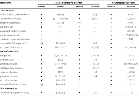
Table 1 Putative mechanisms of neurovascular dysfunction and blood–brain barrier hyperpermeability in major depressive disorder in the context of established mechanisms in various neurological disorders
Mechanisms Major depressive disorder Neurological disorders
Human Sources Animal Sources Human Sources
Oxidative stress
eNOS uncoupling, decreased NO ■ [53-59] ■ [60] ● [61-63]
Increased ROS synthesis ● [10,14-16,64-84] ● [64,85] ● [42,63,86]
Cerebral hypoperfusion ● [87-91] N/A … ● [92-96]
MMP activation ■ [97] ? … ● [39,42,98-101]
Decreased E-cadherin activity ? … ? … ?a [38,102]
Tight junction alteration ? … ? … ● [31,38,41,103-106]
Endothelial cytoskeletal alteration ? … ? … ● [31]
Increased NMDAR expressionb [107-111] ● [40] ● [112]
Mitochondrial alterations ● [65,113-121] ● [65,122] ● [11,123-125]
Neuroinflammation
Astroglial loss ● [20,21,126-133] ● [134-138] ● [139-141]
Decreased AQP4 ● [142] ● [143] ● [144-146]
Microglial activation ● [18,147,148] ● [149-152] ● [42,98,153,154]
Proinflammatory cytokines ● [23,155] ● [156,157] ● [42,98]
Bradykinin alteration ●c [158] ● [159] ● [159,160]
Hyperglutamatergia ● [4,161-163] ● [164] ● [165-167]
Mast cell activation ●c [168,169] ? … ● [170,171]
Increased ICAM-1 and VCAM-1 [172-174] ? … ● [175-178]
Other mechanisms
Increased P-glycoprotein activity ● [179,180] ● [181] ● [179]
Symbol key:●, documented in the central nervous system in major depressive disorder;■, not documented in the central nervous system, but associated with major depressive disorder; ?, insufficient data; , mixed evidence.
Abbreviations:AQP4,aquaporin 4;eNOS, endothelial nitric oxide synthase;ICAM-1, intercellular adhesion molecule 1;NMDAR, N-methyl-D-aspartate receptor;
MMP,matrix metalloproteinases;ROS, reactive oxygen species;VCAM-1, vascular cell adhesion molecule 1. a. Refers to data that has only been shown in animal models.
b. Refers to human data in major depressive disorder refers to increased NMDAR expression that was not specific to the endothelium. Human data of NMDAR subunit composition alteration in neurological disorders was shown in cultured human blood–brain barrier endothelial cells. Animal data refer to increased cerebrovascular endothelial NMDAR subunit 1 (NR1) expression upon exposure to oxidative stress (this was not a depressive-like behavior or chronic stress animal model, though this evidence may be relevant to MDD where oxidative stress is documented).
dysfunction with BBB hyperpermeability and neuronal signaling abnormalities in MDD.
Neurovascular unit dysfunction
The neurovascular unit consists of cerebral microvessels, glial cells (astroglia, microglia, oligodendroglia), and neurons. It is the epicenter of several tightly controlled, dynamic, and complex cellular interactions between glia and neurons, and the coupling of neuronal activity with endothelium-dependent cerebral blood flow [33]. Evi-dence of an association between MDD and neurovascu-lar dysfunction is indirect, deriving primarily from studies assessing peripheral vascular endothelial dysfunc-tion in MDD and from epidemiological data associating MDD with vascular disorders.
One method for evaluating endothelial dysfunction in-volves measuring the relative uptake ratio (RUR) of blood flow in the brachial artery after hyperemic chal-lenge via dynamic nuclear imaging. RUR is a measure of the vascular dilatory response whereby a lower RUR im-plies poorer vascular endothelial function. In a prospect-ive cohort involving 23 patients with MDD, 23 with minor depressive disorder, and 277 non-depressed con-trols, the mean RUR was significantly lower in partici-pants with MDD (unadjusted mean = 3.13, SD = 1.51) or minor depressive disorder (unadjusted mean = 3.38, SD = 1.00) compared with non-depressed controls (un-adjusted mean = 4.22, SD = 1.74) (F = 6.68,P = 0.001) [182]. This effect remained statistically significant after adjusting for age, sex, socioeconomic factors, medical comorbidity, and medications (F = 5.19, P = 0.006) [182]. One study evaluating endothelial proapoptotic ac-tivity, defined as the percentage of apoptotic nuclei in human umbilical vein endothelial cells, found a signifi-cantly increased percentage of proapoptotic nuclei in participants with MDD compared with non-depressed controls (4.4% vs 2.3%, P ≤ 0.001) [183]. This finding remained statistically significant after adjusting for age and cardiovascular comorbidity.
Linking vascular endothelial dysfunction to MDD, epi-demiological studies reveal a strong and bidirectional association between MDD and medical conditions char-acterized by vascular endothelial pathology [184]. A re-cent meta-analysis involving 16,221 study participants found a significantly increased risk of MDD among individuals with major vascular diseases compared with those without vascular disease: diabetes (odds ratio (OR) 1.51, 95% confidence interval (CI) 1.30 to 1.76, P < 0.0005, 15 studies), cardiovascular disease (OR 1.76, 95% CI 1.08 to 1.80, P < 0.0005, 10 studies), and stroke (OR 2.11, 95% CI 1.61 to 2.77, P < 0.0005, 10 studies) [45]. The same meta-analysis also found that MDD was more common among individuals with two or more classic risk factors for vascular disease compared with those
with one or no risk factors (OR 1.49, 95% CI 1.27 to 1.7, P< 0.0005, 18 studies) [45]. These findings remained ro-bust after statistical adjustments for chronic illness and disability. Results from meta-analyses having assessed the association from the reverse direction, indicate that MDD is not only an independent risk factor for cardio-vascular disease (relative risk (RR) 2.69, 95% CI 1.63 to 4.43, P < 0.001, 11 studies) [49], but is also associated with a 3-fold increased cardiovascular disease mortality rate (OR 2.61, 95% CI 1.53 to 4.47, P = 0.0004) [48]. Related studies report similar findings [50-52].
Blood–brain barrier unit hyperpermeability
The BBB consists of the neurovascular endothelium, extra-cellular matrix basal lamina, and astrocytic end-feet pro-cesses. The BBB secures the brain’s immune-privileged status by restricting the entry of peripheral inflammatory mediators (for example, cytokines, antibodies), which can impair neurotransmission [37,168,185,186]. Neurovascular endothelial cells regulate influx of essential nutrients, efflux of toxic substances, ionic homeostasis of brain interstitial fluid, and prevent brain influx of peripheral neuroactive substances, neurotransmitters, and water-soluble molecules [185]. Evidence of an association between BBB hyperper-meability and MDD derives mainly from studies having assessed cerebrospinal fluid (CSF)-to-serum ratios of various molecules, as well as evaluations concerning P-glycoprotein.
Alteration of BBB endothelial expression of P-glycoprotein (a multidrug efflux transporter) is docu-mented in some persons with MDD [192]. Reduced expression or function of P-glycoprotein may facilitate BBB permeability to neurotoxic substances [192]. Posi-tron emission tomography (PET) utilizing the [(11)C]-verapamil radioligand for P-glycoprotein in humans with MDD and in Wistar rats exhibiting depressive-like behavior showed that chronic stress exposure and administration of antidepressants inhibited and en-hanced P-glycoprotein function, respectively [179,181]. A human genetics study (631 MDD, 110 non-depressed con-trols) revealed a significant association between alteration of the P-glycoprotein encoding gene ATP-binding cassette, subfamily B member 1 (ABCB1) and MDD (P= 0.034) [180].
Theoretical integration with oxidative and neuroinflammatory mechanisms
Oxidative stress
Common ROS include superoxide (O2-), hydroxyl radical
(HO-), hydrogen peroxide (H2O2-), and peroxynitrite
(ONOO-). ONOO-is a highly reactive oxidant generated by the reaction of nitric oxide (NO) with O2- [8,15,123].
The brain is particularly susceptible to oxidative stress due to high levels of peroxidizable polyunsaturated fatty acids and transition minerals (reduced form) that induce lipid peroxidation and convert H2O2- to HO-;
addition-ally, the brain’s oxygen demand is particularly high and the presence of antioxidant defense mechanisms is rela-tively limited [8,11,12].
Although ROS can limit injury and promote recovery at low levels, ROS facilitate oxidative injury at high levels by damaging biological macromolecules, such as lipids, proteins, and DNA [8,11,12]. We hypothesize that oxidative stress associated with MDD may impair neuro-vascular function through several mechanisms, with an emphasis on mechanisms that can shift the functional balance between beneficial endothelial nitric oxide synthase (eNOS)-generated NO versus harmful eNOS-generated O2
-(Figure 2 and Table 1).
NO has been termed ‘Janus faced’ owing to its ability to either protect vascular endothelial cell function in some instances, while impairing it in others [193]. These differential effects of NO are primarily determined by its cellular source (non-endothelial vs endothelial) and con-centration (high vs low). NOS isoforms regulate NO syn-thesis in the brain. Of these, one is constitutively expressed in endothelial cells and astrocytes (eNOS) [194,195] (that is, eNOS), and another is expressed in neurons (neuronal NOS (nNOS)).
eNOS regulates vascular smooth muscle tone and nNOS modulates neurotransmission. The expression of
a third NOS isoform, iNOS, occurs in glial and inflamma-tory cells and is induced by pathological inflammainflamma-tory states, such as following trauma [38]. More recently, a fourth NOS isoform was described, mitochondrial (mtNOS), which is an eNOS-like isoform that is constitu-tively expressed in the inner mitochondrial membrane [196,197]. When combined with O2-, NO produced by
non-endothelial cellular sources (as regulated by nNOS, iNOS) can impair the vascular endothelium and disrupt BBB integrity [38,53]. nNOS activity itself is positively reg-ulated by Ca2+influx [198], whereas iNOS activity is posi-tively regulated by proinflammatory cytokine [199] and NFκB signaling [200].
NO produced by endothelial cells (as regulated by eNOS) increases cellular levels of cyclic guanosine monophosphate, which can increase cerebral blood flow via mechanisms involving endothelium-dependent vaso-dilation and platelet aggregation inhibition [38,53,201]. In vitro studies showed that endothelial-derived NO may dilate cerebral vessels by inhibiting the synthesis of 20-hydroxyeicostetranoic acid—an arachidonic acid metabolite that promotes vasoconstriction [202,203]. Endothelial-derived NO can also limit endothelial vascu-lar oxidative stress injury by scavenging free radicals [38,53]. Endothelial eNOS mediates NO synthesis via oxidative conversion of L-arginine to L-citrulline. Activ-ity of eNOS is modulated by several factors, including endothelial levels of Ca2+, arginine (eNOS substrate) [204], as well as tetrahydrobiopterin (BH4) (eNOS
cofac-tor) [53-55,201,205,206] (Figure 2). Downregulation of eNOS activity can decrease endothelial NO levels, po-tentially resulting in (a) reduced cerebral blood flow, (b) increased platelet aggregation, which may contribute to the increased risk of cardiovascular disease in MDD, (c) increased oxidative stress, and (d) decreased vascular re-activity [38,53,201].
Under oxidative conditions, such as those associated with MDD [4,8,11,12,15] (Figure 1), endothelial levels of BH4are decreased due to increased oxidative conversion
of BH4to dihydrobiopterin (BH2). Decreased endothelial
levels of BH4 and increased endothelial levels of BH2
(which can also reduce BH4binding to eNOS) uncouple
L-arginine oxidation from the electron transfer process and shift the eNOS substrate from L-arginine to molecu-lar oxygen (that is, eNOS uncoupling), thereby promot-ing the synthesis of harmful O2- instead of beneficial NO
[53-55,205,207,208]. Once formed, O2- reacts with
re-sidual NO (still being produced at a lower rate) to form ONOO- [205]. ONOO- in turn oxidizes BH4, thereby
further decreasing its levels in a positive feedback loop [54,205] (Figure 2).
worsen neuronal injury [209-213]. In murine models of ischemic stroke, knocking out iNOS and nNOS de-creased the size of infarct while knocking out eNOS ex-panded infracted zone, compared to wild-type mice [214,215]. In animal models of traumatic brain injury, increased levels of endothelial ONOO- are associated with BBB breakdown and neurobehavioral deficits [209]; additionally, treatment with the antioxidant S-nitroso-glutathione enhances neural reparative mechanisms and improves neurovascular unit function by decreasing endothelial ONOO-synthesis [209].
Clinical and experimental studies suggest that eNOS uncoupling can contribute to vascular endothelial
dysfunction in both cardiovascular diseases and MDD [4,53-55,182,205,206,216]. In cardiovascular diseases, eNOS uncoupling-mediated endothelial dysfunction is thought to result from (a) increased O2- synthesis
(through an NAD(P)H oxidase-dependent mechan-ism), (b) increased ONOO- formation, and (c) de-creased BH4levels [54,55,182,206]. In MDD, however,
the potential contribution of eNOS uncoupling to vascular endothelial dysfunction is inferred from less direct evidence. For example, several clinical studies of persons with MDD have shown significant reductions in eNOS activity and NO levels in platelets and sera, respectively [53-57]. In a study of 57 MDD patients
Figure 2Theoretical integration of the human and animal data linking oxidative stress, eNOS uncoupling, low endothelial NO levels, and neuroinflammation to indirect evidence of functional and structural abnormalities of neurovascular unit in major depressive disorder.Adapted with permission from Abbottet al., [185]. This figure describes several putative mechanisms involving neuroinflammation, oxidative stress, endothelial nitric oxide synthase uncoupling, and hyperglutamatergia, as well as their relationships to indirect evidence of neurovascular dysfunction in MDD. Neurovascular endothelial lipofuscin granule accumulation is a marker of endothelial oxidative stress, which we recently documented by ultrastructural analysis of cerebral microvasculature in brain biopsy from a patient with chronic refractory MDD [90]. Abbreviations: AQP4, aquaporin 4; BH2: dihydrobiopterin; BH4, tetrahydrobiopterin; CRH, corticotropin-releasing hormone; eNOS, endothelial nitric
oxide synthase; mGluR, metabotropic glutamate receptor; MDD, major depressive disorder; MMP, matrix metalloproteinase; NAD(P)H, nicotinamide adenosine dinucleotide phosphate; Na+/K+ATPase, sodium-potassium adenosine triphosphatase; NFκB, nuclear factorκB; NMDAR,N-methyl-D-aspartate
receptor; NO, nitric oxide, ONOO-, peroxynitrite; O 2
randomized to either citalopram (n = 36) or placebo (n = 21), a 3-month trial of citalopram was associated with a statistically significant increase in serum NO levels compared to placebo (P = 0.005) [58]. Another study involving a 2-month trial of paroxetine repro-duced similar results [59]. Fluoxetine treatment in a chronic stress mouse model restored previously defi-cient aortic endothelial NO levels [60], suggesting that eNOS uncoupling may not only occur in MDD, but also that eNOS recoupling may be one of the mecha-nisms by which antidepressants exert their therapeutic effects [8,11,14].
The antidepressant effect of L-methylfolate, which can reverse eNOS uncoupling in vitro via upregulating BH4
synthesis [206], suggests that eNOS uncoupling contrib-utes to the neurobiology of MDD. A randomized controlled trial showed that adding L-methylfolate at 15 mg/day, but not at 7.5 mg/day, to a stable regimen of selective serotonin reuptake inhibitors (SSRIs) had su-perior efficacy to SSRIs plus placebo [217]. Although the authors attributed BH4 augmenting the antidepressant
effects of SSRIs to direct activation of the rate-limiting enzymes of monoamine synthesis (serotonin, norepin-ephrine, dopamine), we suggest that these effects may also be related to the ability of BH4 to reverse eNOS
uncoupling.
Although regionally selective (thalamic nuclei, pre-frontal, anterior cingulate, temporal, and occipital corti-ces) cerebral hypoperfusion abnormalities in MDD have traditionally been attributed to depressed mood states and reduced neuronal activity [87-91] [208], these findings may also be related to eNOS uncoupling [65,113,114,218] (Figure 2). Sustained cerebral hypoper-fusion can impair endothelial mitochondrial oxidative function, resulting in increased synthesis of endothelial ROS [219-222]. ROS can in turn promote eNOS un-coupling, leading to reduced vasodilatory endothelial NO levels and cerebral hypoperfusion in a positive feed-back loop [54,55,182,206]. In addition, SSRIs have been shown to induce vasodilation through eNOS-mediated downregulation of NO [223]. We recently reported a case of chronic and refractory MDD with moderately se-vere bifrontal cerebral hypoperfusion (seen via single photon emission tomography (SPECT)) associated with lipofuscin granule accumulation (a marker of oxidative stress [224-228]) (Figure 2) identified exclusively within the neurovascular unit (predominately within the endo-thelium) [90]; restoration of cerebral hypoperfusion in temporal association with intravenous immunoglobulin and minocycline therapy was accompanied with signifi-cant improvement of depressive symptoms, after more than 20 years of refractoriness to conventional psychi-atric treatments [90]. We suggest that eNOS uncoupling may occur in MDD primarily as the result of
non-heritable factors such as oxidative mechanisms. Indeed, sev-eral genetic studies show a non-significant association between eNOS gene polymorphisms and MDD [229,230].
Under oxidative conditions, BBB endothelial cells are not only the source of harmful eNOS uncoupling, but also can be the target of oxidative damage [39]. In neurological disorders associated with neurovascular dysfunction, oxidative stress can also increase BBB per-meability through several mechanisms (Table 1), which include: (a) activation of metalloproteinase (MMP)-2/9 directly or indirectly through proinflammatory cytokines [39]; (b) downregulation of endothelial expression of E-cadherin [38]; (c) alteration of the expression, distribu-tion, and phosphorylation of BBB tight junction proteins (for example, claudin, occluding, ZO proteins) by molecules such as phosphatidylinositol-3-kinaseγ[38,41,103,104]; (d) alteration of endothelial cytoskeletal structure; (e) induction of endothelial NMDAR subunit expression such as NMDA receptor subunit 1 (NR1) subunit, leading endothelial exci-totoxicity [40]; and (f) impairment of vascular endothelial mitochondrial oxidative metabolism [11,123]. The rele-vance of these mechanisms to the neurobiology of MDD, however, remains unclear (Table 1 and Figure 2).
Neuroinflammation
Neuroinflammation may impair neurovascular function and increase BBB permeability in MDD [4,168] (Figure 2 and Table 1). Astroglial cells are an integral part of the neurovascular unit. They are involved in regulating blood flow, BBB permeability, energy metabolism, and neuronal signaling [4,184]. Astroglial loss has been con-sistently documented in functionally relevant areas (pre-frontal and cingulate cortices, amygdala, hippocampus) among persons with MDD [4,142,168,231-236]. Other studies have documented decreased expression of the astroglial end-feet process water channel, aquaporin 4 (AQP4) in the orbitofrontal cortical gray matter (but not white matter) of individuals with MDD relative to non-depressed controls [142]. Animal models of depressive-like behavior also found decreased AQP4 density in as-sociation with oxidative stress [143]. Decreased AQP4 density may impair critical glial-vascular homeostatic pathways within the neurovascular unit and increase BBB permeability (Figure 2). Reduced AQP4 density may also contribute to cerebral perfusion and metabolic abnormalities detected by SPECT and PET imaging in human MDD [184].
MDD [4,17,19], though neuropathological evidence of MAP in the brains of subjects with MDD is inconsistent [4,18,148,239]. One neuropathological study found a positive association between suicidality and both MAP density and microglial quinolinic acid expression [17]. In rats, chronic psychological stress promotes MAP in the prefrontal cortex, amygdala, and hippocampus [19]. Re-cent meta-analysis in MDD patients confirmed elevation of serum levels of proinflammatory cytokines, such as interleukin 6 (IL-6) and TNFα[23,240]. Multiplein vitro studies of various neurological conditions showed that MAP and proinflammatory cytokines could increase BBB permeability [4,38-40,168,184,241] (Figures 1 and 2) (Table 1). BBB hyperpermeability may in turn increase crosstalk between innate and adaptive immunity, thereby resulting in further upregulation of MAP and brain cyto-kine production in a positive feedback loop [242]. MAP can activate iNOS [8,11,26,27], increase ROS synthesis [28], and promote COX2 expression within the neuro-vascular unit [4]; these factors may increase BBB perme-ability in vitro [38,53]. MAP and proinflammatory cytokines can release and activate matrix metalloprotein-ases (MMPs) [38,39,168], which have been shown in vitro to disrupt BBB endothelial tight junction pro-teins and increase BBB opening [38,39,168,184]. Serum MMP-9 levels have been shown to correlate with depres-sive symptom severity in humans (as assessed by the Hamilton Depression Scale) [97]. Highly reproducible in vitro data showed that proinflammatory cytokines (TNFα, IL-1β, interferon γ (IFNγ)) can cause a dose-dependent increase in BBB permeability by inducing ex-pression of intercellular adhesion molecule 1 (ICAM-1) on the luminal surface of BBB endothelial cells in ani-mals [243-249] and humans [250,251]. One neuropatho-logical study found a significant increase in the ICAM-1 expression in the deep white matter of the dorsolateral prefrontal cortex in MDD relative to controls [172]. An-other study showed SSRIs can reduce vascular endothe-lial expression and serum levels of both ICAM-1 and vascular cell adhesion molecule 1 (VCAM-1) [173]. Thus, increased BBB endothelial cell expression of
adhe-sion molecules may be one mechanism by
which BBB hyperpermeability occurs in MDD [174,252] (Figure 2). However, contrary to this interpretation, a separate postmortem study has shown decreased expres-sion of VCAM-1 and ICAM-1 in the orbitofrontal cor-tex in depressed subjects compared with non-depressed controls [174]. Increased TNFα production occurring after acute myocardial infarction is associated with an increased risk of MDD and BBB endothelial hyper-permeability [241]. In vitro animal studies showed that TNFα could reduce mitochondrial density and im-pair mitochondrial oxidative metabolism, leading to in-creased ROS synthesis [11,253]. Several lines of human
[65,113-121] and animal [65,122] evidence implicate mitochondrial abnormalities in MDD.In vitrodata mech-anistically link mitochondrial abnormalities to oxidative injury-related vascular abnormalities [219] (Figures 1 and 2). Thus, proinflammatory cytokines may also induce de-pression and increase BBB permeability by promoting oxi-dative stress and impairing mitochondrial functions. The relevance of these mechanisms to MDD, however, remains unproven.
Bradykinin is a polypeptide that mediates inflamma-tion, vasodilainflamma-tion, and increased capillary permeability. Human data of bradykinin alterations in MDD are lim-ited to evidence of functional single nucleotide polymor-phisms of the bradykinin receptor B2 gene (BDKRB2) [158] (Table 1). LPS-induced depressive-like behavior in mice was associated with upregulation of bradykinin activity and bradykinin B1 receptor expression [159]; further, selective bradykinin B1 receptor antagonists improved depression-like behavior [159]. Activation of bradykinin and its inducible B1 and constitutively expressed B2 receptors induces inflammation, promotes oxidative injury, and increases BBB permeability [160] (Figures 1 and 2). Bradykinin activation can augment the astroglial NFκB pathway-mediated IL-6 production, which may increase BBB permeability [168,184]. Brady-kinin activation can also stimulate phospholipase A2 ac-tivity, which in turn enhances arachidonic acid release and its metabolism, leading to increased malondialde-hyde [12] and NO production [38] that may increase BBB permeability. Activation of B2 receptor increases endothelial Ca2+ influx, which can activate pro-oxidant enzymes involved in ROS synthesis [38,168,184]. In-creased ROS production can increase BBB permeability and its susceptibility to the harmful effects of bradykinin [12]. In vitro human studies showed that inflammation-related upregulation of BBB endothelial bradykinin B1 receptor expression could increase BBB permeability [160].
Glutamatergic hyperfunction may contribute to neuro-vascular dysfunction in MDD (Figure 2 and Table 1). Numerous experimental paradigms such as, brain proton magnetic resonance imaging, postmortem brain investiga-tions, and CSF studies, have documented glutamatergic hyperfunction in persons with MDD [4,161,162]. Neuroin-flammation may contribute to hyperglutamatergia in a posi-tive feedback loop through several potential mechanisms, which include: (a) inhibition and reversal of astroglial exci-tatory amino acid transporter-mediated glutamate reuptake function (this process mediates more than 90% of glutam-ate uptake [254]); (b) stimulation of microglial synthesis of quinolinic acid, which can promote synaptosomal glutam-ate release and increase astroglial glutamglutam-ate and D-serine release; and (c) upregulation of MAP expression of Xc
receptors (NMDARs) subunit expression in the brains of MDD subjects compared with those of non-depressed controls show (a) an increase or no change of NR1 subunit expression in the hippocampus [107-109], (b) an increase of NR2A and NR2B subunit expression in the hippocam-pus [107,108], (c) a decrease or no change in NR1 subunit expression in the prefrontal cortex [110,111], (d) a de-crease of NR2A and NR2B subunit expression in the prefrontal cortex [110], and (e) an increase of NR2A subunit expression in the lateral amygdalae [255]. Binding of excess glutamate to its dysregulated BBB endothelial ionic NMDARs and metabotropic glutamate receptors (mGluRs) can increase intracellular Ca2+ level-dependent oxidative stress and BBB permeability via increasing Ca2+ influx and release from endoplasmic reticulum stores, respectively [38,40,159,256]. Animal data showed that NMDAR activation facilitates free radical production such as ONOO-[38,40,256] (Figures 1 and 2). Administration of glutamate receptor antagonists has been shown to attenu-ate NMDAR-induced oxidative stress [40]. Animal studies showed that oxidative stress in turn can alter cerebral endo-thelial NMDAR subunit composition and upregulate NR1 subunit expression [40], thus setting up a positive feedback loop that increases BBB endothelium vulnerability to both glutamate excitotoxicity and oxidative stress [40]. Alteration of endothelial NMDAR subunit compositions may also reduce cerebral blood flow, as physiologic activation of endothelial NMDAR may activate eNOS and increase endothelial-derived NO [256]. BBB breakdown may also increase CNS glutamate levels via disruption of endothelial-bound glutamate efflux transporters [44]; in turn, hyperglu-tamatergia may heighten BBB susceptibility to the harmful effects of bradykinin. Administration of glutamate receptor antagonists can block bradykinin-induced endothelial Ca2+ rise [38]. Thus, BBB hyperpermeability, increased endothe-lial NMDAR expression, and increased CNS glutamate levels may contribution to neuronal dysfunction in MDD.
Mast cells are tissue-bound granulated cells most com-monly found in the skin and gastrointestinal tract. They, like basophils, contain high levels of histamine and heparin. In the brain, mast cells are particularly abun-dant in the hypothalamic region. Mast cell activation has been associated with MDD [169] (Table 1). Approxi-mately 40% to 70% of persons with mastocytosis, an uncommon and heterogeneous syndrome characterized by increased mast cell density, exhibit depressive symp-toms [257]. Increased corticotropin-releasing hormone (CRH) secretion may contribute to mast cell activation associated with MDD [168,170,171]. Experimental evi-dence suggests that mast cells can cause inflammation [170], modulate BBB permeability [170], and facilitate NMDAR-induced neuronal excitotoxicity [170] (Figure 2). Mast cell activation can release inflammatory substances (for example, IL-6, TNFα, vascular endothelial growth
factor) and stimulate vascular endothelial cell adhesion molecule expression [170]. These molecules can disrupt BBB integrity and enhance inflammatory cell transmigra-tion into the brain [170].
Future Directions
Human and animal studies are needed to evaluate the validity of the BBB dysfunction hypothesis and to ex-plore the mechanistic links between oxidative stress, eNOS uncoupling, and neuroinflammation and neuro-vascular unit dysfunction with BBB hyperpermeability in MDD. Future postmortem studies investigating the relationship between neurovascular unit dysfunction with BBB hyperpermeability and MDD should focus pri-marily on the neuroanatomical regions where astroglial loss and MAP have been documented in MDD brains such as anterior mid/cingulate cortex, prefrontal cortex, amygdala, and white matter [4]. Developing methods with increased sensitivity to detect and quantitate subtle BBB hyperpermeability in MDD are likely to be inform-ative [37]. These methods might utilize fluorescent dyes in animal models of depressive-like behavior similar to those developed forin vivoimaging of specific neurovas-cular elements in animal models of various neurological disorders associated with neurovascular dysfunction [43]: sulforhodamine 101 dye, Ca2+ sensitive dyes, glial fibrillary acidic protein (GFAP), AQP4 (astroglia), CX3C chemokine receptor 1 (CX3CR1) (microglia), dextran-conjugated dyes, alpha SMA-RFPcherry (pericytes), dex-tran dyes, Tie2 (vasculature) and Thy1 (neurons) [43]. A promising neuroimaging modality for visualizing MAP in humans with psychiatric illnesses is PET imaging util-izing microglial peripheral benzodiazepine receptor (also known as translocator protein) C11-PK11195 radioli-gand [4,258-260]. We suspect that various neurovascular processes particularly those promoting endothelial (and potentially astroglial) eNOS dysfunction may emerge as key targets for cellular and molecular research in MDD. Adequately powered randomized controlled trials inves-tigating the effects of inflammatory agents and anti-oxidants in MDD [4,90] should also assess their effects on cerebral microvascular endothelial functions (for ex-ample, by utilizing techniques that measure peripheral vascular dilatory response [182] and cerebral perfusion [90]), as well as the relationship between the extent of endothelial dysfunction and the severity of depressive symptoms.
Conclusions
MDD. Our theoretical integration of the human and animal data links oxidative stress, eNOS uncoupling, low endothe-lial NO levels, and neuroinflammation to putative neuro-vascular and BBB abnormalities in MDD. If future studies confirm their relevance to the pathophysiology of MDD, novel agents correcting these abnormalities may prove to be effective treatment strategies.
Abbreviations
AQP4:Aquaporin 4; BH2: Dihydrobiopterin; BH4: Tetrahydrobiopterin;
CBF: Cerebral blood flow; COX2: Cyclooxygenase 2; CRH: Corticotropin-releasing hormone; CSF: Cerebrospinal fluid; CT: Computed tomography; EEG: Electroencephalogram; eNOS: Endothelial nitric oxide synthase; EAAT: Excitatory amino acid transporter; Fc: Immunoglobulin constant region;
H2O2: Hydrogen peroxide; HO-: Hydroxyl radical; ICAM-1: Intercellular
adhesion molecule 1; IL: Interleukin; iNOS: Inducible nitric oxide synthase; MAP: Microglial activation and proliferation; MDD: Major depressive disorder; MRI: Magnetic resonance imaging; mGluR: Metabotropic glutamate receptor; MMPs: Matrix metalloproteinases; NAD(P)H: Nicotinamide adenosine dinucleotide phosphate; Na+/K+ATPase: Sodium-potassium adenosine
triphosphates; NFκB: Nuclear factorκB; NMDAR:N-methyl-D-aspartate
receptor; NO: Nitric oxide; ONOO-: Peroxynitrite; O 2
-: Superoxide;
PET: Positron emission tomography; PLA2: Phospholipase A2; RNS: Reactive nitrogen species; ROS: Reactive oxygen species; RUR: Relative uptake ratio; SOD-1: Superoxide dismutase 1; SPECT: Single photon emission computed tomography; SSRI: Selective serotonin reuptake inhibitor; Th: T helper; TNFα: Tumor necrosis factorα; TReg: CD4+CD25+FOXP3+T regulatory;
VCAM-1: Vascular cell adhesion molecule 1.
Competing interests
The authors declare that they have no competing interests.
Authors’contributions
SN, DMP conceived and designed the research; SN, DMP wrote the manuscript; SN, DMP, AN, OD, DZ, revised the manuscript for important content; SN, DMP, AN, OD, DZ, performed literature searches and gathered data for the review; all authors read and approved the final version of the manuscript for submission.
Author details
1Department of Neurology, Neuroinflammation Research Group, Epilepsy
Center Division, NYU School of Medicine, New York, NY 10016, USA. 2Department of Neurology, NYU Comprehensive Epilepsy Center, NYU
School of Medicine, New York, NY 10016, USA.3The Dartmouth Institute for Health Policy and Clinical Practice, Geisel School of Medicine at Dartmouth, Lebanon, NH 03766, USA.4Department of Pathology, Division of Neuropathology, NYU School of Medicine, New York, NY 10016, USA. 5
Department of Neurosurgery, NYU School of Medicine, New York, NY 10016, USA.
Received: 15 August 2013 Accepted: 15 November 2013 Published: 1 December 2013
References
1. Murray CJ, Vos T, Lozano R, Naghavi M, Flaxman AD, Michaud C, Ezzati M, Shibuya K, Salomon JA, Abdalla S, Aboyans V, Abraham J, Ackerman I, Aggarwal R, Ahn SY, Ali MK, Alvarado M, Anderson HR, Anderson LM, Andrews KG, Atkinson C, Baddour LM, Bahalim AN, Barker-Collo S, Barrero LH, Bartels DH, Basáñez MG, Baxter A, Bell ML, Benjamin EJ,et al: Disability-adjusted life years (DALYs) for 291 diseases and injuries in 21 regions, 1990–2010: a systematic analysis for the Global Burden of Disease Study 2010.Lancet2012,380:2197–2223.
2. Trivedi MH, Rush AJ, Wisniewski SR, Nierenberg AA, Warden D, Ritz L, Norquist G, Howland RH, Lebowitz B, McGrath PJ, Shores-Wilson K, Biggs MM, Balasubramani GK, Fava M:STAR*D Study Team: Evaluation of out-comes with citalopram for depression using measurement-based care in STAR*D: implications for clinical practice.Am J Psychiatry2006,163:28–40. 3. Insel TR, Wang PS:The STAR*D trial: revealing the need for better
treatments.Psychiatr Serv2009,60:1466–1467.
4. Najjar S, Pearlman DM, Alper K, Najjar A, Devinsky O:Neuroinflammation and psychiatric illness.J Neuroinflammation2013,10:43.
5. Belmaker RH, Agam G:Major depressive disorder.N Engl J Med2008,358:55–68. 6. Papakostas GI, Shelton RC, Kinrys G, Henry ME, Bakow BR, Lipkin SH, Pi B,
Thurmond L, Bilello JA:Assessment of a multi-assay, serum-based biological diagnostic test for major depressive disorder: a pilot and replication study.Mol Psychiatry2013,18:332–339.
7. Ozcan ME, Gulec M, Ozerol E, Polat R, Akyol O:Antioxidant enzyme activities and oxidative stress in affective disorders.Int Clin Psychopharmacol2004,19:89–95.
8. Ng F, Berk M, Dean O, Bush AI:Oxidative stress in psychiatric disorders: evidence base and therapeutic implications.Int J Neuropsychopharmacol 2008,11:851–876.
9. Berk M, Copolov DL, Dean O, Lu K, Jeavons S, Schapkaitz I, Anderson-Hunt M, Bush AI:N-acetyl cysteine for depressive symptoms in bipolar disorder - A double-blind randomized placebo-controlled trial.Biol Psychiatry2008,
64:468–475.
10. Maes M, Ruckoanich P, Chang YS, Mahanonda N, Berk M:Multiple aberrations in shared inflammatory and oxidative & nitrosative stress (IO&NS) pathways explain the co-association of depression and cardiovascular disorder (CVD), and the increased risk for CVD and due mortality in depressed patients.Prog Neuropsychopharmacol Biol Psychiatry 2011,35:769–783.
11. Scapagnini G, Davinelli S, Drago F, De Lorenzo A, Oriani G:Antioxidants as antidepressants: fact or fiction?CNS Drugs2012,26:477–490.
12. Maes M, Mihaylova I, Kubera M, Leunis JC, Geffard M:IgM-mediated autoimmune responses directed against multiple neoepitopes in depression: new pathways that underpin the inflammatory and neuroprogressive pathophysiology.J Affec Disord2011,135:414–418. 13. Galecki P, Szemraj J, Bienkiewicz M, Florkowski A, Galecka E:Lipid
peroxidation and antioxidant protection in patients during acute depressive episodes and in remission after fluoxetine treatment.
Pharmacol Rep2009,61:436–447.
14. Gibson SA, Korade Z, Shelton RC:Oxidative stress and glutathione response in tissue cultures from persons with major depression.
J Psychiatr Res2012,46:1326–1332.
15. Maes M, Galecki P, Chang YS, Berk M:A review on the oxidative and nitrosative stress (O&NS) pathways in major depression and their possible contribution to the (neuro)degenerative processes in that illness.Prog Neuropsychopharmacol Biol Psychiatry2011,35:676–692. 16. Gawryluk JW, Wang JF, Andreazza AC, Shao L, Young LT:Decreased levels
of glutathione, the major brain antioxidant, in post-mortem prefrontal cortex from patients with psychiatric disorders.Int J
Neuropsychopharmacol2011,14:123–130.
17. Steiner J, Bogerts B, Sarnyai Z, Walter M, Gos T, Bernstein HG, Myint AM:
Bridging the gap between the immune and glutamate hypotheses of schizophrenia and major depression: potential role of glial NMDA receptor modulators and impaired blood–brain barrier integrity.World J Biol Psychiatry2012,7:482–492.
18. Steiner J, Bielau H, Brisch R, Danos P, Ullrich O, Mawrin C, Bernstein HG, Bogerts B:Immunological aspects in the neurobiology of suicide: elevated microglial density in schizophrenia and depression is associated with suicide.J Psychiatr Res2008,42:151–157.
19. Frick LR, Williams K, Pittenger C:Microglial dysregulation in psychiatric disease.Clin Dev Immunol2013,2013:608654.
20. Gosselin RD, Gibney S, O'Malley D, Dinan TG, Cryan JF:Region specific decrease in glial fibrillary acidic protein immunoreactivity in the brain of a rat model of depression.Neuroscience2009,159:915–925.
21. Banasr M, Duman RS:Glial loss in the prefrontal cortex is sufficient to induce depressive-like behaviors.Biol Psychiatry2008,64:863–870. 22. Haroon E, Raison CL, Miller AH:Psychoneuroimmunology meets
neuropsychopharmacology: translational implications of the impact of inflammation on behavior.Neuropsychopharmacology2012,37:137–162. 23. Liu Y, Ho RC, Mak A:Interleukin (IL)-6, tumour necrosis factor alpha
(TNF-alpha) and soluble interleukin-2 receptors (sIL-2R) are elevated in patients with major depressive disorder: a meta-analysis and meta-regression.J Affect Disord2012,139:230–239.
24. Raison CL, Lowry CA, Rook GA:Inflammation, sanitation, and consternation: loss of contact with coevolved, tolerogenic microorganisms and the pathophysiology and treatment of major depression.Arch Gen Psychiatry2010,
25. Hong M, Zheng J, Ding ZY, Chen JH, Yu L, Niu Y, Hua YQ, Wang LL:
Imbalance between Th17 and Treg cells may play an important role in the development of chronic unpredictable mild stress-induced depression in mice.Neuroimmunomodulation2013,20:39–50. 26. Anderson G, Berk M, Dodd S, Bechter K, Altamura AC, Dell'osso B, Kanba S,
Monji A, Fatemi SH, Buckley P, Debnath M, Das UN, Meyer U, Müller N, Kanchanatawan B, Maes M:Immuno-inflammatory, oxidative and nitrosative stress, and neuroprogressive pathways in the etiology, course and treatment of schizophrenia.Prog Neuropsychopharmacol Biol Psychiatry2013,42:1–4.
27. Salim S, Chugh G, Asghar M:Inflammation in anxiety.Adv Protein Chem Struct Biol2012,88:1–25.
28. Block ML, Zecca L, Hong JS:Microglia-mediated neurotoxicity: uncovering the molecular mechanisms.Nat Rev Neurosci2007,8:57–69.
29. Liu JY, Thom M, Catarino CB, Martinian L, Figarella-Branger D, Bartolomei F, Koepp M, Sisodiya SM:Neuropathology of the blood–brain barrier and pharmaco-resistance in human epilepsy.Brain2012,135:3115–3133. 30. Khatri R, McKinney AM, Swenson B, Janardhan V:Blood–brain barrier, reperfusion injury, and hemorrhagic transformation in acute ischemic stroke.Neurology2012,79:S52–S57.
31. Cristante E, McArthur S, Mauro C, Maggioli E, Romero IA, Wylezinska-Arridge M, Couraud PO, Lopez-Tremoleda J, Christian HC, Weksler BB, Malaspina A, Solito E:Identification of an essential endogenous regulator of blood– brain barrier integrity, and its pathological and therapeutic implications.
Proc Natl Acad Sci U S A2013,110:832–841.
32. Carmeliet P, De Strooper B:Alzheimer's disease: a breach in the blood– brain barrier.Nature2012,485:451–452.
33. Abbott NJ, Friedman A:Overview and introduction: the blood–brain barrier in health and disease.Epilepsia2012,53(Suppl 6):1–6.
34. Garbuzova-Davis S, Rodrigues MC, Hernandez-Ontiveros DG, Tajiri N, Frisina-Deyo A, Boffeli SM, Abraham JV, Pabon M, Wagner A, Ishikawa H, Shinozuka K, Haller E, Sanberg PR, Kaneko Y, Borlongan CV:Blood–brain barrier alterations provide evidence of subacute diaschisis in an ischemic stroke rat model.PLoS ONE2013,8:e63553.
35. Lund H, Krakauer M, Skimminge A, Sellebjerg F, Garde E, Siebner HR, Paulson OB, Hesse D, Hanson LG:Blood–brain barrier permeability of normal appearing white matter in relapsing-remitting multiple sclerosis.
PLoS ONE2013,8:e56375.
36. Correale J, Villa A:The blood–brain-barrier in multiple sclerosis: functional roles and therapeutic targeting.Autoimmunity2007,40:148–160. 37. Friedman A, Kaufer D:Blood–brain barrier breakdown and blood–brain
communication in neurological and psychiatric diseases.Cardiovasc Psychiatry Neurol2011,2011:431470.
38. Pun PB, Lu J, Moochhala S:Involvement of ROS in BBB dysfunction.
Free Radic Res2009,43:348–364.
39. Lehner C, Gehwolf R, Tempfer H, Krizbai I, Hennig B, Bauer HC, Bauer H:
Oxidative stress and blood–brain barrier dysfunction under particular consideration of matrix metalloproteinases.Antioxid Redox Signal2011,
15:1305–1323.
40. Betzen C, White R, Zehendner CM, Pietrowski E, Bender B, Luhmann HJ, Kuhlmann CR:Oxidative stress upregulates the NMDA receptor on cerebrovascular endothelium.Free Radic Biol Med2009,47:1212–1220. 41. Jin R, Song Z, Yu S, Piazza A, Nanda A, Penninger JM, Granger DN, Li G:
Phosphatidylinositol-3-kinase gamma plays a central role in blood– brain barrier dysfunction in acute experimental stroke.Stroke2011,
42:2033–2044.
42. Wang Q, Tang XN, Yenari MA:The inflammatory response in stroke.
J Neuroimmunol2007,184:53–68.
43. Merlini M, Davalos D, Akassoglou K:In vivo imaging of the neurovascular unit in CNS disease.IntraVital2012,1:87–94.
44. Shlosberg D, Benifla M, Kaufer D, Friedman A:Blood–brain barrier breakdown as a therapeutic target in traumatic brain injury.Nat Rev Neurol2010,6:393–403.
45. Valkanova V, Ebmeier KP:Vascular risk factors and depression in later life: a systematic review and meta-analysis.Biol Psychiatry2013,73:406–413. 46. Yapislar H, Aydogan S, Ozum U:Biological understanding of the
cardiovascular risk associated with major depression and panic disorder is important.Int J Psychiatry Clin Pract2012,16:27–32.
47. Le Melledo JM, Mahil N, Baker GB:Nitric oxide: a key player in the relation between cardiovascular disease and major depressive disorder?J Psychiatry Neurosci2004,29:414–416.
48. Barth J, Schumacher M, Herrmann-Lingen C:Depression as a risk factor for mortality in patients with coronary heart disease: a meta-analysis.
Psychosom Med2004,66:802–813.
49. Rugulies R:Depression as a predictor for coronary heart disease. a review and meta-analysis.Am J Prev Med2002,23:51–61.
50. Carney RM, Freedland KE, Miller GE, Jaffe AS:Depression as a risk factor for cardiac mortality and morbidity: a review of potential mechanisms.J Psychosom Res2002,53:897–902.
51. Van der Kooy K, van Hout H, Marwijk H, Marten H, Stehouwer C, Beekman A:Depression and the risk for cardiovascular diseases: systematic review and meta analysis.Int J Geriatr Psychiatry2007,22:613–626.
52. Ford DE, Mead LA, Chang PP, Cooper-Patrick L, Wang NY, Klag MJ:Depression is a risk factor for coronary artery disease in men: the precursors study.
Arch Intern Med1998,158:1422–1426.
53. Stuehr DJ, Santolini J, Wang ZQ, Wei CC, Adak S:Update on mechanism and catalytic regulation in the NO synthases.J Biol Chem2004,
279:36167–36170.
54. Chen CA, Wang TY, Varadharaj S, Reyes LA, Hemann C, Talukder MA, Chen YR, Druhan LJ, Zweier JL:S-glutathionylation uncouples eNOS and regulates its cellular and vascular function.Nature2010,468:1115–1118. 55. Chen W, Druhan LJ, Chen CA, Hemann C, Chen YR, Berka V, Tsai AL, Zweier
JL:Peroxynitrite induces destruction of the tetrahydrobiopterin and heme in endothelial nitric oxide synthase: transition from reversible to irreversible enzyme inhibition.Biochemistry2010,49:3129–3137. 56. Luiking YC, Ten Have GA, Wolfe RR, Deutz NE:Arginine de novo and nitric
oxide production in disease states.Am J Physiol Endocrinol Metab2012,
303:E1177–E1189.
57. Ikenouchi-Sugita A, Yoshimura R, Hori H, Umene-Nakano W, Ueda N, Nakamura J:Effects of antidepressants on plasma metabolites of nitric oxide in major depressive disorder: comparison between milnacipran and paroxetine.Prog Neuropsychopharmacol Biol Psychiatry2009,33:1451–1453. 58. van Zyl LT, Lesperance F, Frasure-Smith N, Malinin AI, Atar D, Laliberte MA,
Serebruany VL:Platelet and endothelial activity in comorbid major depression and coronary artery disease patients treated with citalopram: the Canadian Cardiac Randomized Evaluation of Antidepressant and Psychotherapy Efficacy Trial (CREATE) biomarker sub-study.J Thromb Thrombolysis2009,27:48–56.
59. Lara N, Archer SL, Baker GB, Le Melledo JM:Paroxetine-induced increase in metabolic end products of nitric oxide.J Clin Psychopharmacol2003,
23:408–412.
60. Isingrini E, Belzung C, Freslon JL, Machet MC, Camus V:Fluoxetine effect on aortic nitric oxide-dependent vasorelaxation in the unpredictable chronic mild stress model of depression in mice.Psychosom Med2012,74:63–72. 61. Jeynes B, Provias J:Significant negative correlations between capillary
expressed eNOS and Alzheimer lesion burden.Neurosci Lett2009,
463:244–248.
62. Szolnoki Z, Havasi V, Bene J, Komlósi K, Szöke D, Somogyvári F, Kondacs A, Szabó M, Fodor L, Bodor A, Gáti I, Wittman I, Melegh B:Endothelial nitric oxide synthase gene interactions and the risk of ischaemic stroke.
Acta Neurol Scand2005,111:29–33.
63. Chrissobolis S, Miller AA, Drummond GR, Kemp-Harper BK, Sobey CG:
Oxidative stress and endothelial dysfunction in cerebrovascular disease.
Front Biosci (Landmark Ed)2011,16:1733–1745.
64. Michel TM, Pulschen D, Thome J:The role of oxidative stress in depressive disorders.Curr Pharm Des2012,18:5890–5899.
65. Tobe EH:Mitochondrial dysfunction, oxidative stress, and major depressive disorder.Neuropsychiatr Dis Treat2013,9:567–573. 66. Frank MG, Hendricks SE, Bessette D, Johnson DR, Wieseler Frank JL, Burke
WJ:Levels of monocyte reactive oxygen species are associated with reduced natural killer cell activity in major depressive disorder.
Neuropsychobiology2001,44:1–6.
67. Wolkowitz OM, Mellon SH, Epel ES, Lin J, Dhabhar FS, Su Y, Reus VI, Rosser R, Burke HM, Kupferman E, Compagnone M, Nelson JC, Blackburn EH:
Leukocyte telomere length in major depression: correlations with chronicity, inflammation and oxidative stress - preliminary findings.
PLoS ONE2011,6:e17837.
68. Szuster-Ciesielska A, Slotwinska M, Stachura A, Marmurowska-Michalowska H, Dubas-Slemp H, Bojarska-Junak A, Kandefer-Szerszen M:Accelerated apoptosis of blood leukocytes and oxidative stress in blood of patients with major depression.Prog Neuropsychopharmacol Biol Psychiatry2008,
69. Shungu DC, Weidschat N, Mao X, Pillemer S, Murrough JW, Mathew SJ:
In vivo neuroimaging evidence of oxidative stress in major depressive disorder.Eur Psychiatry2012,27:1.
70. Shelton RC, Gibson SA, Korade Z:Elevation of oxidative stress in tissue cultures from persons with major depression.Neuropsychopharmacology 2011,36:S443–S444.
71. Selek S, Dalkilic A, Kaya MC, Savas HA, Bez Y, Celik H, Erel O, Kaptanoglu B, Herken H:The relationship of oxidative metabolism to treatment response in major depression: a biological basis for treatment duration.
Neurol Psychiatry Brain Res2012,18:15–18.
72. Sarandol A, Sarandol E, Eker SS, Erdinc S, Vatansever E, Kirli S:Major depressive disorder is accompanied with oxidative stress: short-term antidepressant treatment does not alter oxidative - Antioxidative systems.Hum Psychopharmacol2007,22:67–73.
73. Rawdin BJ, Mellon SH, Dhabhar FS, Epel ES, Puterman E, Su Y, Burke HM, Reus VI, Rosser R, Hamilton SP, Nelson JC, Wolkowitz OM:Dysregulated relationship of inflammation and oxidative stress in major depression.
Brain Behav Immun2013,31:143–152.
74. Rawdin B, Mellon S, Dhabhar F, Epel E, Su Y, Rosser R, Burke H, Reus V, Hamilton S, Nelson C, Wolkowitz O:Inflammatory and oxidative stress are highly correlated in unmedicated major depression.Brain Behav Immun2012,26:S47. 75. Pasco JA, Nicholson GC, Ng F, Henry MJ, Williams LJ, Kotowicz MA, Hodge
JM, Dodd S, Kapczinski F, Gama CS, Berk M:Oxidative stress may be a common mechanism linking major depression and osteoporosis.
Acta Neuropsychiatrica2008,20:112–116.
76. Nunes SOV, Vargas HO, Prado E, Barbosa DS, de Melo LP, Moylan S, Dodd S, Berk M:The shared role of oxidative stress and inflammation in major depressive disorder and nicotine dependence.Neurosci Biobehav Rev 2013,37:1336–1345.
77. Mathew S, Murrough J, Mao X, Pillemer S, Shungu D:Proton magnetic resonance spectroscopy measurement of brain glutathione supports increased oxidative stress in major depressive disorder.
Neuropsychopharmacology2010,35:S151.
78. Maes M, Mihaylova I, Kubera M, Uytterhoeven M, Vrydags N, Bosmans E:
Increased 8-hydroxy-deoxyguanosine, a marker of oxidative damage to DNA, in major depression and myalgic encephalomyelitis/chronic fatigue syndrome.Neuroendocrinol Lett2009,30:715–722.
79. Leonard B, Maes M:Mechanistic explanations how cell-mediated immune activation, inflammation and oxidative and nitrosative stress pathways and their sequels and concomitants play a role in the pathophysiology of unipolar depression.Neurosci Biobehav Rev2012,
36:764–785.
80. Kotan VO, Sarandol E, Kirhan E, Ozkaya G, Kirli S:Effects of long-term antidepressant treatment on oxidative status in major depressive disorder: a 24-week follow-up study.Prog Neuropsychopharmacol Biol Psychiatry2011,35:1284–1290.
81. Khanzode SD, Dakhale GN, Khanzode SS, Saoji A, Palasodkar R:
Oxidative damage and major depression: the potential antioxidant action of selective serotonin-re-uptake inhibitors.Redox Rep2003,
8:365–370.
82. Herken H, Gurel A, Selek S, Armutcu F, Ozen ME, Bulut M, Kap O, Yumru M, Savas HA, Akyol O:Adenosine deaminase, nitric oxide, superoxide dismutase, and xanthine oxidase in patients with major depression: impact of antidepressant treatment.Arch Med Res2007,38:247–252. 83. Ghodake SR, Suryakar AN, Kulhalli PM, Padalkar RK, Shaikh AK:A study of
oxidative stress and influence of antioxidant vitamins supplementation in patients with major depression.Curr Neurobiol2012,3:107–111. 84. Stefanescu C, Ciobica A:The relevance of oxidative stress status in first
episode and recurrent depression.J Affect Disord2012,143:34–38. 85. Zafir A, Ara A, Banu N:Invivo antioxidant status: a putative target of
antidepressant action.Prog Neuropsychopharmacol Biol Psychiatry2009,
33:220–228.
86. Brea D, Roquer J, Serena J, Segura T, Castillo J, Artico S:Oxidative stress markers are associated to vascular recurrence in non-cardioembolic stroke patients non-treated with statins.BMC Neurol2012,12:65. 87. Orosz A, Jann K, Federspiel A, Horn H, Hofle O, Dierks T, Wiest R, Strik W,
Muller T, Walther S:Reduced cerebral blood flow within the default-mode network and within total gray matter in major depression.Brain Connect 2012,2:303–310.
88. Smith DJ, Cavanagh JT:The use of single photon emission computed tomography in depressive disorders.Nucl Med Commun2005,26:197–203.
89. Vlassenko A, Sheline YI, Fischer K, Mintun MA:Cerebral perfusion response to successful treatment of depression with different serotoninergic agents.J Neuropsychiatry Clin Neurosci2004,16:360–363.
90. Najjar S, Pearlman D, Hirsch S, Friedman K, Strange J, Reidy J, Khoukaz M, Ferrell R, Devinsky O, Najjar A, Zagzag D:Brain biopsy findings link major depressive disorder to neuroinflammation, oxidative stress, and neurovascular dysfunction: a case report.Biol Psychiatry2013. doi: 10.1016/j.biopsych.2013.07.041. [Epub ahead of print].
91. Nagafusa Y, Okamoto N, Sakamoto K, Yamashita F, Kawaguchi A, Higuchi T, Matsuda H:Assessment of cerebral blood flow findings using 99mTc-ECD single-photon emission computed tomography in patients diagnosed with major depressive disorder.J Affect Disord2012,140:296–299. 92. Johnson NA, Jahng GH, Weiner MW, Miller BL, Chui HC, Jagust WJ,
Gorno-Tempini ML, Schuff N:Pattern of cerebral hypoperfusion in Alzheimer disease and mild cognitive impairment measured with arterial spin-labeling MR imaging: initial experience.Radiology2005,234:851–859.
93. Ruitenberg A, den Heijer T, Bakker SL, van Swieten JC, Koudstaal PJ, Hofman A, Breteler MM:Cerebral hypoperfusion and clinical onset of dementia: the Rotterdam Study.Ann Neurol2005,57:789–794.
94. de la Torre JC:Critically attained threshold of cerebral hypoperfusion: the CATCH hypothesis of Alzheimer's pathogenesis.Neurobiol Aging2000,
21:331–342.
95. Waldemar G, Bruhn P, Kristensen M, Johnsen A, Paulson OB, Lassen NA:
Heterogeneity of neocortical cerebral blood flow deficits in dementia of the Alzheimer type: a [99mTc]-d, l-HMPAO SPECT study.J Neurol Neurosurg Psychiatry1994,57:285–295.
96. Catafau AM:Brain SPECT in clinical practice.Part I: perfusion. J Nucl Med 2001,42:259–271.
97. Yoshida T, Ishikawa M, Niitsu T, Nakazato M, Watanabe H, Shiraishi T, Shiina A, Hashimoto T, Kanahara N, Hasegawa T, Enohara M, Kimura A, Iyo M, Hashimoto K:Decreased serum levels of mature brain-derived neurotrophic factor (BDNF), but not its precursor proBDNF, in patients with major depressive disorder.PLoS ONE2012,7:e42676.
98. Grammas P:Neurovascular dysfunction, inflammation and endothelial activation: implications for the pathogenesis of Alzheimer's disease.
J Neuroinflammation2011,8:26.
99. Kurzepa J, Bielewicz J, Grabarska A, Stelmasiak Z, Stryjecka-Zimmer M, Bartosik-Psujek H:Matrix metalloproteinase-9 contributes to the increase of tau protein in serum during acute ischemic stroke.J Clin Neurosci2010,17:997–999. 100. Manso H, Krug T, Sobral J, Albergaria I, Gaspar G, Ferro JM, Oliveira SA,
Vicente AM:Variants of the Matrix Metalloproteinase-2 but not the Matrix Metalloproteinase-9 genes significantly influence functional outcome after stroke.BMC Med Genet2010,11:40.
101. Ramos-Fernandez M, Bellolio MF, Stead LG:Matrix metalloproteinase-9 as a marker for acute ischemic stroke: a systematic review.J Stroke Cerebrovasc Dis2011,20:47–54.
102. Agiostratidou G, Muros RM, Shioi J, Marambaud P, Robakis NK:The cytoplasmic sequence of E-cadherin promotes non-amyloidogenic degradation of A beta precursors.J Neurochem2006,96:1182–1188. 103. Haorah J, Knipe B, Leibhart J, Ghorpade A, Persidsky Y:Alcohol-induced
oxidative stress in brain endothelial cells causes blood–brain barrier dysfunction.J Leukoc Biol2005,78:1223–1232.
104. Lochhead JJ, McCaffrey G, Quigley CE, Finch J, DeMarco KM, Nametz N, Davis TP:Oxidative stress increases blood–brain barrier permeability and induces alterations in occludin during hypoxia-reoxygenation.J Cereb Blood Flow Metab2010,30:1625–1636.
105. Matsuzaki M, Takahashi R, Nakayama T, Shishikura K, Suzuki H, Hirayama Y, Osawa M, Oda H:Disruption of endothelial tight junctions in a patient with mitochondrial encephalomyopathy, lactic acidosis and stroke-like episodes (MELAS).Neuropediatrics2010,41:72–74.
106. Kirk J, Plumb J, Mirakhur M, McQuaid S:Tight junctional abnormality in multiple sclerosis white matter affects all calibres of vessel and is associated with blood–brain barrier leakage and active demyelination.
J Pathol2003,201:319–327.
107. Oh DH, Park SC, Park YC, Kim SH:Excessive activation of the loop between the NR2B subunit of the N-methyl-D-aspartate receptor and glycogen synthase kinase-3beta in the hippocampi of patients with major depressive disorder.Acta Neuropsychiatrica2012,24:26–33. 108. Oh D, Kim SH, Park YC:The biological pathway underlying dysregulation
of hippocampal 5HT1A-NR2BGSK-3beta function in major depression.
109. Toro C, Deakin JF:NMDA receptor subunit NRI and postsynaptic protein PSD-95 in hippocampus and orbitofrontal cortex in schizophrenia and mood disorder.Schizophr Res2005,80:323–330.
110. Feyissa AM, Chandran A, Stockmeier CA, Karolewicz B:Reduced levels of NR2A and NR2B subunits of NMDA receptor and PSD-95 in the prefrontal cortex in major depression.Prog Neuropsychopharmacol Biol Psychiatry2009,33:70–75. 111. Beneyto M, Meador-Woodruff JH:Lamina-specific abnormalities of NMDA
receptor-associated postsynaptic protein transcripts in the prefrontal cortex in schizophrenia and bipolar disorder.Neuropsychopharmacology 2008,33:2175–2186.
112. Neuhaus W, Freidl M, Szkokan P, Berger M, Wirth M, Winkler J, Gabor F, Pifl C, Noe CR:Effects of NMDA receptor modulators on a blood–brain barrier in vitro model.Brain Res2011,1394:49–61.
113. Moreno J, Gaspar E, Lopez-Bello G, Juarez E, Alcazar-Leyva S, Gonzalez-Trujano E, Pavon L, Alvarado-Vasquez N:Increase in nitric oxide levels and mitochondrial membrane potential in platelets of untreated patients with major depression.Psychiatry Res2013,209:447–452.
114. Gardner A, Pagani M, Wibom R, Nennesmo I, Jacobsson H, Hallstom T:
Alterations of rCBF and mitochondrial dysfunction in major depressive disorder: a case report.Acta Psychiatr Scand2003,107:233–238. 115. Rollins B, Martin MV, Sequeira PA, Moon EA, Morgan LZ, Watson SJ,
Schatzberg A, Akil H, Myers RM, Jones EG, Wallace DC, Bunney WE, Vawter MP:Mitochondrial variants in schizophrenia, bipolar disorder, and major depressive disorder.PLoS ONE2009,4:e4913.
116. McPhie DL, Logan D, Sargent L, Berry JT, Ravichandran C, Carpenter A, Cohen B:Detecting disease-specific differences in mitochondrial morphology and distribution in fibroblasts from patients with bipolar, schizophrenic, and major depressive disorders.Neuropsychopharmacology 2012,38:S287.
117. Koene S, Kozicz TL, Rodenburg RJT, Verhaak CM, de Vries MC, Wortmann S, van de Heuvel L, Smeitink JAM, Morava E:Major depression in adolescent children consecutively diagnosed with mitochondrial disorder.J Affect Disord2009,114:327–332.
118. Gardner A, Johansson A, Wibom R, Nennesmo I, Von Dobeln U, Hagenfeldt L, Hallstrom T:Alterations of mitochondrial function and correlations with personality traits in selected major depressive disorder patients.J Affect Disord2003,76:55–68.
119. Gardner A, Boles RG:Beyond the serotonin hypothesis: mitochondria, inflammation and neurodegeneration in major depression and affective spectrum disorders.Prog Neuropsychopharmacol Biol Psychiatry2011,
35:730–743.
120. Ben-Shachar D, Karry R:Neuroanatomical pattern of mitochondrial complex I pathology varies between schizophrenia, bipolar disorder and major depression.PLoS ONE2008,3:e3676.
121. Abdallah CG, Mason GF, De Feyter H, Fasula M, Kelmendi B, Simen A, Jiang L, Krystal JH, Rothman DL, Sanacora G:Reduced mitochondrial energy production in major depressive disorder: associations with the serotonin transporter and glutamine synthetase genes.Neuropsychopharmacology 2012,38:S127–S128.
122. Della FP, Abelaira HM, Réus GZ, Antunes AR, Dos Santos MA, Zappelinni G, Steckert AV, Vuolo F, Galant LS, Dal-Pizzol F, Kapczinski F, Quevedo J:Tianeptine exerts neuroprotective effects in the brain tissue of rats exposed to the chronic stress model.Pharmacol Biochem Behav2012,103:395–402. 123. Enciu AM, Gherghiceanu M, Popescu BO:Triggers and effectors of
oxidative stress at blood–brain barrier level: relevance for brain ageing and neurodegeneration.Oxid Med Cell Longev2013,2013:297512. 124. Kimura A, Sakurai T, Yamada M, Koumura A, Hayashi Y, Tanaka Y, Hozumi I,
Ohtaki H, Chousa M, Takemura M, Seishima M, Inuzuka T:Antibodies against the tom40 subunit of the translocase of the outer mitochondrial membrane complex and cognitive impairment in Alzheimer's disease.
J Alzheimers Dis2012,29:373–377.
125. Mecocci P, MacGarvey U, Beal MF:Oxidative damage to mitochondrial DNA is increased in Alzheimer's disease.Ann Neurol1994,36:747–751. 126. Miguel-Hidalgo JJ, Baucom C, Dilley G, Overholser JC, Meltzer HY,
Stockmeier CA, Rajkowska G:Glial fibrillary acidic protein
immunoreactivity in the prefrontal cortex distinguishes younger from older adults in major depressive disorder.Biol Psychiatry2000,48:861–873. 127. Altshuler LL, Abulseoud OA, Foland Ross L, Bartzokis G, Chang S, Mintz J,
Hellemann G, Vinters HV:Amygdala astrocyte reduction in subjects with major depressive disorder but not bipolar disorder.Bipolar Disord2010,
12:541–549.
128. Webster MJ, Knable MB, Johnston-Wilson N, Nagata K, Inagaki M, Yolken RH:
Immunohistochemical localization of phosphorylated glial fibrillary acidic protein in the prefrontal cortex and hippocampus from patients with schizophrenia, bipolar disorder, and depression.Brain Behav Immun2001,
15:388–400.
129. Doyle C, Deakin JFW:Fewer astrocytes in frontal cortex in schizophrenia, depression and bipolar disorder.Schizophr Res2002,53:106.
130. Johnston-Wilson NL, Sims CD, Hofmann JP, Anderson L, Shore AD, Torrey EF, Yolken RH:Disease-specific alterations in frontal cortex brain proteins in schizophrenia, bipolar disorder, and major depressive disorder. The Stanley Neuropathology Consortium.Mol Psychiatry2000,5:142–149. 131. Tseng PT, Lee Y, Lin PY:Age-associated decrease in serum glial cell
line-derived neurotrophic factor levels in patients with major depressive disorder.Prog Neuropsychopharmacol Biol Psychiatry2013,40:334–339. 132. Ordway GA, Szebeni A, Chandley MJ, Stockmeier CA, Xiang L, Newton SS,
Turecki G, Duffourc MM, Zhu MY, Zhu H, Szebeni K:Low gene expression of bone morphogenetic protein 7 in brainstem astrocytes in major depression.Int J Neuropsychopharmacol2012,15:855–868. 133. Diniz BS, Teixeira AL, Miranda AS, Talib LL, Gattaz WF, Forlenza OV:
Circulating Glial-derived neurotrophic factor is reduced in late-life depression.J Psychiatr Res2012,46:135–139.
134. Li B, Dong L, Wang B, Cai L, Jiang N, Peng L:Cell type-specific gene expression and editing responses to chronic fluoxetine treatment in the in vivo mouse brain and their relevance for stress-induced anhedonia.
Neurochem Res2012,37:2480–2495.
135. Araya-Callis C, Hiemke C, Abumaria N, Flugge G:Chronic psychosocial stress and citalopram modulate the expression of the glial proteins GFAP and NDRG2 in the hippocampus.Psychopharmacology (Berl)2012,
224:209–222.
136. Sun JD, Liu Y, Yuan YH, Li J, Chen NH:Gap junction dysfunction in the prefrontal cortex induces depressive-like behaviors in rats.
Neuropsychopharmacology2012,37:1305–1320.
137. Allaman I, Fiumelli H, Magistretti PJ, Martin JL:Fluoxetine regulates the expression of neurotrophic/growth factors and glucose metabolism in astrocytes.Psychopharmacology (Berl)2011,216:75–84.
138. Ye Y, Wang G, Wang H, Wang X:Brain-derived neurotrophic factor (BDNF) infusion restored astrocytic plasticity in the hippocampus of a rat model of depression.Neurosci Lett2011,503:15–19.
139. Hayakawa K, Pham LD, Katusic ZS, Arai K, Lo EH:Astrocytic high-mobility group box 1 promotes endothelial progenitor cell-mediated neurovascular remodeling during stroke recovery.Proc Natl Acad Sci USA2012,
109:7505–7510.
140. Heinemann U, Kaufer D, Friedman A:Blood–brain barrier dysfunction, TGFbeta signaling, and astrocyte dysfunction in epilepsy.Glia2012,
60:1251–1257.
141. Merlini M, Meyer EP, Ulmann-Schuler A, Nitsch RM:Vascular beta-amyloid and early astrocyte alterations impair cerebrovascular function and cerebral metabolism in transgenic arcAbeta mice.Acta Neuropathol2011,
122:293–311.
142. Rajkowska G, Hughes J, Stockmeier CA, Javier Miguel-Hidalgo J, Maciag D:
Coverage of blood vessels by astrocytic endfeet is reduced in major depressive disorder.Biol Psychiatry2013,73:613–621.
143. Liu L, Lu Y, Kong H, Li L, Marshall C, Xiao M, Ding J, Gao J, Hu G:
Aquaporin-4 deficiency exacerbates brain oxidative damage and memory deficits induced by long-term ovarian hormone deprivation and D-galactose injection.Int J Neuropsychopharmacol2012,1:55–68. 144. Takahashi T, Fujihara K, Nakashima I, Misu T, Miyazawa I, Nakamura M,
Watanabe S, Ishii N, Itoyama Y:Establishment of a new sensitive assay for anti-human aquaporin-4 antibody in neuromyelitis optica.Tohoku J Exp Med2006,210:307–313.
145. Takahashi T, Fujihara K, Nakashima I, Misu T, Miyazawa I, Nakamura M, Watanabe S, Shiga Y, Kanaoka C, Fujimori J, Sato S, Itoyama Y: Anti-aquaporin-4 antibody is involved in the pathogenesis of NMO: a study on antibody titre.Brain2007,130:1235–1243.
146. Tanaka K, Tani T, Tanaka M, Saida T, Idezuka J, Yamazaki M, Tsujita M, Nakada T, Sakimura K, Nishizawa M:Anti-aquaporin 4 antibody in selected Japanese multiple sclerosis patients with long spinal cord lesions.
Mult Scler2007,13:850–855.