R E S E A R C H
Open Access
Klotho expression is a prerequisite for
proper muscle stem cell function and
regeneration of skeletal muscle
Hellen E. Ahrens
1†, Judith Huettemeister
1,2†, Manuel Schmidt
1, Christoph Kaether
1and Julia von Maltzahn
1*Abstract
Background:Klotho is a well-known anti-aging hormone, which serves as a suppressor of aging through a variety of mechanisms. Aging of skeletal muscle is concomitant with a decrease in muscle stem cell function resulting in impaired regeneration.
Methods:Here we investigate the functional role of the anti-aging hormone Klotho for muscle stem cell function after cardiotoxin-induced injury of skeletal muscle using a klotho hypomorphic mouse line, which is characterized by a premature aging phenotype. Furthermore, we perform floating single myofiber cultures with their adjacent muscle stem cells to investigate the interplay between canonical Wnt signaling and Klotho function.
Results:We demonstrate that muscle stem cell numbers are significantly decreased in klotho hypomorphic mice. Furthermore, we show that muscle stem cell function is also severely impaired upon loss of klotho expression, in culture and during regeneration in vivo. Moreover, we demonstrate that addition of recombinant Klotho protein inhibits aberrant excessive Wnt signaling in aged muscle stem cells thereby restoring their functionality.
Conclusions:The anti-aging hormone Klotho counteracts aberrant canonical Wnt signaling in muscle stem cells and might be one of the naturally occurring inhibitors of canonical Wnt signaling in skeletal muscle.
Keywords:Skeletal muscle, Regeneration, Klotho, Myogenesis, Muscle stem cell, Aging, Canonical Wnt signaling, Wnt3a
Background
Skeletal muscle is one of the tissues with the highest ability to regenerate [1]. Muscle stem cells, also named satellite cells, are a prerequisite for functional regeneration [2, 3]. Under resting conditions they are quiescent and located under the basal lamina [4]. After injury, muscle stem cells become activated, proliferate, and differentiate to form new myotubes [5]. Muscle stem cell functionality can be affected by intrinsic changes in the cells themselves and changes in the environment—extrinsic changes [1,4]. Diseases such as muscular dystrophy and other conditions such as natural aging are major factors influencing muscle stem cell func-tion [6–8]. However, during aging, the regenerative capacity of skeletal muscle decreases dramatically [7,9–12]. Further-more, muscle stem cell number and functionality decline
with age caused by intrinsic and extrinsic changes [8]. Inter-estingly, several studies demonstrated constant upregula-tion of a number of developmental signaling pathways such as JAK/STAT and Hox signaling in aged muscle stem cells [7, 11, 12]. Additionally it is known that systemic factors and changes in the muscle stem cell niche can influence re-generation of skeletal muscle in the aged [10].
Klotho is a well-known anti-aging gene coding for the
αklotho protein, which serves as a suppressor of aging through a variety of mechanisms [13]. Two transcripts can arise from the klotho gene through alternative spli-cing. Thereby, a membrane protein (mKlotho) and a se-creted protein are generated, of which the latter one is lacking the transmembrane domain [14]. Furthermore, mKlotho can be cleaved and shedded by secretases, and this form of klotho is referred to as the soluble klotho. The secreted protein and the soluble klotho are here both referred to as sKlotho. Importantly, the different forms of klotho (soluble and membrane bound) inhibit
* Correspondence:julia.vonmaltzahn@leibniz-fli.de
†Hellen E. Ahrens and Judith Huettemeister contributed equally to this work.
1Leibniz-Institute on Aging–Fritz-Lipmann-Institute, Beutenbergstrasse 11,
07745 Jena, Germany
Full list of author information is available at the end of the article
or activate different signaling pathways [13, 14]. mKlotho serves as an obligate co-receptor for fibroblast growth factor 23 (FGF23) in many tissues, e.g., the kid-ney [15]. There, FGF23 signaling inactivates 1,25-dihy-droxyvitamin D3 synthesis and inhibits phosphate reabsorption via ion channel NaPi2a, thus regulating mineral homeostasis [16]. The soluble form of Klotho is mostly shed into the circulation where it interacts with different signaling pathways in the target organs. For in-stance, sKlotho is known to inhibit insulin/IGF-1 signal-ing [17]. Furthermore it was demonstrated that sKlotho can inhibit Wnt/β-catenin and TGF-β signaling and might serve as a potential tumor suppressor [18].
Interestingly, serum levels of soluble Klotho decline with age in mice and men [19, 20]. This is in line with reports on klotho hypomorphic mice (ΔKlotho), a well-established model of premature aging [21]. Those mice are genetically characterized by an insertional mu-tation in the promoter of theklothogene leading to a se-vere hypomorphic variant through reduced transcription of theklothogene [21]. Mice, which are homozygous for this mutation develop multiple signs of aging including reduced life span, kyphosis, osteoporosis, and arterio-sclerosis. Klotho hypomorphic mice are indistinguishable from their wild type littermates until weaning (p21, post-natal day 21) but then rapidly develop a premature aging phenotype with reduced growth, kyphosis, and osteopor-osis. Around postnatal day 40 (p40), the aging phenotype is fully developed [21]. Conversely, klotho overexpres-sion leads to an increased lifespan in mice by up to ~ 20–30% [22]. Klotho is predominantly expressed in the kidney, the parathyroid gland, and the cerebral choroid plexus, but also in other organs including skeletal muscle [23].
So far, little is known about the expression and func-tion of klotho in the skeletal muscle. mRNA transcript was detected in lysates from the whole skeletal muscle [21] while the cell type/cell types expressing klotho and its function are still unknown. A study by Phelps et al. in 2013 demonstrated that muscle strength and running endurance are significantly decreased in klotho hypo-morphic mice when compared to wildtype littermates [24]. So far, the underlying cause of this decline in muscle strength still needs to be identified. The process of muscle regeneration is fine-tuned and depending on muscle stem cells, which are affected by intrinsic factors in muscle stem cells themselves as well as by systemic effects and factors coming from the stem cell niche [1]. One of the signaling pathways affecting regeneration of skeletal muscle is canonical Wnt signaling described to be increased in aged skeletal muscle [25]. sKlotho is a known inhibitor of canonical Wnt signaling. Therefore, we investigated the effect of Klotho on regeneration of the skeletal muscle, muscle stem cell function, and the
interplay between canonical Wnt signaling and sKlotho in muscle stem cells.
We show that klotho hypomorphic mice display dis-turbed muscle stem cell function as well as reduced re-generative capacity. Furthermore, we identify sKlotho as one of the modulators of muscle stem cell function and thereby regeneration of skeletal muscle, potentially by inhibiting aberrant canonical Wnt signaling, e.g., in the context of aging.
Methods
Mice
Klotho deficient (ΔKlotho) mice used in this study were the original hybrid klotho mutant mice backcrossed to 129Sv inbred mice for more than nine generations as de-scribed previously [21]. Wildtype and heterozygous lit-termates served as controls. The C57BL/6J mice used for myofiber culture experiments were obtained from Janvier. Mice were kept in an SPF facility with food and water ad libitum and a fixed 12-h day/night light cycle. All animal experiments were performed in accordance with the German Animal Welfare Act and approved by the responsible local authority of Thuringia (TLV), TVA no.: 03-11/14.
Muscle injury
Mice were anesthetised with isoflurane. The right hind limb was shaved and disinfected before 50μl cardiotoxin (10μM in 0.9% NaCl, Sigma) were injected into the tibi-alis anterior muscle using a 29 gauge needle as described previously [26]. Analgesics (meloxicam 1 mg/kg body weight) were applied for 3 days. Animals were sacrificed 10 or 21 days after muscle injury.
Immunofluorescence and immunoblot analyses
Tibialis anterior (TA) and extensor digitorum longum (EDL) muscles were isolated, embedded in OCT (Tissue Tec) containing 10% sucrose and snap-frozen in liquid nitrogen.
anti-MHC type IIa (mouse IgG1, SC-71, undiluted, DSHB), anti-MHC type IIx (mouse IgM, 6HI, undiluted, DSHB), anti-MHC type IIb (mouse IgM, BF-F3, undiluted, DSHB), anti-developmental MHC (devMHC) (mouse IgG1, 1:500, BF-45, DSHB), and anti-CD68 (rat, 1:500, Biorad MCA1957). No fixation with PFA was used for devMHC stainings.
The following secondary antibodies were used: anti-rabbit IgG (Alexa-Fluor 488), anti-rabbit IgG (Alex-a-Fluor 647), anti-mouse IgG1 (Alex(Alex-a-Fluor 546 and Alexa 488), anti-mouse IgG2b (Alexa-Fluor 488), and anti-mouse IgM (Alexa-Fluor 488 and Alexa-Fluor 546) (all obtained from Life Technologies, all diluted 1:1000). Nuclei were counterstained with DAPI (1:5000) before mounting with Permafluor (Thermo Scientific).
Immunoblot analyses were performed using the follow-ing antibodies: MuRF1/2/3 (rabbit, 1:1000, ab172479, AbCam), Atrogin/Fbxo32 (rabbit, 1:1000, ab168372, AbCam), non-phospho (active) β-Catenin (Ser33/37/ Thr41) (D13A1) (rabbit, 1:1000, 8814S, Cell Signaling), phospho (inactive) β-Catenin (Ser33/37/Thr41) (rabbit, 1:1000, 9561S, Cell Signaling), Klotho (goat, 1:2000, AF1819, R&D Systems), Tubulin (mouse, 1:500, T9026, Sigma-Aldrich), GAPDH (G-9) (mouse, 1:200, sc-365062, Santa Cruz), peroxidase goat anti-rabbit (1:1000, P0448, Dako), peroxidase rabbit anti-goat (1:1000, P0449, Dako), and peroxidase goat anti-mouse (1:1500, P0447, Dako), in-cluding Pierce™ ECL Western blotting substrate (32106, Thermo Scientific) and Immobilon™ Western chemilu-minescent HRP substrate (WBKLS0500, Millipore). Images were acquired with a MyECL Imager (Thermo Scientific).
Histochemistry
Hematoxylin-Eosin (H&E) stainings of TA cross-sections were performed automatically with the Leica stainer XL as described earlier [26]. Sirius Red, Oil RedO, and Alizarin Red stainings were performed as described earlier [26].
Muscle stem cell isolation
Complete hind limb musculature was minced and incu-bated with collagenase B (2.5 g/ml, Roche) and dispase II (1 g/ml, Roche) for 30 min at 37 °C. The digested muscles were further homogenized by trituration and filtered through 74-μm cell filters. Isolated cells were pelleted at 450×gand gently resuspended in 500μl Magnetic-activated cell sorting (MACS) buffer (0.5% BSA, 2 mM EDTA in PBS) followed by addition of 60-μl microbeads coupled to monoclonal antibodies against non-target cells according to instructions provided by the manufacturer (Satellite Cell Isolation Kit, Miltenyi Biotec GmbH). After incubation for 30 min on ice, cells were filtered through a 50-μm filter and loaded on a MACS column (Miltenyi Biotec). The flow-through was collected, washed, and plated on
collagen-coated 10-cm cell culture dishes. Staining for Pax7 was performed to check for purity of the isolated cells.
Cell culture
Primary myoblasts were kept in Ham’s F-10 Nutrient Mix supplemented with 20% fetal bovine serum, 2% penicillin/ streptomycin, and 2.5 ng/ml bFGF (Gibco) at 37 °C and 5% CO2. For the proliferation assay, cells were washed in PBS, trypsinized, and seeded onto collagen-coated 24-well plates (25,000 cells per well). After 48 h, cells were fixed with 2% PFA for 5 min followed by subsequent immuno-fluorescence staining. For differentiation assays cells (33,000 cells per 24-well) were cultured for 3 or 5 days in DMEM containing 2% horse serum and 2% penicillin/ streptomycin. Concentration of cell culture supernatants was performed using Amicon columns with a cut-off of 30 kDA as described earlier [27].
EDL single fiber culture
Single myofibers were isolated from EDL muscle by di-gestion in DMEM with 0.2% collagenase (from
Clostrid-ium hystolyticum, Sigma) and cultured in DMEM
supplemented with 20% FBS and 1% chick embryo ex-tract (US biological) as described previously [27].
Myofibers of old (22–24 months) and young (2– 4 months) C57BL6/J mice were incubated with recombin-ant human soluble Klotho protein (100 ng/ml, tebu-bio, Cat# enz-369), recombinant mouse Wnt3a (100 ng/ml, R&D Systems, # P27467), recombinant mouse Dkk1 (100 ng/ml, R&D Systems, # O54908), or different combi-nations of those.
RNA isolation, cDNA synthesis, and quantitative real-time PCR (qRT-PCR)
method: ΔCt = Ct (αKlotho)−Ct (GAPDH), relative expression = 2(−ΔCt).
Digital image acquisition and processing
Images were acquired with the Axio Imager and Axio Observer.Z1 (Carl Zeiss). Immunofluorescence images from whole muscle cross-sections were generated by using the tile mode at 20-fold magnification with the ZEN software (Carl Zeiss). Representative image de-tails were selected from whole cross-section tiled im-ages. The minimal fiber feret of myofibers was determined as the closest distance between two paral-lel tangents of the fiber [28].
Statistical analysis
Data were analyzed with the GraphPad Prism soft-ware. Statistical significance was determined by an unpaired, one-tailed Student’s t test. Results are pre-sented as mean ± standard error of the mean. The calculated P values are shown as *, **, and *** which
represent P< 0.05, P< 0.01, and P< 0.001, respectively.
Results
Muscle stem cell numbers are reduced inΔKlotho mice Several reports suggested that stem cell function in dif-ferent organs is perturbed in ΔKlotho mice, e.g., adipo-genic stem cells [29]. We first asked whether the muscle stem function is also affected in ΔKlotho mice. There-fore, we investigated if the number of muscle stem cells shows differences when comparing muscles from
ΔKlotho mice and their littermate controls. Indeed, we found a reduction in the number of muscle stem cells in
ΔKlotho mice at p56 and p14 while numbers were comparable at p21, a time point when skeletal muscle matures (Fig. 1a–d). Furthermore, we found an age-dependent difference in myofiber size between
ΔKlotho and control mice (Additional file 1: Figure S1A–C). Of note, at p14, the size of myofibers of
ΔKlotho and control littermates did not differ. With age,
D
C
A
B
p56
Control ΔKlotho
Pax7
DNA
Laminin
Pax7
p21
Control ΔKlotho
Pax7
DNA
Laminin
Pax7
p14
Control ΔKlotho
Pax7
DNA
Laminin
Pax7
Fig. 1Muscle stem cell numbers are reduced inΔKlotho mice.a–cRepresentative immunofluorescence images of TA muscle cross-sections from
ΔKlotho and control mice stained for DAPI (DNA, blue) and Laminin (green), and Pax7 (red) atap56,bp14, andcp21. Scale bar = 50μm.d
Quantification of Pax7-positive SCs on whole TA cross-sections fromΔKlotho and control mice at p14, p21, and p56.n= 3 mice per genotype
the average fiber feret in muscles of control mice in-creased at p21 and p56 as expected. In contrast, the average fiber feret from ΔKlotho mice did not increase with age (Additional file1: Figure S1B). Accordingly, the size of tibialis anterior muscle cross sectional area dem-onstrated differences at p56 between ΔKlotho and con-trol littermates (Additional file 1: Figure S1C). These age-dependent changes in myofiber size could be either due to a defect in muscle growth or due to a premature aging phenotype which would be concomitant with in-creased expression of atrophy markers, e.g., MuRF1-3 and Fbxo32/Atrogin/MAFbx [30–33]. Therefore, we in-vestigated the expression of the ubiquitin ligases MuRF1-3 and Fbxo32/Atrogin/MAFbx in ΔKlotho and age-matched control littermates (Additional file1: Figure S1D). Here, we found that expression of all ubiquitin li-gases was increased in ΔKlotho muscles compared to age-matched littermate controls (Additional file1: Figure S1E). Furthermore, TA muscles are lighter in ΔKlotho mice (Additional file 1: Figure S1F) also when the muscle weight is normalized to the tibia length (Add-itional file 1: Figure S1G). This is suggesting that mus-cles from ΔKlotho mice are more likely undergoing a sarcopenia-like muscle wasting than a developmental de-fect, while also a combination of both is possible. When analyzing the fiber type distribution in EDL muscles be-tween ΔKlotho and control littermates with an age of p56, we did not observe any differences (Additional file2: Figure S2A–C). The reduced myofiber size in ΔKlotho mice at an age of p56 concomitant with the increased expression of the ubiquitin ligases (MuRF1-3 and Fbxo32/Atrogin/MAFbx) in muscles fromΔKlotho mice suggest thatΔKlotho mice display an age-dependent re-duction in muscle mass, which is reminiscent of sarco-penia. This decreased muscle mass is accompanied by reduced numbers of muscle stem cells, which might lead to impaired regeneration of skeletal muscle.
Regeneration of skeletal muscle is severely impaired in
ΔKlotho mice
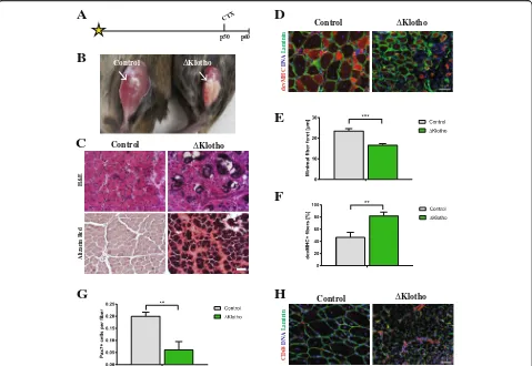
Impaired muscle stem cell function generally coincides with decreased regenerative capacity of skeletal muscle [4, 34]. To analyze the role of klotho during regener-ation, we injured the tibialis anterior muscles ofΔKlotho and control mice at an age of p50 and analyzed the re-generation 10 days after injury (Fig.2a). Intriguingly, we already observed obvious impairments in the regener-ation process in muscles from ΔKlotho mice on the macroscopic level (Fig. 2b). Muscles from control litter-mates after 10 days of regeneration showed the expected degree of regeneration (d10) (compare [34]). In contrast,
ΔKlotho mice exhibited signs of fibrosis, calcification, and scarring suggesting major impairments in regener-ation, confirmed by histological analyses (Fig. 2c). To
quantify regeneration, we measured the minimal fiber feret as a measure of myofiber size. Thereby, we found significantly smaller myofibers after d10 of regeneration in ΔKlotho mice (control 23.58 ± 1.08 μm vs. ΔKlotho 16.70 ± 0.71 μm) (Fig. 2d, e). This suggests either im-paired differentiation of muscle stem cells or loss of self-renewal capacity thereby limiting the number of stem cells available for formation and growth of newly formed fibers. To further investigate the differentiation process in vivo, we quantified the number of devMHC (developmental myosin heavy chain) positive myofibers (Fig.2d, f), a well-known marker for newly formed myo-fibers. Importantly, during maturation of myofibers, ex-pression of devMHC decreases [35]. We found that 82% of myofibers in ΔKlotho mice still expressed devMHC while only 47% of myofibers in the control littermates were positive for devMHC at day 10 after CTX injury. This indicates that differentiation/maturation of myofi-bers is delayed/inhibited inΔKlotho muscles (Fig.2d, f). Muscle stem cells can divide asymmetrically during the regeneration process thereby generating progenitor cells to build new myofibers or self-renew to replenish the pool of stem cells [25]. To investigate the self-renewal potential of muscle stem cells, we counted the number of muscle stem cells (Pax7 positive) at day 10 after in-jury. Here, we found a dramatic reduction of 69% in muscle stem cell numbers in ΔKlotho muscles (control 0.200 ± 0.016 Pax7+ cells/fiber vs.ΔKlotho 0.062 ± 0.033 Pax7+ cells/fiber; Fig.2g) while numbers of macrophages were increased inΔKlotho mice (Fig. 2h). These results suggest that differentiation/maturation of newly formed myofibers and self-renewal of muscle stem cells are se-verely compromised.
To further address the self-renewal ability of muscle stem cells in vivo, we investigated regeneration of skel-etal muscle 21 days after injury (Fig. 3a), a time point when the regeneration process is nearly completed and muscle stem cells start to become quiescent again [4]. Gross morphological impairments in muscle regener-ation were also obvious 21 days after injury inΔKlotho mice (Fig. 3b), again showing scarring, aberrant calcifi-cation, accumulation of lipids, and signs of fibrosis as observed 10 days after injury (Fig. 2b, c). Histological analyses demonstrated that the regeneration process in
1.62μm; Fig. 3d). To get further insights into the mat-uration process of newly formed myofibers, we counted the number of devMHC-positive myofibers at day 21 after injury (Fig. 3f). In control muscles, we hardly found any devMHC-positive myofibers as expected and described in the literature [35]. Consistent with our previous observations, the percentage of devMHC myo-fibers inΔKlotho muscles was increased by nearly two-fold (control 6.49 ± 1.73% vs. ΔKlotho 12.08 ± 5.083%; Fig. 3f) suggesting impaired differentiation/maturation of myofibers in ΔKlotho mice after injury. When we counted the number of Pax7-positive muscle stem cells 21 days after regeneration, we found a dramatic de-crease inΔKlotho mice compared to littermate controls (60% difference; Fig. 3g) indicating severe impairments in self-renewal of muscle stem cells when klotho
expression is lost. Furthermore, numbers of macro-phages were still increased at day 21 after regeneration inΔKlotho mice (Fig.3h).
Loss of mklotho does not affect myogenic proliferation or differentiation
In the in vivo model, we used the expression of both forms of klotho (mKlotho and sKlotho) which are ubiquitously perturbed. Furthermore, it is not possible to investigate the role of mKlotho independently of systemic effects including reduced serum levels of sKlotho, which arise from loss of klotho expression in the whole body. To gain information about expression of klotho mRNA (detecting mRNA for mKlotho and sKlotho) in myogenic cells, we performed ex-pression analyses of primary myoblasts and myotubes (Fig. 4a). We can detect klotho mRNA in myoblasts (d0),
A
CTXp50 p60
C
B
Control ΔKlothoE
G
F
devMHC
DNA
Laminin
Control ΔKlotho
D
H
Control ΔKlotho
H&E
Aliz
arin Red
ΔKlotho
Control
CD68
DNA
Laminin
Fig. 2Early regeneration of skeletal muscle is impaired inΔKlotho mice.aThe right TA muscle fromΔKlotho and control mice was injured by
injection of cardiotoxin (CTX) at p50 and isolated at p60.bTA muscles (arrows) show extensive fibrosis inΔKlotho mice 10 days (d10) after injury.
cRepresentative H&E stainings and Alizarin Red stainings of TA muscle cross-sections at d10 after injury. Necrotic tissue, fatty vacuoles, and
massive cell invasion are visible inΔKlotho TA muscles, while control TA muscles show mostly regenerating myofibers. Scale bar = 100μm.d
Immunostainings of cross-sections of TA muscles at d10 after injury with antibodies directed against Laminin (green) and developmental (dev)
MHC (red), nuclei are stained with DAPI (DNA, blue). Scale bar = 50μm.eMinimal fiber feret measured on whole cross-sections fromΔKlotho
and control TA muscles at d10 after injury. (ΔKlothon= 4 mice, controln= 5 mice). Proportion offdevMHC-positive fibers andgPax7-positive
cells per myofiber on whole cross-sections at d10 after CTX-induced injury (ΔKlothon= 3 mice, controln= 4 mice).hRepresentative images of
stainings for CD68 (in red) and laminin (in green) showing increased infiltration with macrophages in regenerating muscles fromΔKlotho mice.
where expression decreases following differentiation sug-gesting that expression of klotho mRNA is most important in undifferentiated cells. Furthermore, we asked whether myoblasts secrete sKlotho (Fig.4b, Additional file3: Figure S3E, F). Indeed, we found that myoblasts isolated from wt animals secrete small amounts of sKlotho (Additional file3: Figure S3E), this is lost in primary myoblasts fromΔKlotho mice—as expected (Fig. 4b). We then isolated primary myoblasts from ΔKlotho and control littermates and ana-lyzed their proliferation and differentiation potential (Fig.4c–g). Importantly, by using this approach, we exclude systemic factors, e.g., changes in the mineral homeostasis of
ΔKlotho mice and sKlotho secreted by the kidney and transported to the muscle via the blood stream.
Furthermore, we exclude factors from the muscle stem cell niche, e.g., changes in the extracellular matrix. The only dif-ference between the myogenic cells fromΔKlotho and con-trol mice is the expression of mKlotho and secretion of sKlotho by myoblasts or myotubes themselves. Interest-ingly, no differences in proliferation (Fig.4c, d) or differen-tiation (Fig. 4e–g and Additional file 3: Figure S3 A–D) were observed between myogenic cells isolated from
ΔKlotho and control littermates implying that expression of mKlotho and secretion of sKlotho in myoblasts does not affect their proliferation and differentiation. Therefore, we can conclude that endogenous expression of mKlotho is dispensable for proper proliferation and differentiation of myoblasts.
A
CTXp39 p60
C
B
Control ΔKlotho Control ΔKlothoD
E
G
F
ΔKlotho
Control
devMHC
DNA
Laminin
ΔKlotho
Control
CD68
DNA
Laminin
H
ΔKlotho
Control
H&E
Alizarin Red
Sirius Red
Oil Red O
Fig. 3Late regeneration of skeletal muscle is impaired inΔKlotho mice.aThe right TA muscle fromΔKlotho and control mice was injured by
injection of cardiotoxin (CTX) at p39 and isolated at p60.bTA muscles (arrows) show signs of impaired regeneration by H&E staining and extensive
fibrosis inΔKlotho mice at day 21 after injury (c), but not in control littermates as demonstrated by Sirius Red staining. Alizarin Red marks calcifications,
visible in regenerating muscles fromΔKlotho mice as well as lipid accumulations shown by Oil Red O staining. Scale bar = 100μm.dMinimal fiber
feret measured on whole cross-sections of TA muscles fromΔKlotho and control at d21 after injury. (ΔKlothon= 4 mice, controln= 4 mice).e
Immunostainings of cross-sections of TA muscles at d21 after injury with antibodies directed against laminin (green) and developmental (dev) MHC
(red), nuclei are stained with DAPI (DNA, blue). Scale bar = 50μm. Proportion of (f) devMHC-positive fibers (ΔKlothon= 2 mice, controln= 4 mice)
and (g) Pax7-positive satellite cells per myofiber on whole cross-sections at d21 after CTX-induced injury. (ΔKlothon= 4 mice, controln= 4 mice).h
Representative images of stainings for CD68 (in red) and laminin (in green) showing increased infiltration with macrophages in regenerating muscles
Muscle stem cell function is severely impaired in adult
ΔKlotho mice
To investigate the muscle stem cell function in their en-dogenous niche but without the influence of systemic factors, we performed single myofiber cultures from
EDL muscles [36]. In this culture system, the muscle stem cells are located in their endogenous niche adjacent to the muscle fiber and can undergo multiple rounds of divisions under controlled culture conditions. Therefore, we first compared the number, activation, and
D
F
Control ΔKlothoDNA
MHC
Merge
Myogenin
C
Control ΔKlothoDNA
Merge
Ki67
E
G
A
B
WT ΔKlotho
sKlotho 130kDa (full length) sKlotho 72kDa (cleaved) supernatant
Fig. 4Proliferation and differentiation are not affected in myoblasts fromΔKlotho mice in vitro.aαklothomRNA expression during myogenic
differentiation of myoblasts d0, growth medium (myoblasts); d1, day1 of differentiation (myocytes); d3 and d5, days 3 and 5 of differentiation
(myotubes). (n= 3 mice per time point). All data are presented as means ± SEM. *p< 0.05, **p< 0.01. Expression was normalized to GAPDH.b
Immunoblot analysis showing expression of sKlotho in the supernatant from primary myoblasts isolated from wt mice but not in the supernatant
fromΔKlotho mice. An image of the Ponceau stained membrane can be found in Additional file3: Figure S3F.cPrimary myoblasts fromΔKlotho
and control mice were immunostained for the proliferation marker Ki67 (green) and DAPI (DNA, blue) after 48 h of proliferation time in normal
culture medium. Arrow heads mark Ki67 positive cells. Scale bar = 100μm.dThe proportion of Ki67-positive cells was counted in 15 randomly
chosen regions of interest per condition (ΔKlothon= 3 mice, controln= 4 mice).eThe mean myotube diameter was measured in six randomly
chosen regions of interest per condition (ΔKlothon= 3 mice, controln= 4 mice).fRepresentative images of immunostainings from myotubes
after 5 days of differentiation. DNA (DAPI, blue), MHC (green), myogenin (red). Scale bar = 100μm.gThe fusion index was calculated as the ratio
of nuclei in myotubes in relation to the total number of nuclei in 6 randomly chosen regions of interest per condition (ΔKlothon= 3 mice,
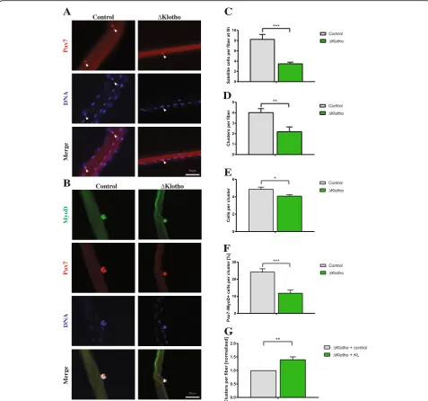
differentiation potential of muscle stem cells ofΔKlotho and control mice. We observed a dramatic reduction (58%) in the average number of muscle stem cells per myofiber inΔKlotho mice directly after isolation (Fig.5a, c). Importantly, the reduction of muscle stem cells
observed in the EDL was similar to the one observed in the TA muscle (Fig. 1a, d). To investigate the prolifera-tive capacity, we determined the number of clusters formed from a single muscle stem cell. Notably, we also observed a dramatic reduction in the average number of
A
DNA
Merge
Pax7
Control ΔKlotho
B
C
D
E
F
DNA
Merge
Pax7
MyoD
Control ΔKlotho
Pax7
DNA
Merge
MyoD
G
Fig. 5Muscle stem cell function is impaired inΔKlotho mice.aMyofibers with their adjacent muscle stem cells were isolated from EDL muscles
of p42 old mice and directly fixed and stained with antibodies directed against Pax7 (red) and DNA (DAPI, blue). Arrows point at Pax7-positive
muscle stem cells. Scale bar = 50μm.bRepresentative images of clusters of muscle stem cells on their adjacent EDL myofibers isolated at p42
and cultured for 72 h and then stained with antibodies directed against MyoD (green), Pax7 (red), and DNA (DAPI, blue). Scale bar = 50μm.c
The number of Pax7-positive muscle stem cells per myofiber was quantified from p42ΔKlotho and control mice. (ΔKlothon= 5 mice, controln=
7 mice).dThe number of clusters per myofiber isolated from p42 animals and cultured for 72 h of culture was enumerated. (ΔKlothon= 5 mice,
controln= 7 mice).eThe number of cells per cluster per myofiber isolated from p42 animals and cultured for 72 h was determined. (ΔKlothon
= 5 mice, controln= 7 mice).fThe composition of clusters shows a reduction in the proportion of further differentiated cells (Pax7−/MyoD+) in
ΔKlotho compared to control mice at p42. (ΔKlothon= 5 mice, controln= 7 mice).gAddition of recombinant sKlotho to fiber cultures from
ΔKlotho mice results in an increase in cluster number per myofiber (n= 3). All data are presented as means ± SEM. *p< 0.05,
clusters per myofiber in ΔKlotho mice compared to lit-termate controls (Fig. 5b, d) as well as in the average number of cells per cluster (Fig.5e). This indicates that proliferation and/or differentiation of muscle stem cells are affected in ΔKlotho mice. However, we did not observe differences in the activation potential (Add-itional file4: Figure S4A). After muscle injury or culture, muscle stem cells are activated resulting in the expres-sion of MyoD and acquisition of a progenitor status. Subsequent downregulation of Pax7 (Pax7−/MyoD+) is followed by differentiation [37, 38]. Indeed, we found a reduction in the percentage of cells, which only express MyoD in clusters from ΔKlotho mice (Fig. 5f; Add-itional file 4: Figure S4B–D). This is suggestive of im-paired differentiation of muscle stem cells in ΔKlotho mice. Interestingly, the same loss of differentiation can be seen in muscle stem cells cultured on floating myofi-bers isolated from aged mice [7]. To investigate if re-combinant sKlotho (KL) is able to rescue the decrease in cluster formation observed in ΔKlotho mice, we applied sKlotho recombinant protein to fiber cultures from
ΔKlotho mice (Fig. 5g). Indeed, addition of sKlotho re-sults in an increase in the number of clusters per fiber of ΔKlotho mice suggesting that sKlotho and not mKlotho is important for muscle stem cell function.
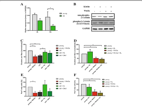
Wnt3a antagonizes Klotho function in muscle stem cells Canonical Wnt is one of the secreted factors, which play an important role in stem cell maintenance and stem cell proliferation [39]. In muscle stem cells, canonical Wnt sig-naling controls differentiation of muscle stem cells; for in-stance, it was demonstrated that a switch from Notch signaling to canonical Wnt signaling is a prerequisite for the differentiation of muscle stem cells [25, 40, 41]. Al-though necessary for differentiation, exogenous induction of canonical Wnt signaling during early regeneration of skeletal muscle leads to premature differentiation of pro-genitor cells thereby depleting the muscle stem cell pool [40]. Furthermore, it was described that addition of ca-nonical Wnt3a to adult skeletal muscle leads to increased amounts of connective tissue resulting in a skeletal muscle, which resembles aged skeletal muscle after regen-eration. sKlotho is a known inhibitor of canonical Wnt signaling [42]. The KL1 domain of sKlotho was recently mapped as the interaction domain of sKlotho with Wnt3a [43]. This evidence prompted us to speculate if sKlotho could be an inhibitor of canonical Wnt signaling in muscle stem cells.
Therefore, we first evaluated the expression levels of klotho mRNA in isolated myofibers with their adjacent muscle stem cells isolated from young and old C57BL/6J mice (Fig.6a). Interestingly, expression of klotho mRNA is reduced in myofibers with adjacent muscle stem cells from old mice directly after isolation and after 72 h of culture
when the number of muscle stem cells on the myofibers is increased. This let us speculate that less sKlotho is secreted from aged myofibers and their adjacent muscle stem cells thereby impeding muscle stem cell function. To demon-strate that sKlotho can also inhibit canonical Wnt signaling in myogenic cells, we treated primary myoblasts with Wnt3a, sKlotho, or a combination of both (Fig. 6b). Im-portantly, Wnt3a-mediated activation of canonical Wnt sig-naling is inhibited by addition of sKlotho suggesting that sKlotho is also an inhibitor of canonical Wnt signaling in myogenic cells (Fig.6b, Additional file5: Figure S5A).
Supplementation with sKlotho rejuvenates aged muscle stem cells
We speculated that sKlotho could be one of the natur-ally occurring inhibitors of canonical Wnt signaling in muscle stem cells in the young and that loss of klotho expression in the aged might be one of the causes for in-creased canonical Wnt signaling in aged muscle stem cells. Therefore, we cultured muscle stem cells on float-ing fibers from young and old mice with different com-binations of sKlotho protein (KL), Wnt3a and Dkk1, a well-known inhibitor of canonical Wnt signaling (Fig.6c). Interestingly, we found that addition of sKlotho protein to fiber cultures obtained from old C57BL/6J mice (22– 24 months of age) resulted in an increase in the number of clusters per fiber formed after 72 h of culture and also an increased activation potential (Additional file 5: Figure S5C). Importantly, the number of clusters per fiber was similar to the number of clusters per fiber in young animals (4 months of age) suggesting that sKlotho rejuvenates old muscle stem cells (Fig.6c). The effect of sKlotho on cluster numbers on aged fiber cultures was similar to the effect of Dkk1. Importantly, addition of sKlotho and Dkk1 to old myofibers did not lead to an increased cluster number compared to old myofibers treated with either sKlotho or Dkk1 alone indicating that sKlotho and Dkk1 are both acting by inhibition of ca-nonical Wnt signaling (Additional file 5: Figure S5B). Addition of Wnt3a to young fibers resulted in a decrease in cluster numbers on young fibers as expected (Fig.6c). Interestingly, the number of clusters was then similar to the ones on old myofibers. However, if we simultan-eously added sKlotho and Wnt3a to the myofiber cul-tures from young mice, this sKlotho-mediated increase in the number of clusters per fiber was completely blocked suggesting that sKlotho and Wnt3a antagonize each other in muscle stem cells (Fig.6d).
cells in the aged to numbers comparable to muscle stem cells on young fibers. Of note, Wnt3a and sKlotho are also counteracting each other regarding the self-renewal of young muscle stem cells (Fig. 6f) and a combined treatment with sKlotho and Dkk1 does not lead to in-creased self-renewal compared to treatment with either sKlotho or Dkk1 alone (Additional file5: Figure S5D).
sKlotho might be an effective natural inhibitor of canonical Wnt signaling in muscle stem cells in the young and loss of klotho expression in the aged might be one of the causes for increased canonical Wnt signaling and thereby impeded muscle stem cell functionality in the aged. This, together
with the data on supplementation of sKlotho in aged muscle stem cells, suggests that muscle stem cells and myofibers se-crete sKlotho thereby inhibiting canonical Wnt signaling, a mechanism which is particularly hampered in the aged.
Discussion
Klotho hypomorphic mice display several aberrations in muscle function, for instance, reduced muscle strength and running endurance [24]. They also demonstrate re-duced serum levels of sKlotho. Interestingly, a study by Semba et al. [44] found that reduced serum levels of sKlotho in older humans is correlated with reduced grip
C
A
D
B
Klotho
Wnt3a
phospho-β-Catenin (S33/37/Thr41)
-+
-+ +
+
GAPDH
non-phospho-β-Catenin
E
F
Fig. 6sKlotho antagonizes aberrant Wnt3a function in aged muscle stem cells.aαKlothomRNA expression in myofibers with their adjacent
muscle stem cells directly after isolation or after 72 h of culture from young and old C57BL/6J mice (young mice:n= 4, old mice:n= 5).b
Addition of sKlotho to primary myoblasts reduces induction of canonical Wnt signaling evoked by addition of recombinant Wnt3a.cMyofibers
with their adjacent muscle stem cells from young (4 months) and old (22–24 months) mice were cultured for 72 h with normal medium,
recombinant soluble klotho (KL) protein, recombinant Wnt3a, or recombinant Dkk1. (n= 6 mice (young),n= 5 (old)). The number of clusters per
myofiber was normalized to young control.dMyofibers with their adjacent muscle stem cells from young (4 months) mice were cultured for
72 h with normal medium, recombinant soluble klotho (KL) protein, recombinant Wnt3a, or a combination of both (n= 6 mice). The number of
clusters per myofiber was normalized to young control.eAddition of sKlotho recombinant protein (KL) increases the proportion of Pax7+/MyoD−
cells per fiber located in a cluster. The number of Pax7+/MyoD−cells (located in a cluster) per myofiber was normalized to young control (n= 6
mice (young),n= 5 (old)).fMyofibers with their adjacent muscle stem cells from young (4 months) mice were cultured for 72 h with normal
medium, recombinant soluble klotho (KL) protein, recombinant Wnt3a, or a combination of both (n= 6 mice). The number of Pax7+/MyoD−cells
strength further strengthening the importance of con-stant klotho expression for maintenance and functional output of skeletal muscle. So far, nothing was known re-garding effects of klotho expression on muscle stem cell function and differentiation of myoblasts.
Notably, in the current study, we observed that muscle stem cell function in klotho hypomorphic mice is massively perturbed. Furthermore, numbers of muscle stem cells are significantly decreased inΔKlotho mice (Figs.1and5a, c). Concomitantly, loss of klotho expression leads to a dra-matic reduction in the regenerative capacity of skeletal muscle, which reminded us of the phenotype we observed during natural aging (Figs.2 and3; [7,11]). Klotho hypo-morphic mice are not only characterized by the loss of klotho gene expression but also by a disturbed mineral homeostasis caused by impaired FGF23 signaling in the kidney [45]. Since we could not rule out the effects of this disturbed mineral homeostasis in ΔKlotho mice, we per-formed experiments using cultures of muscle stem cells on single floating myofibers. In this system, we can exclude direct effects from the mineral homeostasis but not the fact that those changes might have led to changes in the epige-nome or activity of signaling pathways before isolation. Such changes were described for naturally aged muscle stem cells [7, 11]. Of note, Wehling-Henricks and col-leagues described that epigenetic silencing of klotho in muscular dystrophy contributes to the disturbed regener-ation and fibrosis in mdx mice, the mouse model for Du-chenne muscular dystrophy [46].
In our floating fiber culture system, we found that loss of klotho expression reduces the number of clusters formed and the number of cells per cluster suggesting that loss of klotho expression negatively influences proliferation and self-renewal of muscle stem cells (Fig.5). Furthermore, dif-ferentiation of muscle stem cells on fibers is affected when expression of klotho is lost (Fig. 5 and Additional file 4: Figure S4) implying also a function of klotho in differenti-ation of muscle stem cells. In adipose-derived stem cells, loss of klotho expression hinders proliferation and differen-tiation [29]. Application of recombinant sKlotho protein on the other hand restored the proliferative capacity as well as the ability to differentiate. In the skin and intestine, loss of klotho results in exhaustion of the stem cell pool [42].
Canonical Wnt signaling plays an important role in muscle stem cell function [25]. Differentiation of muscle stem cells is controlled by canonical Wnt signaling, espe-cially by signaling through the ligand Wnt3a. Although necessary for the differentiation process, aberrant canon-ical Wnt signaling results in increased fibrosis of skeletal muscle, for instance, in the context of aging or muscular dystrophy [40,47]. Aged skeletal muscle has been shown to display increased canonical Wnt signaling resulting in a conversion of muscle stem cells from a myogenic to a fibrogenic lineage [40]. sKlotho is a known antagonist of
canonical Wnt signaling [42]. Therefore we investigated if addition of sKlotho protein counteracts aberrant canonical Wnt signaling, here in the context of aging. Indeed, we found that Wnt3a antagonizes sKlotho function in myo-blasts and old muscle stem cells (Fig.6c–f). Furthermore we could demonstrate that inhibition of aberrant canon-ical Wnt signaling in the aged by Dkk1 had very similar ef-fects compared to addition of sKlotho further supporting the notion that sKlotho is a naturally occurring inhibitor of canonical Wnt signaling in muscle stem cells.
We consider it likely that the large quantity of fibrotic tis-sue during impaired regeneration and the depletion of muscle stem cells in ΔKlotho mice can be traced back to chronic canonical Wnt stimulation as observed during nat-ural aging [40]. Transforming growth factor beta (TFG-β1) signaling is another pro-fibrotic pathway increased in skel-etal muscle during aging resulting in reduced regenerative capacity [48]. Interestingly, sKlotho is a direct inhibitor of TGF-β1 signaling by binding to the type-II TGF-βreceptor thereby preventing fibrosis [49]. Therefore, it is likely that sKlotho not only inhibits canonical Wnt signaling but also TFG-β1 signaling in aged skeletal muscle thereby maintain-ing muscle stem cell function.
Importantly, restoration of sKlotho levels in the aged muscle stem cell niche improves muscle stem cell function in the aged. This suggests that acute sKlotho availability is dominant over intrinsic changes in aged muscle stem cells, which have been acquired over life time [7,11,12].
Conclusions
We conclude that reduction of sKlotho is an important contributor to loss of muscle stem cell function in the aged, most likely through loss of inhibition of canonical Wnt signaling. Therefore, replenishing the levels of sKlotho is a promising therapeutic approach for stimula-tion of muscle regenerastimula-tion in the aged.
Additional files
Additional file 1:Figure S1.ΔKlotho mice display signs of sarcopenia.
(A) Immunofluorescence of cross-sections of TA muscles fromΔKlotho
and control mice stained for DAPI (DNA, blue) and Laminin (green) at
p14, p21, and p56. Scale bar = 50μm. (B) Minimal fiber feret measured
on the whole cross-sections fromΔKlotho and control TA muscles at
p14, p21, and p56. (p14, p56n≥3 mice per genotype, p21n= 3 mice
per genotype). (C) Quantification of the cross-section area of the
mid-belly region of TA muscles determined from sections fromΔKlotho and
control at p14, p21, and p56. (p14, p56n≥3 mice per genotype, p21n=
3 mice per genotype). (D) Immunoblot analyses of atrophy associated
ubiquitin ligases in TA muscles fromΔKlotho and control littermates at
different ages. (E) Fold expression of different ubiquitin ligases as shown
in (D), values forΔKlotho are normalized to control animal of the same
age. (F) Muscle weight of tibialis anterior (TA) muscles from adultΔKlotho
(n= 6) and control mice,n= 8 (G) Muscle weight of tibialis anterior (TA)
muscles normalized to the length of the tibia bone from adultΔKlotho
(n= 6) and control mice (n= 8). All data are presented as means ± SEM.
Additional file 2:Figure S2.Fiber types are not changed inΔKlotho mice. (A) Representative immunofluorescence stainings of cross-sections
from EDL of p56ΔKlotho and control mice stained for DAPI (DNA, blue),
laminin (white), MHCIIa and MHCIIb, or MHCIIa and MHCIIx, respectively.
Scale bar = 50μm. (B) Percentage of MHC type IIa and IIb positive
myofi-bers on EDL cross-sections fromΔKlotho and control mice at p56.
(ΔKlothon= 3 mice, controln= 7 mice). (C) Percentage of MHC type IIa
and IIx positive myofibers on EDL cross-sections fromΔKlotho and
con-trol mice at p56. (ΔKlothon= 3 mice, controln= 6 mice). All data are
presented as means ± SEM. (PDF 8407 kb)
Additional file 3:Figure S3.The differentiation of myoblasts from
ΔKlotho mice is not affected in vitro. (A) Quantification of the average
number of nuclei per myotube counted on 6 random regions of interest
per condition after 5 days of differentiation (ΔKlothon= 3 mice, controln=
4 mice). (B) Percentage of myogenin-positive nuclei of all nuclei within
myo-tubes after 5 days of differentiation (ΔKlothon= 3 mice, controln= 4 mice).
(C) Differentiation index (percentage of myotubes with more than three
nu-clei) after 5 days of differentiation (ΔKlothon= 3 mice, controln= 4 mice).
(D) Distribution of classes of myotubes after 5 days of differentiation
(ΔKlothon= 3 mice, controln= 4 mice). (E) Immunoblot analyses of
super-natants from primary myoblasts and lysate from a kidney from wt animals using an antibody directed against klotho showing sKlotho in the super-natant and in whole kidney lysates (as expected). (F) Ponceau stained mem-brane showing similar loading of concentrated supernatants from primary
myoblasts isolated fromΔKlotho and control mice. All data are presented as
means ± SEM. (PDF 7378 kb)
Additional file 4:Figure S4.Muscle stem cell function is impaired in
adultΔKlotho mice. (A) The activation potential (number of clusters per
myofiber after 72 h of culture divided by the number of muscle stem
cells per myofiber directly after isolation) (ΔKlothon= 5 mice, controln
= 7 mice). (B) Percentage of Pax7+/MyoD- cells within a cluster on
myofibers isolated from p42 old mice. (ΔKlothon= 5 mice, controln= 7
mice). (C) Percentage of Pax7+/MyoD+ cells within a cluster on myofibers
isolated from p42 old mice. (ΔKlothon= 5 mice, controln= 7 mice). (D)
Percentage of Pax7−/MyoD−cells within clusters on myofibers isolated
from p42 old mice. (ΔKlothon= 5 mice, controln= 7 mice). All data are
presented as means ± SEM. *p< 0.05. (PDF 511 kb)
Additional file 5:Figure S5.Addition of recombinant sKlotho protein rejuvenates aged muscle stem cells. (A) Addition of sKlotho to primary myoblasts reduces canonical Wnt signaling induced by addition of recombinant Wnt3a as evidenced by measuring levels of
phosho-beta-catenin and non-phospho-beta-phosho-beta-catenin, densitometric analysis of Fig.6b
after normalization to GAPDH, values are shown as normalization to con-trol. (B) Myofibers with their adjacent muscle stem cells from young
(4 months) and old (22–24 months) mice were cultured for 72 h with
medium, recombinant soluble klotho (KL) protein, recombinant Dkk1 or a combination of both. The number of clusters per myofiber was
normal-ized to young control. (n= 4 mice (young),n= 3 (old)). (C) The activation
potential is increased in old mice when sKlotho protein (KL) is added (n
= 4). (D) Myofibers with their adjacent muscle stem cells from young
(4 months) and old (22–24 months) mice were cultured for 72 h with
medium, recombinant soluble klotho (KL) protein, recombinant Dkk1 or a
combination of both. The number of Pax7+/MyoD−cells per myofiber
was normalized to young control. (n= 4 mice (young),n= 3 (old)). All
data are presented as means ± SEM. *p< 0.05. (PDF 616 kb)
Abbreviations
devMHC:Developmental myosin heavy chain; Dkk1: Dickkopf-related protein 1; EDL: Extensor digitorum longum; Fbxo32: F-Box Protein 32;
FGF23: Fibroblast growth factor 23; Hox: Homeobox; IGF1: Insulin-like growth factor 1; JAK: Janus kinase; mKlotho: Membrane Klotho; MuRF: Muscle RING-finger protein; MyoD: Myogenic differentiation 1; Pax7: Paired box protein 7; sKlotho: Soluble Klotho; SPF: Specific pathogen free; STAT: Signal Transducer and Activator of Transcription proteins; TA: Tibialis anterior;
TGF-β: Transforming growth factorβ; Wnt: Wingless-related integration site
Acknowledgements
We thank Christine Poser, Christina Picker, Sabine Landmann, and Daniela Reichenbach for their excellent technical assistance and the FLI Core
Facilities histology (L. Rothenburger) and imaging and the mouse facility (J. Brüchert, C. Maisch) for their services. We thank M. Kuro-o for providing the klotho hypomorphic mice.
Funding
J.V.M. and M.S. were supported by a grant from the German Research Foundation (DFG, MA-3975/2-1). HEA was supported by a grant from the Leibniz association (SAW2015).
Availability of data and materials
All datasets generated and analyzed during the current study are available from the corresponding author upon reasonable request.
Authors’contributions
HEA and JH performed most experiments, analyzed the data, and interpreted the results. MS performed the qPCR and immunoblot analyses. HEA and JVM designed and performed the experiments, analyzed the data, interpreted the results, and wrote the manuscript. CK provided support and suggestions for the experiments concerning klotho hypomorphic mice. All authors read and approved the final version of the manuscript.
Ethics approval
All animal experiments were performed in accordance with the German Animal Welfare Act and approved by the responsible local authority of Thuringia (TLV), TVA no.: 03-11/14.
Competing interests
The authors declare that they have no competing interest.
Publisher’s Note
Springer Nature remains neutral with regard to jurisdictional claims in published maps and institutional affiliations.
Author details
1Leibniz-Institute on Aging–Fritz-Lipmann-Institute, Beutenbergstrasse 11,
07745 Jena, Germany.2Present address: Max-Delbrück-Center for Molecular Medicine, Robert-Rössle-Str. 10, 13125 Berlin, Germany.
Received: 29 January 2018 Accepted: 21 June 2018
References
1. Bentzinger CF, von Maltzahn J, Rudnicki MA. Extrinsic regulation of satellite
cell specification. Stem Cell Res Ther. 2010;1:27.
2. Lepper C, Partridge TA, Fan CM. An absolute requirement for Pax7-positive
satellite cells in acute injury-induced skeletal muscle regeneration.
Development. 2011;138:3639–46.
3. Murphy MM, Lawson JA, Mathew SJ, Hutcheson DA, Kardon G. Satellite
cells, connective tissue fibroblasts and their interactions are crucial for
muscle regeneration. Development. 2011;138:3625–37.
4. Bentzinger CF, Wang YX, Dumont NA, Rudnicki MA. Cellular dynamics in the
muscle satellite cell niche. EMBO Rep. 2013;14:1062–72.
5. von Maltzahn J, Bentzinger CF, Rudnicki MA. Characteristics of satellite cells and
multipotent adult stem cells in the skeletal muscle. Dordrecht: Springer; 2013.
6. Dumont NA, Wang YX, von Maltzahn J, Pasut A, Bentzinger CF, Brun CE,
Rudnicki MA. Dystrophin expression in muscle stem cells regulates their
polarity and asymmetric division. Nat Med. 2015;21:1455–63.
7. Price FD, von Maltzahn J, Bentzinger CF, Dumont NA, Yin H, Chang NC,
Wilson DH, Frenette J, Rudnicki MA. Inhibition of JAK-STAT signaling
stimulates adult satellite cell function. Nat Med. 2014;20:1174–81.
8. Sousa-Victor P, Garcia-Prat L, Serrano AL, Perdiguero E, Munoz-Canoves P.
Muscle stem cell aging: regulation and rejuvenation. Trends Endocrinol
Metab. 2015;26:287–96.
9. Chakkalakal JV, Jones KM, Basson MA, Brack AS. The aged niche disrupts
muscle stem cell quiescence. Nature. 2012;490:355–60.
10. Lukjanenko L, Jung MJ, Hegde N, Perruisseau-Carrier C, Migliavacca E, Rozo
M, Karaz S, Jacot G, Schmidt M, Li L, Metairon S, Raymond F, Lee U, Sizzano F, Wilson DH, Dumont NA, Palini A, Fassler R, Steiner P, Descombes P, Rudnicki MA, Fan CM, von Maltzahn J, Feige JN, Bentzinger CF. Loss of fibronectin from the aged stem cell niche affects the regenerative capacity
11. Schworer S, Becker F, Feller C, Baig AH, Kober U, Henze H, Kraus JM, Xin B, Lechel A, Lipka DB, Varghese CS, Schmidt M, Rohs R, Aebersold R, Medina KL, Kestler HA, Neri F, von Maltzahn J, Tumpel S, Rudolph KL. Epigenetic stress responses induce muscle stem-cell ageing by Hoxa9 developmental
signals. Nature. 2016;540:428–32.
12. Sousa-Victor P, Gutarra S, Garcia-Prat L, Rodriguez-Ubreva J, Ortet L,
Ruiz-Bonilla V, Jardi M, Ballestar E, Gonzalez S, Serrano AL, Perdiguero E, Munoz-Canoves P. Geriatric muscle stem cells switch reversible quiescence into
senescence. Nature. 2014;506:316–21.
13. Xu Y, Sun Z. Molecular basis of Klotho: from gene to function in aging.
Endocr Rev. 2015;36:174–93.
14. Bian A, Neyra JA, Zhan M, Hu MC. Klotho, stem cells, and aging. Clin Interv
Aging. 2015;10:1233–43.
15. Kurosu H, Ogawa Y, Miyoshi M, Yamamoto M, Nandi A, Rosenblatt KP, Baum
MG, Schiavi S, Hu MC, Moe OW, Kuro-o M. Regulation of fibroblast growth
factor-23 signaling by klotho. J Biol Chem. 2006;281:6120–3.
16. Quarles LD. Endocrine functions of bone in mineral metabolism regulation.
J Clin Invest. 2008;118:3820–8.
17. Utsugi T, Ohno T, Ohyama Y, Uchiyama T, Saito Y, Matsumura Y, Aizawa H,
Itoh H, Kurabayashi M, Kawazu S, Tomono S, Oka Y, Suga T, Kuro-o M, Nabeshima Y, Nagai R. Decreased insulin production and increased insulin sensitivity in the klotho mutant mouse, a novel animal model for human
aging. Metabolism. 2000;49:1118–23.
18. Abramovitz L, Rubinek T, Ligumsky H, Bose S, Barshack I, Avivi C, Kaufman B,
Wolf I. KL1 internal repeat mediates klotho tumor suppressor activities and inhibits bFGF and IGF-I signaling in pancreatic cancer. Clin Cancer Res. 2011;
17:4254–66.
19. Pedersen L, Pedersen SM, Brasen CL, Rasmussen LM. Soluble serum Klotho
levels in healthy subjects. Comparison of two different immunoassays. Clin
Biochem. 2013;46:1079–83.
20. Sato S, Kawamata Y, Takahashi A, Imai Y, Hanyu A, Okuma A, Takasugi M,
Yamakoshi K, Sorimachi H, Kanda H, Ishikawa Y, Sone S, Nishioka Y, Ohtani N, Hara E. Ablation of the p16(INK4a) tumour suppressor reverses ageing phenotypes of klotho mice. Nat Commun. 2015;6:7035.
21. Kuro-o M, Matsumura Y, Aizawa H, Kawaguchi H, Suga T, Utsugi T, Ohyama
Y, Kurabayashi M, Kaname T, Kume E, Iwasaki H, Iida A, Shiraki-Iida T, Nishikawa S, Nagai R, Nabeshima YI. Mutation of the mouse klotho gene
leads to a syndrome resembling ageing. Nature. 1997;390:45–51.
22. Kurosu H, Yamamoto M, Clark JD, Pastor JV, Nandi A, Gurnani P, McGuinness
OP, Chikuda H, Yamaguchi M, Kawaguchi H, Shimomura I, Takayama Y, Herz J, Kahn CR, Rosenblatt KP, Kuro-o M. Suppression of aging in mice by the
hormone Klotho. Science. 2005;309:1829–33.
23. Olauson H, Mencke R, Hillebrands JL, Larsson TE. Tissue expression and
source of circulating alphaKlotho. Bone. 2017;100:19–35.
24. Phelps M, Pettan-Brewer C, Ladiges W, Yablonka-Reuveni Z. Decline in
muscle strength and running endurance in klotho deficient C57BL/6 mice.
Biogerontology. 2013;14:729–39.
25. von Maltzahn J, Chang NC, Bentzinger CF, Rudnicki MA. Wnt signaling in
myogenesis. Trends Cell Biol. 2012;22:602–9.
26. von Maltzahn J, Jones AE, Parks RJ, Rudnicki MA. Pax7 is critical for the
normal function of satellite cells in adult skeletal muscle. Proc Natl Acad Sci
U S A. 2013;110:16474–9.
27. von Maltzahn J, Zinoviev R, Chang NC, Bentzinger CF, Rudnicki MA. A
truncated Wnt7a retains full biological activity in skeletal muscle. Nat Commun. 2013;4:2869.
28. Briguet A, Courdier-Fruh I, Foster M, Meier T, Magyar JP. Histological
parameters for the quantitative assessment of muscular dystrophy in the
mdx-mouse. Neuromuscul Disord. 2004;14:675–82.
29. Fan J, Sun Z. The antiaging gene klotho regulates proliferation and
differentiation of adipose-derived stem cells. Stem Cells. 2016;34:1615–25.
30. Rom O, Reznick AZ. The role of E3 ubiquitin-ligases MuRF-1 and MAFbx in
loss of skeletal muscle mass. Free Radic Biol Med. 2016;98:218–30.
31. Gumucio JP, Mendias CL. Atrogin-1, MuRF-1, and sarcopenia. Endocrine.
2013;43:12–21.
32. Bodine SC, Latres E, Baumhueter S, Lai VK, Nunez L, Clarke BA,
Poueymirou WT, Panaro FJ, Na E, Dharmarajan K, Pan ZQ, Valenzuela DM, DeChiara TM, Stitt TN, Yancopoulos GD, Glass DJ. Identification of ubiquitin ligases required for skeletal muscle atrophy. Science. 2001;294:
1704–8.
33. Gomes MD, Lecker SH, Jagoe RT, Navon A, Goldberg AL. Atrogin-1, a
muscle-specific F-box protein highly expressed during muscle atrophy. Proc
Natl Acad Sci U S A. 2001;98:14440–5.
34. Bentzinger CF, Wang YX, Rudnicki MA. Building muscle: molecular
regulation of myogenesis. Cold Spring Harb Perspect Biol. 2012;4:1-16.
35. Schiaffino S, Rossi AC, Smerdu V, Leinwand LA, Reggiani C. Developmental
myosins: expression patterns and functional significance. Skelet Muscle. 2015;5:22.
36. Pasut A, Oleynik P, Rudnicki MA. Isolation of muscle stem cells by fluorescence
activated cell sorting cytometry. Methods Mol Biol. 2012;798:53–64.
37. Zammit PS, Golding JP, Nagata Y, Hudon V, Partridge TA, Beauchamp JR.
Muscle satellite cells adopt divergent fates: a mechanism for self-renewal? J
Cell Biol. 2004;166:347–57.
38. Cooper RN, Tajbakhsh S, Mouly V, Cossu G, Buckingham M, Butler-Browne
GS. In vivo satellite cell activation via Myf5 and MyoD in regenerating
mouse skeletal muscle. J Cell Sci. 1999;112 ( Pt 17:2895–901.
39. Ring A, Kim YM, Kahn M. Wnt/catenin signaling in adult stem cell
physiology and disease. Stem Cell Rev. 2014;10:512–25.
40. Brack AS, Conboy MJ, Roy S, Lee M, Kuo CJ, Keller C, Rando TA. Increased
Wnt signaling during aging alters muscle stem cell fate and increases
fibrosis. Science. 2007;317:807–10.
41. Brack AS, Conboy IM, Conboy MJ, Shen J, Rando TA. A temporal switch
from notch to Wnt signaling in muscle stem cells is necessary for normal
adult myogenesis. Cell Stem Cell. 2008;2:50–9.
42. Liu H, Fergusson MM, Castilho RM, Liu J, Cao L, Chen J, Malide D, Rovira D
II, Schimel CJ, Kuo JS, Gutkind PM, Hwang TF. Augmented Wnt signaling in
a mammalian model of accelerated aging. Science. 2007;317:803–6.
43. Mirza SB, Ekhteiari Salmas R, Fatmi MQ, Durdagi S. Discovery of Klotho
peptide antagonists against Wnt3 and Wnt3a target proteins using combination of protein engineering, protein-protein docking, peptide docking and molecular dynamics simulations. J Enzyme Inhib Med Chem.
2017;32:84–98.
44. Semba RD, Cappola AR, Sun K, Bandinelli S, Dalal M, Crasto C, Guralnik JM,
Ferrucci L. Relationship of low plasma klotho with poor grip strength in older community-dwelling adults: the InCHIANTI study. Eur J Appl Physiol.
2012;112:1215–20.
45. Kuro-o M. Klotho as a regulator of fibroblast growth factor signaling and
phosphate/calcium metabolism. Curr Opin Nephrol Hypertens. 2006;15:437–41.
46. Wehling-Henricks M, Li Z, Lindsey C, Wang Y, Welc SS, Ramos JN, Khanlou
N, Kuro OM, Tidball JG. Klotho gene silencing promotes pathology in the mdx mouse model of Duchenne muscular dystrophy. Hum Mol Genet.
2016;25:2465–82.
47. Trensz F, Haroun S, Cloutier A, Richter MV, Grenier G. A muscle resident cell
population promotes fibrosis in hindlimb skeletal muscles of mdx mice through
the Wnt canonical pathway. Am J Phys Cell Physiol. 2010;299:C939–47.
48. Carlson ME, Hsu M, Conboy IM. Imbalance between pSmad3 and Notch
induces CDK inhibitors in old muscle stem cells. Nature. 2008;454:528–32.
49. Doi S, Zou Y, Togao O, Pastor JV, John GB, Wang L, Shiizaki K, Gotschall R,
Schiavi S, Yorioka N, Takahashi M, Boothman DA, Kuro-o M. Klotho inhibits transforming growth factor-beta1 (TGF-beta1) signaling and suppresses