ABSTRACT
The Suprascapular notch is situated in the lateral part of the superior border of the scapula, just adjacent to the base of Coracoid process. The notch is bridged by the superior transverse scapular ligament (STSL) which some time ossifies and is attached laterally to the root of the coracoid process and medially to the limit of the notch. A number of variations occur in the shape of suprascapular notch, from a discrete notch to "J" shaped, "V" shaped, "U" shaped or "O" shaped (i.e. as a complete foramen).
To study morphological and morphometric variations of suprascapular notch of Indian population.
We studied 140 dried scapula bone and measurements of SSN were done
using digital vernier calipers. We used the Rengachary classification for this study. The following measurements were The superior transverse diameter - maximum distance between superior most edges of suprascapular notch (SSN). The inferior transverse diameter - maximum distance between the edges of the curved arch at the base of the SSN. The results of our study were: J-shaped -28%, U-shaped-26%, V-shaped -15%, Partial-ossification-7%, Indentation-10%, Absent-9%, Complete ossification- 2%. Type IV supra scapular notch was found to be the most prevalent type amongst all shapes. We also found that the characteristics of the scapula (dimensions) are related to the characteristics of the supra scapular notch (type and dimensions) and there is a distinct difference between right and left side scapula.
Vibhash Kumar Vaidya, Geetanjali Srivastava, Tehsin Munsif, VineetaTewari, P. K. Sharma
Department of Anatomy
Era’s Lucknow Medical College & Hospital, Sarfarazganj, Lucknow, U.P., India 226003
-INTRODUCTION
The scapula is a large flattened and triangular bone which lies on the postero- lateral aspect of the thorax, against second to the seventh ribs. The Suprascapular notch is situated in the lateral part of the superior border of the scapula, just adjacent to the base of Coracoid process (1).
The notch is bridged by the superior transverse scapular ligament (STSL) which some time ossifies and is attached laterally to the root of the coracoid process and medially to the limit of the notch (2, 3). A number of variations occur in the shape of suprascapular notch, from a discrete notch to "J" shaped, "V" shaped , "U" shaped or "O" shaped (i.e. as a complete foramen).Various authors have classified the suprascapular notch into different types. Authors like Olivier (4) have divided it into 5 types and like Rengachary (5) has divided into 6 types.
In all these types the notch can be more or less open, narrower or wider. Extrinsic compression or traction on the suprascapular nerve may result in suprascapular neuropathy.
The compression of this nerve may occur at two
distinct locations: the suprascapular notch and the spinoglenoid notch.
MATERIALS AND METHOD
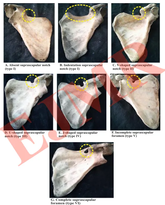
A study includes 140 dried scapula bone irrespective to sex and age which are based on inclusion and exclusion criteria. We used Rengachary classification of morphologically different variations of suprascapular notches. The measurements of SSN were made using digital vernier calipers and these were recorded in millimeters (resolution of 0.01 mm).. The following measurements were taken: The superior transverse diameter - maximum distance between superior most edges of suprascapular notch (SSN). The inferior transverse diameter - maximum distance between the edges of the curved arch at the base of the SSN. The data was analyzed statistically by using ANOVA- test. OBSERVATIONS AND RESULTS
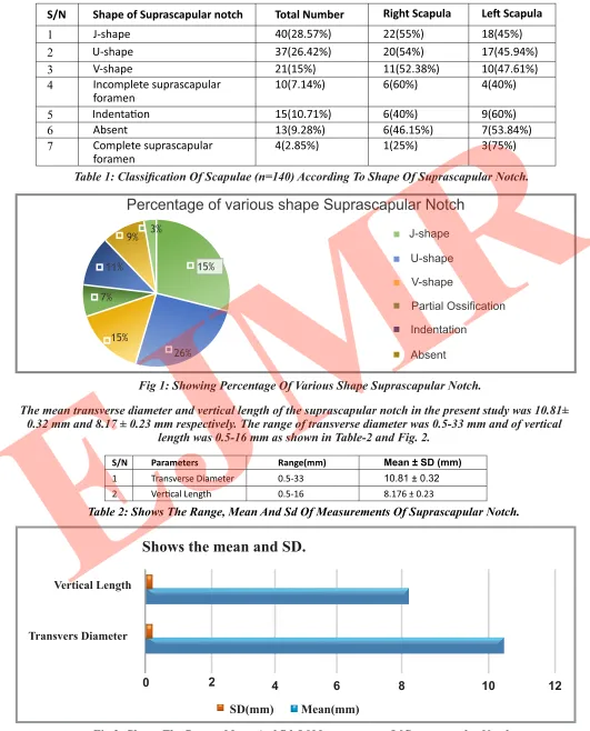
Table-1 shows varies type of supra scapular notches we found in our study: J-shaped -28%, U-shaped-26%, V-shaped -15%, Partial-ossification-7%, Indentation-10%, Absent-9%, Complete ossification- 2% as shown in Table-1, along with the percentages of right and left side scapula.
ERA’S JOURNAL OF MEDICAL RESEARCH
THE MORPHOLOGICAL AND MORPHOMETRIC STUDY OF
SUPRASCAPULAR NOTCH AND ITS VARIATIONS
KEYWORDS: Supra scapular notch, Scapula, Superior transverse diameter, Vertical diameter, Supra scapular ligament. VOL.5 NO.1
Address for correspondence Dr. Vibhash Kumar Vaidya
Department of Anatomy Era’s Lucknow Medical College &
Hospital, Lucknow-226003. Email:[email protected]
Contact no: +91-8077409384
Received on : 28-03-2018 Accpected on : 01-05-2018
Original Article
Page: 23
Indentation J-shape
Partial Ossification
Absent U-shape
V-shape
15%
26% 15% 3%
9%
11%
7%
Percentage of various shape Suprascapular Notch
S/N Shape of Suprascapular notch Total Number Right Scapula Le Scapula
1 J-shape 40(28.57%) 22(55%) 18(45%)
2 U-shape 37(26.42%) 20(54%) 17(45.94%)
3 V-shape 21(15%) 11(52.38%) 10(47.61%)
4 Incomplete suprascapular foramen
10(7.14%) 6(60%) 4(40%)
5 Indenta on 15(10.71%) 6(40%) 9(60%)
6 Absent 13(9.28%) 6(46.15%) 7(53.84%)
7 Complete suprascapular foramen
4(2.85%) 1(25%) 3(75%)
Table 1: Classification Of Scapulae (n=140) According To Shape Of Suprascapular Notch.
Fig 1: Showing Percentage Of Various Shape Suprascapular Notch.
The mean transverse diameter and vertical length of the suprascapular notch in the present study was 10.81± 0.32 mm and 8.17 ± 0.23 mm respectively. The range of transverse diameter was 0.5-33 mm and of vertical
length was 0.5-16 mm as shown in Table-2 and Fig. 2.
S/N Parameters Range(mm) Mean ± SD (mm)
1 Transverse Diameter 0.5-33 10.81 ± 0.32
2 Ver cal Length 0.5-16 8.176 ± 0.23
Table 2: Shows The Range, Mean And Sd Of Measurements Of Suprascapular Notch.
Shows the mean and SD.
Vertical Length
Transvers Diameter
SD(mm) Mean(mm)
0 2 4 6 8 10 12
A. Absent suprascapular notch (type I)
B. Indentation suprascapular notch (type I)
C. V-shaped suprascapular notch (type II)
D. U-shaped suprascapular notch (type III)
E. J-shaped suprascapular notch (type IV)
F. Incomplete suprascapular foramen (type V)
G. Complete suprascapular foramen (type VI)
Fig 3: Showing The Various Shapes Of Suprascapular Notch.
ERA’S JOURNAL OF MEDICAL RESEARCH VOL.5 NO.1
DISCUSSION
In the present study, the incidence of morphological variations in the suprascapular notch is Type IV>Type III>Type I>Type II> Type V> Type VI. This finding is very close to the findings of Paolo Albino et al (11). Also in the present study, Type IV i.e. J- shaped suprascapular notch, has the highest rate i.e. 28.57% which is similar to the findings of Paolo Albino et al (6, 11). and Apurba Patra et al (25). i.e. 31% and 39.09% but Rengachary et al.(5), Sinkeet et al.(9) Vandana R. et al(10)., Usha Kannan et al (12, 13)., Udayasree L et al (14, 22)., G.H.E.S. Hassanein et al (23)., Manmeet kour et al (24, 31)., S. Vedha et al (32)., Reddy et al (28). described the highest rate of Type III suprascapular notch and Iqbal k. et al (8)., Krishna Gopal et al (18). and Manikum et al (22). described the highest rate of Type II suprascapular notch whereas in the present study the incidence rate of Type III, Type I and Type II is 26.42%, 20% and 15% and Type V and Type VI is 7.14% and 2.85%. Type III described the
second highest rate and Type VI described the lowest incidence rate among all finding of the present study. CONCLUSION
Type IV supra scapular notch was found to be the most prevalent type amongst all shapes. This study also found that the characteristics of the scapula (dimensions) are related to the characteristics of the supra scapular notch (type and dimensions). Our findings demonstrated that there is a distinct difference between right and left side scapula. Type III was the second most common type and the least was found to be type VI. Though, it was a small study, but on the basis of the conclusions drawn, further research into this area of classification is required to investigate different shapes of the suprascapular notch. A larger sample size for better external validity and also the knowledge of anatomical variations of SSN is required for better understanding of the location and source of entrapment syndrome So that these
Author Population Type IV Type III Type II Type I Type V Type VI Present study North India 28.57% 26.42% 15% 20% 7.14% 2.85% Rengachary et al
(1979) (5)
America 3% 48% 31% 8% 6% 4%
Sinkeet et al (2010) (9) Kenya 5% 29% 21% 22% 18% 4% Iqbal k. et al(2010) (8) Pakistan _ 13% 20% 18% _ _ Vandana R. et al
(2013) (10)
South India _ 35% 5.2% 4.5% 3% 12.6%
Paolo Albino et al (2013) (11)
Italy 31% 22.8% 19.8% 12.4% 10% 3%
Usha Kannan et al (2014) (13)
South India 4% 52% 10% 20% 4% 10%
Udayasree L et al. (2014) (14)
South India 21.4% 47.6% 4.7% 11.9% 4.7% 9.5%
Krishna Gopal et al. (2014) (18)
North India 12.5% 25% 41.7% 15.8% 1.7% 3.3%
Manikum et al (2015) (22)
South Africa 18% 5% 65% 5% 7% _
G.H.E.S. Hassanein et al. (2015) (23)
Egypt 31.58% 60.53% 7.89% 8.24% 2.35%
Manmeet kour et al (2016) (24)
India _ 46.6% 8.24% 13.3% _ 3.2%
Apurba Patra et al. (2016) (25)
India 39.09% 31.81% 9.09% 11.81% 3.63% 4.54%
S. Vedha et al
(2017) (32)
South India 5.2% 37.2% 5.6% 21.2% 5.2% 9.2%
Reddy et al. (2017)(28) South India _ 44.3% 41.5% 6.6% 4.7% 2.8% _
Table 3: Comparison With The Previous Studies
variations could be kept in mind during surgical or arthroscopic shoulder procedures to prevent supra scapular nerve injuries.
REFERENCES
th
1. Standring Gray' Anatomy, 4 edition. P. No. 1441-1445
2. Standring S, Borley NR, Collins P, Crossman AR, Gatzoulis MA, Healy JC. Section 6 – Pectoral girdle and upper limb Chapter 46 -Pectoral Girdle, Shoulder region and Axilla. In: Gray's Anatomy the Anatomical Basis of Clinical Practice. 40th ed., Churchill Livingstone,2008:791-822
3. Gamal Hamed El-Syed Hassanein, Mohammad Bahgat Ali. International journal of anatomy and research, Int J Anat Res. 2015; 3(4):1536-1542. 4. Polguj M, Sibinski M, Grzegorzewski A.
Variation in morphology of suprascapular notch as a factor of suprascapular entrapment. Intl Orthop. 2013; 37(11): 2185-2192.
5. Rengachary SS, Burr D, Lucas S, et al. Suprascapular entrapment neuropathy: a clinical, anatomical, & comparative study. Part 2: anatomical study. Neurosurgery, 1979; 5(4):447-551.
6. Kopell HP, Thompson WA. Pain and the frozen shoulder. Surg Gynecol Obstet. 1959;109: 92-96. 7. Antonoiou J, Tae SK, Wiliams GR, Bird S,
Ramsey MJ, Iannotti JP. Suprascapular neuropathy. Variability in the diagnosis, treatment, and outcome. Clin Orthop Rel Res. 2001; 386:131-138.
8. Iqbal, K.1, Iqbal, R. and Khan, SG. Anatomical variations in shape of suprascapular notch of scapula.J. Morphol. Sci. 2010; 27(1): 1-2.
9. S.R. Sinkeet, K.O. Awori, P.O. Odula, J.A. Ogeng'o, P.M. Mwachaka. The suprascapular notch: its morphology and distance from the glenoid cavity in a Kenyan population. Folia Morphol. 2010; 69( 4); 241–245 .
10. Vandana R, SudhaPatil. Morphometric study of suprascapular notch. National Journal of clinical Anatomy, 2013; 2 (3):140-144.
11. Paolo Albino, Stefano Carbone, Vittorio Candela, Valerio Arceri, Anna Rita Vestri and Stefano Gumina. Morphometric of the suprascapular notch: correlation with scapular dimensions and c l i n i c a l r e l e v a n c e . A l b i n o e t a l . B M C Musculoskeletal Disorders. 2013; 14:172. 12. D. Toneva, S. Nikolova. Morphology of
suprascapular notch in medieval skeletons from
Bulgaria, Folia Morphol. 2014; 73(2): 210–215. 13. Usha Kannan, N.S.Kannan, J. Anbalagan, Sudha
Rao. Morphometric Study of Suprascapular Notch in Indian Dry Scapulae with Specific Reference to the Incidence of Completely Ossified Superior Transverse Scapular Ligament. Journal of Clinical and Diagnostic Research, 2014; 8(3): 7-10.
14. Udayasree L, Siva Prasad G. V, Lakshmi V.,Study of anatomical variations in the shape of suprascapular notch in dried human scapulae and its clinical significance: J of Evolution of Med and Dent Sci. 2004 June; 3(22): 6054-6057.
15. Dr Girish V. Patil, Dr Shishirkumar, Dr Apoorva D, Dr Thejeswari, Mr. Sushanth N.K., Study of Morphological Variations of Suprascapular Notch in Human Dry Scapulae of South Indians. International Journal of Scientific and Research Publications, 2014 Sep; 4(9) 2250-3153.
16. Dushyant Agrawal, Brijendra Singh1, Gitika Arya Agrawa. Human Scapulae: Supra Scapular Notch, Morphometry and Variations. Indian Journal of Clinical Anatomy and Physiology, 2014 Oct; 1(1) : 1-7.
17. Dr. Md. Jawed Akhtar, Dr. Premjeet Kumar Madhukar. A Study on Complete Absence of the Suprascapular Notch. International Journal of Science and Research (IJSR). 2012 Nov; 3(11): 411-415.
18. Krishna Gopal, Alok Kumar Choudhary, Jolly Agarwal, Virendra Kumar. Variations in suprascapular notch morphology and its clinical importance. International Journal of Research in Medical Sciences Gopal K et al. Int J Res Med Sci. 2015; 3(1): 301-306.
19. Dr. Nagaraj S, Dr. M. Krishnaiah, Praveen Kumar M, Dr. Anil R Sher. Study of Morphological Variation of Suprascapular Notch. IOSR Journal of Dental & Medical Science, 2014; 13(6): 121-123. 20. S. Cirpan, N. Gocmen-Mas, F. Aksu, M. Edizer,
S. Karabekir, A.O. Magden. Suprascapular foramen: a rare variation caused by ossified suprascapular ligaments. Folia Morphol. 2015 Sep;75(1):21–26.
21. Ritika Sharma, Rajan Sharma, Rajan Kumar Singla, Jagdev Singh Kullar, Tripta Sharma. Suprascapular notch: a morphometric and morphologic study in north Indian population. International Journal of Anatomy and Research, Int J Anat Res 2015; 3(3):1306-1311.
22. Manikum. C, Rennie, C. Naidu. E.C.S.
ERA’S JOURNAL OF MEDICAL RESEARCH VOL.5 NO.1
&Azu.O.A Morphological Study of the Suprascapular Notch in a Sample of Scapulae at the University of Kwazulu Natal. Int. J. Morphol. 2015; 33(4):1365-1370.
23. GamalHamed El-Syed Hassanein, Mohammad Bahgat Ali. Variations of suprascapular notch in adult Egyptian scapulae. International Journal of Anatomy and Research, Int J Anat Res. 2015; 3(4):1536-1542.
24. Manmeet Kour, Sangeeta Gupta. Morphology of Suprascapular Notch of Scapula and its Clinical Implications. Jk Science. 2016 March;18(1):31-34. 25. Apurba Patra, Manjeet Singh, Harsimarjit Kaur.
Variations in the Shape and Dimension of the Suprascapular Notch in Dried Human Scapula-An Osteological Study with its Clinical Implications. International Journal of Anatomy, Radiology and Surgery, 2016 Apr; 5(2): 1-5. 26. Neeta Chhabra, Suraj Prakash, and MS Ahuja.
Morphometry and morphology of supra scapular notch: its importance in suprascapular nerve entrapment. International Journal of Anatomy and Research, Int J Anat Res. 2016; 4(3): 2536-2541. 27. Hamzah M. Hafezji. An osteological study of
measurement of safe zone to preventiatrogenic suprascapular nerve injury and its correlation with type of suprascapular notch. International Journal of Research in Medical Sciences Hafezji HM. Int. J Res Med Sci. 2016 Nov; 4(11): 5034-5040
28. G. Manoj Kumar Reddy, C. Siddaramulu. Morphological Variations of the Human Suprascapular Notch in the Rayalseema Zone of South India and its Surgical Implications. International Journal of Contemporary Medical Research. 2017 Feb; 4(2):361-363
29. Rubi Saikia, Rupak Jyoti Baishya, Banani Deka. Variations in the Shape of the Suprascapular Notch in Dry Human Scapula: An Anatomical Study. International Journal of Scientific Study | 2017 April; 5(1): 187-190.
30. Nafees Fatima, Shamir Rahman, Bipin Kumar. A n a t o m y A M o r p h o l o g i c a l S t u d y o f Suprascapular Notch in Population of Bihar. Annals of International Medical and Dental Research. 2017 May; 3(4):1-5.
31. Roopali D Nikumbh, Dhiraj B Nikumbh, Anjali N Wanjari. Morphological variations of the suprascapular notch: clinical relevance in suprascapular neuropathy vis-a-vis ossified s u p e r i o r t r a n s v e r s e s c a p u l a r l i g a m e n t . International Journal of Anatomy and Research, Int. J Anat. Res 2017; 5 (3.1):4168-4172.
32. S. Vedha, K. Vidulatha. A Morphological Study of Suprascapular Notch and Incidence of Ossification of Superior Transverse Scapular Ligament in South Indian Dry Scapula. Int.J Cur Res Rev. 2017 Jul; 9(13):45-49.
How to cite this article : Vaidya K.V, Srivastava G, Munsif T, Tewari V, Sharma P.K, The Morphological And Morphometric Study Of Suprascapular Notch And Its Variations. EJMR2018;5(1):22-27.