www.fm.viamedica.pl
Address for correspondence: J. Wysocki, MD, PhD, DSci, Department of Vertebrate Morphology, B. Prusa 14, 08–110 Siedlce, Poland, tel./fax: +48 25 643 11 86, e-mail: jwysocki@ib.amwaw.edu.pl
The size of selected human skull foramina in
relation to skull capacity
J. Wysocki
1, 2, J. Reymond
3, H. Skarżyński
2, B. Wróbel
41Department of Vertebrate Morphology, Academy of Podlasie, Siedlce, Poland
2Center of Excellence PROKSIM, Institute of Physiology and Pathology of Hearing, Warsaw, Poland 3Department of Maxillofacial Surgery, Radom Regional Hospital, Radom, Poland
4Department of Otolaryngology Head and Neck Surgery, University of Southern California, Los Angeles, CA, USA
[Received 18 May 2006; Revised 8 September 2006; Accepted 8 September 2006]
An anatomical study was undertaken in order to investigate whether the sizes of selected human skull foramina with significant venous compartments correlated significantly with skull capacity. A total of 100 macerated human skulls were examined to determine the diameter of the foramina and the skull capacity. Measurements of the surface area of the foramina were made using a comput-erised digital analysis system.
Only the size of the hypoglossal canal and jugular foramen were found to corre-late significantly with the capacity of the skull. This correlation, together with the considerable size of the hypoglossal canal, indicated its important role in the venous drainage of the brain.
There was considerable centralisation of venous outflow from the brain, with 60% of the area of all venous foramina of the skull occupied by jugular foramina. Asymmetry between the right and left jugular foramina was identified, with an average ratio of 1.6 (ranging between 1 and 3.47). In the case of right-sided domination the correlation between the skull capacity and the size of both jug-ular foramina was negative (the larger the skull cavity, the less the asymmetry), while in the case of left-sided domination the correlation was positive. Perhaps the left-sided domination is less advantageous for the haemodynamics of blood outflow, as the left brachiocephalic vein is longer and is often compressed by the sternum and aortic arch.
Key words: human skull, skull capacity, foramina, emissary veins, surface area, anatomy
INTRODUCTION
It is well-known that the jugular foramina are es-sential for venous drainage of the brain, but the rela-tive contribution of the other foramina with consider-able venous compartments still needs to be determined. The role of the venous emissaries is better understood after a review of comparative studies on the evolution of human cranial blood drainage and may be crucial in the process of skull enlargement [10].
As far as jugular foramina are concerned, asym-metry is seen in 2/3 of the cases, with the right jug-ular foramen reported as being larger [6, 8, 9, 15, 17, 18, 21, 24]. The purpose of this asymmetry still remains unclear.
and the capacity of the skull. Of special interest was the contribution of the jugular foramina to the total venous outflow of the skull.
MATERIAL AND METHODS
A total of 100 macerated adult human skulls (50 male and 50 female) dated to the 13th century and
representing the population of the region of Kielce, Poland, were examined. The skulls were derived from the Polish population and from an anthropological point of view represented a homogenous sub-Nor-dic type [22, 28]. The gender of each skull was de-termined on the basis of obvious gender-specific morphological characteristics. The selected skull fo-ramina with considerable venous components were visually identified. Each foramen was examined by a probing method with a steel wire 0.5 mm in diam-eter to demonstrate direct communication between the foramen and the skull cavity. The capacity of each skull was measured within 10 cm3
by filling the skull with 2 mm diameter lead shot. The surface area of each venous foramen of the skull was measured by a digital microscope image analysis system utilising MultiScan. Systematic error was minimised by mak-ing three consecutive measurements of the each fo-ramen and calculating the mean values, although the error was calculated as being about 10% from measurement of a surface of a figure of known area. The data analysis was performed with several sta-tistical methods. Student’s t-test and analysis of varia-nce ANOVA (with F-Snedecor distribution) were used to analyse differences between foramina with two main factors (gender and side). A test for depen-dent pairs was utilised in analysis of the right and left variation of paired foramina. Non-metric data was analysed with the c2
test. The Pearson test was used to calculate the correlation. The step sion model of Efroymson was used for the regres-sion analysis. A p value of 0.05 was considered sta-tistically significant.
RESULTS
In the skulls studied the jugular and hypoglossal foramina and foramina ovalia were present in every case. The condylar canal was identified in 81% of the skulls, a mastoid foramen in 94%, a postglenoid foramen in 7%, a parietal foramen unilaterally or bilaterally in 60%, a foramen of Vesalius in 17%, a frontal emissary foramen in 3%, an occipital fora-men in 3% and a temporal forafora-men in 5.5%. A much more thorough analysis of the variability and inci-dence of the condylar and hypoglossal canals,
Ve-salian foramina and foramina ovalia can be found in our previous publications based on the same mate-rial [25–27].
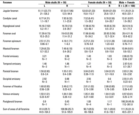
The results of the measurements of the surface area of the foramina are displayed in Table 1. The data are divided according to gender and side of the body. For many of the foramina p values of 0.01 and 0.001 were obtained. There were four statisti-cally significant differences between the results ob-tained for pairs of foramina (left versus right) or for gender (male versus female).
1. The surface area of the right and left jugular fo-ramina summarised for all skulls (male and fe-male) and for the male skulls only (there was no statistically significant difference in the female skulls): the right foramen was statistically sig-nificantly larger then the left for the material as a whole and for the male skulls.
2. The surface area of the left mastoid foramen by gender: the female skulls had a smaller surface area of the left mastoid foramen.
3. The total surface area of all right-sided and left--sided foramina for combined genders (male and female) and for male skulls (there was no statis-tically significant difference in the female skulls). 4. The total combined surface area of all foramina excluding the jugular foramen, both right-sided and left-sided for combined genders (male and female) and for male skulls (there was no statis-tically significant difference in the female skulls): right side mean value: 42.5 ± 14.9 mm2; left side
mean value 36.4 ± 16.9 mm2
.
The average of the total combined surface area (the sum of the right and left sides) of all venous foramina for the human skull was 190.3 ± 46.1 mm2. There was
the coefficients of asymmetry (Table 2). The surface area of the larger foramen was divided by the surface area of the smaller foramen, and results close to 1.0 (in the range between 0.91 and 1.09, as a “manual” error of about 10% was calculated) were eliminated to better represent their actual coefficients of asym-metry. The abbreviation J/j is the ratio of the larger jugular foramen to the smaller one, while the abbre-viation A/a represents the ratio of the total surface area of all the foramina on the dominant side to the total surface area of the smaller foramina. There were no statistically significant differences in the coefficients of asymmetry calculated.
Next, the capacity (V) of the skulls was calculated. Female skulls had an average of 1279.17 ± 126.15 cm3
with a range of between 1100 and 1600 cm3, and male
skulls had an average of 1421.67 ± 143.28 cm3
with
Table 1. Area of venous foramina of the human skull with reference to gender and body side, all measurements given in mm2.
Mean values with standard deviation (in parentheses) and the range given below are set out in the appropriate columns
Foramen Male skulls (N = 50) Female skulls (N = 50) Male + Female
L R L R R + L (N = 100)
Jugular foramen 51.11 (23.77) 57.53 (17.86) 53.83 (21.33) 59.84 (21.03) 111.35 (31.3) 21–126.2 12.5–94.9 15.8–112.7 25.6–121.7 40.5–221.1
Condylar canal 6.77 (4.31) 7.92 (6.32) 7.52 (6.41) 8.78 (5.56) 12.61 (8.97) 1.1–19.7 1.1–23.8 1.5–28.2 0.4–20.7 1.9–39.2
Hypoglossal canal 19.16 (7.92) 18.70 (7.0) 17.96 (8.75) 18.65 (6.70) 36.9 (14.5) 6.2–41.1 3.9–39.7 6.5–46.1 8.5–37.3 9.1–77.3
Foramen oval 17.39 (4.73) 19.43 (5.59) 17.66 (6.46) 20.92 (5.56) 26.4 (11.9) 10.3–25.3 11.4–31.2 10–34.2 12.7–32.4 10.9–62.2
Foramen spinosum 2.51 (1.21) 4.18 (1.71) 2.27 (1.23) 3.12 (1.28) 3.65 (2.62) 0.86–6.1 1–6.3 0.76–5.8 1.23–6.0 0.76–11.1
Mastoid foramen 7.29 (8.23) 7.48 (6.10) 4.10 (3.30) 6.19 (3.95) 10.94 (9.81) 0.7–32.2 0.4–28.2 0.6–12.4 0.6–15.6 0.6–53.2
Frontal emissary 3.21 2.06 1.40 1.83 2.06 (0.73)
N=1 N=2 N=3 N=3 0.56–2.67
Temporal emissary 1.46 1.86 1.27 1.49 2.97 (5.4)
N=3 N=1 N=4 N=3 0.7–28.7
Parietal foramen 1.50 (0.92) 1.55 (1.01) 3.04 (3.67) 3.04 (5.57) 2.03 (1.24) 0.8–3.4 0.4–3.99 0.38–11.9 0.7–16.8 0.6–2.92
Occipital emissary 2.92 0.98 2.56 0.6 2.55 (1.47)
N=3 N=1 N=1 N=1 0.23–6.5
Foramen of Vesalius 1.60 (0.93) 2.49 (2.17) 2.81 (1.02) 2.86 (0.80) 2.17 (1.63) 0.56–3.26 0.23–6.5 2.15–3.98 1.78–3.65 0.26–6.47
Venous emissary 1.93 (1.61) 1.84 (1.04) 1.85 (1.30) 1.84 (1.62) 0.97 (0.61) 0.23–6.47 0.26–4.3 0.27–4.68 0.3–6.01 0.25–2.14
Postglenoid foramen 0.9 0.43 1.08 1.17 190.28 (45.9)
N=1 N=1 N=4 N=1 112–361.8
Sum of areas of all foramina 81.8 (29.7) 100.96 (25.7) 90.7 (29.5) 101.1 (28.8) 111.35 (31.3) 44.9–164.3 51.6–185.9 40–166.6 41.6–192.1 40.5–221.1
Figure 1. Part of the individual foramina in their total
cross-sec-tion area; J — jugular foramen, H — hypoglossal canal, Ov — foramen ovale, Cond — condylar canal, M — mastoid foramen, Sp — spinous foramen, Al — other venous foramina of the hu-man skull.
0% 60%
50%
40%
30%
20% 59.58%
19.14%
10.32%
4.86% 3.18%
1.48% 1.43% 10%
a range between 1060 cm3 and 1720 cm3. There was
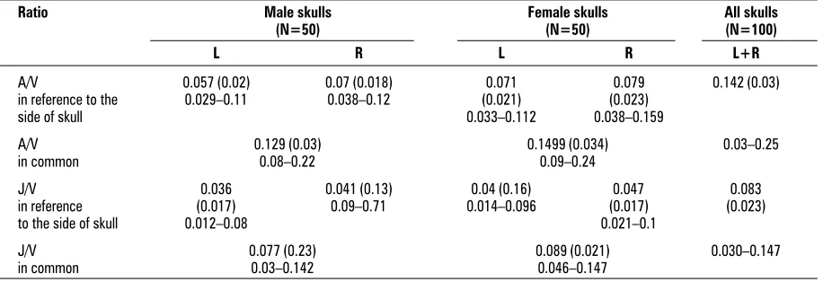
a statistically significant difference for gender. An index number (ratio) of the total surface area of the venous foramina in relation to the capacity of the skull was calculated, the index representing the ra-tio of surface area. A separate index was calculated for gender and body sides (Table 3). The abbrevia-tion A/V is the ratio of the surface area of the emis-sary foramina to the skull capacity while J/V is the ratio of the surface area of the jugular foramen to the skull capacity. In female skulls the surface area of the foramina examined was relatively large com-pared with the male skulls. There were statistically significant differences for gender and sides of the
body. The index (ratio) of the surface area of right--sided foramina related to the skull capacity was greater than that for the left-sided foramina. These results are indicative of a relative dominance of the right-sided foramina over the left.
The correlations between the sizes of individual venous foramina were also calculated. Of all the cor-relations studied only six were statistically significant differences:
1. The total surface areas of the jugular foramina (the sum of the right and left jugular foramina) in relation to the capacity of the skull.
2. The total surface areas of the hypoglossal fora-mina in relation to the capacity of the skull.
Table 2. Reciprocal proportions of the areas of individual venous foramina of the human skull. J/j — the ratio of the area
of the greater jugular foramen to the smaller one. A/a — the ratio of the total surface area of all the foramina on the domi-nant side to the total surface area of the smaller foramina. Mean values with standard deviation (in parentheses) and the range given below are set out in the appropriate columns
Foramen Ratio or number of skulls Male skulls (N = 50) Female skulls (N = 50) All skulls (N = 100)
Jugular J/j 1.62 (0.49) 1.63 (0.61) 1.62 (0.55)
1.0–3.35 1.0–3.47 3.47
L > R 11 16 27
L < R 0 23 53
L = R 9 9 18
Sum of all A/a 1.42 (0.31) 1.40 (0.42) 1.41 (0.37)
foramina 1.0–2.22 1.0–2.77 1.0–2.77
L > R 16 13 29
L < R 26 25 51
L = R 10 10 20
Table 3. The relationship between the surface area of the foramina and the skull capacity. An index represents the ratio of
the surface area of foramen (foramina) to skull capacity for the sum of all foramina (A/V) and for the jugular foramina ex-clusively (J/V) with reference to gender and side of the skull. Mean values with standard deviation (in parentheses) and the range given below are set out in the appropriate columns
Ratio Male skulls Female skulls All skulls
(N=50) (N=50) (N=100)
L R L R L+R
A/V 0.057 (0.02) 0.07 (0.018) 0.071 0.079 0.142 (0.03)
in reference to the 0.029–0.11 0.038–0.12 (0.021) (0.023)
side of skull 0.033–0.112 0.038–0.159
A/V 0.129 (0.03) 0.1499 (0.034) 0.03–0.25
in common 0.08–0.22 0.09–0.24
J/V 0.036 0.041 (0.13) 0.04 (0.16) 0.047 0.083
in reference (0.017) 0.09–0.71 0.014–0.096 (0.017) (0.023)
to the side of skull 0.012–0.08 0.021–0.1
J/V 0.077 (0.23) 0.089 (0.021) 0.030–0.147
3. The ratio of asymmetry J/j and A/a in relation to the capacity of the skull. This correlation was statistically significant for both right-side inance and left-side dominance (right-side dom-inance was negative and left-side domdom-inance was positive).
4. The total surface area of the jugular foramina (the sum of the right and left jugular foramina) com-pared to the total surface area of the hypoglos-sal canals (the sum of the left and right hypoglo-ssal canals).
5. There was a correlation between the total surface area of the left side and the right side for the hy-poglossal and condylar canals, the foramen ovale and the mastoid and parietal foramina.
6. There was a correlation between the surface area of the hypoglossal canal and the surface areas of the condylar canals, foramen ovale and the mas-toid, parietal and venous foramina. The correla-tion was statistically significant in relacorrela-tion to the surface area of the individual foramina as well as the sum of the surface area of all foramina, both right and left, on the same and opposite sides. By means of step regression for determining the factors with which the skull capacity might corre-late, it was demonstrated that the skull capacity cor-related most closely with gender. The skull capacity also correlated with the individual total surface ar-eas of the jugular foramina, the hypoglossal canals, and the right mastoid foramen, but these correla-tions were weaker.
DISCUSSION
It is well known that morphological differences exist between the skulls of different human popula-tions. Much research interest has been focused on anatomical studies of the asymmetry of the skull foramina, especially the jugular foramen (Table 4). No data has previously been made available on com-prehensive anatomical studies of the venous emis-saries in Polish human skulls. The data from the work of Schelling [18] with 210 examined skulls, Hatibo-glu and Antil [14] with 300 skulls and Sturrock [19] with 156 skulls provide important information about the size of skull foramina. The results of these au-thors are the closest to our data, utilising traditional methods of measurement to analyse macerated skulls. The results of Adams et al. [1], especially after averaging the results for right and left handed indi-viduals, are based on computer tomography scan analysis of skulls. The author, however, did not per-form specific measurements of the foramina but
rather made a subjective estimation of the size of the right and left foramina. The results of Idowu [15] are also close to ours, although these are based upon only 20 skulls.
The size of each individual emissary vein foramen as identified in our study, its morphometry, surface area and relation to skull capacity as well as its hae-modynamic role in the venous outflow from the human skull are now discussed and compared with the findings of other investigators.
The jugular foramina were identified as the main venous outflow in all the Polish skulls. In 54% of the skulls there was right-sided size dominance of the jugular foramen compared to 27% of the skulls with a left-sided dominance. In the remaining 19% of skulls there was symmetry with no size dominance. The right-sided dominance was statistically signifi-cant for all skulls and for the male skulls separately. These findings indicate that there is a clear right-sided dominance in the size of the jugular foramen at least in male Polish skulls. Perhaps the size-domi-nance was not seen in female Polish skulls because of the limited number (48) of skulls examined. There was no significant statistical difference in jugular foramina size for gender alone, as has been report-ed by other investigators [1, 14, 18, 19].
Only the results of Idowu [15] are straightforwardly comparable to our investigations. According to this author, the mean area of the jugular foramen on the right was 437.49 mm2 and that on the left 419.48 mm2.
These values are much greater than ours but are some-what dubious, as they do not match the linear mea-surements also given by this author [15].
Because of differences in methodology it is diffi-cult to make an exact comparison between the surface
Table 4. Comparison of data regarding asymmetry of
areas of jugular foramina obtained by various authors, including data from the present study
Author, year L > P L < P L = P
Rhoton, 1975 20% 68% 12%
Schelling, 1978 32.5% 65% 2.5%
Sturrock, 1988 23% 69% 18%
Hatiboglu 1992 26% 61.6% 12.4%
Adams, Right-handed 22% 65% 12% 1997 Left-handed 35% 52% 12%
Idowu, 2004 25% 55% 25%
areas of the jugular foramen in the Polish skulls with the reports of other investigators. Some of the pre-vious studies were based on plain X-rays or CT scan measurements and others on traditional measure-ments in macerated skulls. Our methodology utilised the MULTISCAN digital analysis to determine the exact surface area of each of the venous foramina. In order to convert other investigators’ foramina measurements to a surface area, the foramen mea-surement of length (a) and width (b) were used in the ellipse formula (A = 1/4 p a × b). In data report-ed with only the length (a) of a foramen, the width (b) was calculated as one-half the length.
According to Schelling’s data [18], the jugular foramen on the right side measured 7.2 mm at its greatest diameter and on the left side 6.3 mm. The surface area calculated using the ellipse formula (the smaller diameter as half the greater) for the right jugular foramen is 20.34 mm2 and for the left is 15.56
mm2
. The calculated values are much smaller then our measurements. However, according to the re-sults of Di Chiro et al. [8], the jugular foramen was measured as 15 mm in length and 10 mm in width, and the compartment of the pars nervosa had a max-imum width of 5 mm. From Di Chiro’s data the calcu-lated jugular foramen surface area was 117.75 mm2
, which is close to our results.
The calculated coefficient of asymmetry for the jugular foramen was 1.62 in the Polish skulls, similar to the 1.6 value calculated by Schelling [18]. These values also correlated with the results from our pre-vious investigation based on cadaver analysis of the jugular foramen [23]. The calculated coefficient of asymmetry of the jugular foramina was not statisti-cally significant for gender.
An extreme one-sided domination exceeding 200% was found in 8% (8/100, 4 male and 4 fe-male). One of the male skulls had an extreme asym-metry exceeding 300%, with a calculated coefficient of 3.47. Woodall [21] used the term “large dispro-portion” for extreme asymmetry of 300% of a jugu-lar foramen. Some of these cases may be document-ed in radiography [9]. Perhaps the occurrence of ex-treme asymmetry of the jugular foramina should be considered an anatomical anomaly or congenital defect associated with excessive development of the jugular bulb and internal jugular vein. Extreme asym-metry of the jugular foramen has been reported as the cause of both pulsatile tinnitus and venous hum from excessive venous turbulence [12–14, 19]. It may also be an important obstacle in ligation of one of the jugular veins.
To date only Schelling [18] has analysed the cor-relation between the size of the jugular foramen and the size of the brain. He concluded that, according to data from anthropometric tables, the 8% differ-ence in size between the male and female jugular foramina was directly correlated with a gender-re-lated size difference of the brains.
In the Polish skulls the index ratio of the jugular foramen surface area to skull capacity (J/V) was sta-tistically significant both for gender and side. In the case of left-sided domination the index was posi-tive, while in right-sided domination the index was negative. In left-side dominant skulls there was a direct relationship between larger asymmetry and a larger skull capacity. In right-side domination the degree of asymmetry was less meaningful for the venous outflow.
The different anatomy of the jugular and bra-chiocephalic veins may explain these findings. On the right the distance travelled by the venous blood through the jugular vein to the superior vena cava and the heart is shorter than that on the left. The anatomically longer left venous outflow is via the jugular vein to the brachiocephalic vein and then to the superior vena cava and the heart. The brachio-cephalic vein is partially compressed by the surround-ing structures of the sternum and aorta [20]. Ac-cording to Hagen-Poiseuille’s Law, the size of a ves-sel lumen is inverves-sely proportional to the resistance of the flow. In left-sided jugular domination the more prominent the foramen size, the larger the jugular and brachiocephalic veins. These larger vessels have improved venous blood flow because of a decreased lumen resistance.
In the Polish skulls, the average surface area of the hypoglossal canal was 18.45 mm2
. The exter-nal foramen of the hypoglossal caexter-nal reported by Braun and Tournade [6] was approximately 20 mm2
. Berlis reported linear measurements (length 9 mm, width 5 mm) for the hypoglossal canal, with an ellipse-calculated surface area of 35.33 mm2 [4].
canal had a small surface area of 6.6 mm2 and no
significant role in the venous outflow.
The average surface area of the condylar canal in our material was 6.3 mm2
. Other investigators have reported the surface area of the condylar canal on the right side as 6.9–9 mm2
and on the left side as 4.3–5.2 mm2 [5, 18]. Schelling [18] described
a condylar emissary compensation phenomenon to the jugular foramen. He found that larger jugular foramina were associated with larger condylar fo-ramina. We found no statistical correlation between the size of the jugular and condylar foramina.
Because the mastoid emissaries are frequently multiple, the task of analysing the total surface area was a difficult one. In the Polish skulls, the total sur-face area ranged from 4.1 mm2
to 7.48 mm2
, depend-ing on the side and gender. The mastoid foramen was larger on the right side, especially in women. The right-sided domination of the mastoid foramen has been reported previously by other investigators [5]. Bauer [2] found mastoid foramen asymmetry only in women. The right-sided mastoid domination corre-lates with our data and was statistically significant in the female skulls. Schelling [18] reported that the av-erage surface area of a single mastoid foramen on the right-side was 3.35 mm2
and on the left-side 3.33 mm2
. Abnormally large mastoid foramina are rare [11].
We found no evidence for compensation between the total combined areas of the mastoid foramen and the condylar canal, and the area of the jugular foramen as suggested in previous studies [3]. On the contrary, the results in the Polish skulls showed a statistically significant positive correlation in the surface areas of these foramina.
The foramen ovale was identified as a single struc-ture in the majority of the skulls studied. It was charac-terised by significant morphological variability, with uneven edges of the foramen in many skulls. It is doubt-ful whether these findings were due to post-mortem damage. Berlis et al. [4] reported the foramen ovale divided by an osseous bridge in 12.5% and as a dou-ble structure in 0.8%. We identified similar variations in the morphology of the foramen ovale in the Polish skulls. No data was available in the literature with re-gard to the surface area of the foramen ovale. Howev-er, multiple investigators have reported the linear mea-surements of the foramen ovale with an average length of 6.7 mm (ranging between 4.5 and 11.3 mm) and an average width of 3.9 mm (ranging between 2.4 and 6.7 mm) [4, 7, 18]. By means of these linear measure-ments the surface area was calculated with the ellipse formula. The results obtained ranged from 8.48 to
59.4 mm2, with an average of 20.51 mm2, and were
close to those in the skulls studied.
The postglenoid foramina identified in the skulls studied were rather small, with a surface area of between 0.9 mm2 and 1.17 mm2. Other
investiga-tors have reported larger sizes of the postglenoid foramen in the range of 0.5 mm to 5 mm [5, 18]. Using the linear measurements of the postglenoid foramina, the calculated surface area (ellipse formu-la) from the Boyd and Schelling studies was from 0.098 mm2 to 9.81 mm2. These surface areas are
larg-er than in the Polish skulls but, of course, extremely large foramina are very rare.
The diameter of the parietal foramen according to the reports of other authors measures between 1 and 2 mm and in extreme cases can even reach 7 mm [3, 5, 18]. In general, the smaller sized parietal foramen has only a small haemodynamic role [18]. However, in on female Polish skull the parietal fora-men measured 11.9 mm2
on the left side, and 16.8 mm2
on the right. The capacity of this skull was 1520 cm3
and it had very small jugular foramina (36.4 mm2
on the left and 25.2 mm2 on the right side). In this skull
the small jugular foramina was compensated for by a larger parietal foramina. In the Polish skulls overall, although reciprocal compensation in terms of diam-eter was observed between the jugular and parietal foramina, it was not statistically significant.
The surface area of the foramen of Vesalius in the skulls studied measured between 1.6 and 2.86 mm2, depending on the gender and body side,
without statistically significant difference. Accord-ing to others, the foramen of Vesalius measured between 0.5 and 2.5 mm in diameter [4, 5, 18]. The surface area calculated from these data (as the area of a circle) ranged from 0.2 mm2
to 4.9 mm2
and correlates with the results in the Polish skulls.
The smaller values of the surface area of the venous foramen (1.84–1.93 mm2 depending on the
gender and side) classified this foramen among the smaller emissaries of the skull. In the Polish skulls the significant combined size of the venous foramen and foramen of Vesalius may supplement the venous outflow of the cavernous sinus.
CONCLUSIONS
1. There is asymmetry among the venous emis-sary foramina of the human skull.
2. The jugular foramen is the most important venous foramen of the skull and the hypoglossal canal the second most important. Only these two foramina correlate significantly with skull capacity. For the jugular foramen it was 0.76, and for the hy-poglossal canal 0.62.
3. The asymmetry in the size of the jugular fo-ramina is related to the capacity of the skull and is statistically significant. The asymmetry in the size of the jugular foramina ranges from 1 to 3.47.
4. Skulls with an extreme one-sided jugular dom-ination exceeding 200% are not rare anatomical vari-ants. In these cases ligation of the internal jugular vein may be contraindicated, particularly in cases with an extreme left jugular domination. In these cases compensation of cut-down venous outflow from the skull is difficult to achieve
5. There was no evidence for reciprocal compen-sation between the mastoid foramen or condylar canal, and the jugular foramen.
REFERENCES
1. Adams WM, Jones RL, Chavda SV, Pahor AL (1997) CT assessment of jugular foramen dominance and its as-sociation with hand preference. J Laryngol Otol, 111: 290–292.
2. Bauer U (1971) Anatomische Varianten des Sinus sig-moideus, des Foramen jugulare und der Vena jugu-laris. Z Anat Entwickl Gesch, 135: 35–42.
3. Bekov DB (1965) Atlas venoznoi sistemy golovnogo mozga tscheloveka. Medicina, Moskwa.
4. Berlis A, Putz R, Schumacher M (1992) Direct CT mea-surements of canals and foramina of the skull base. Br J Radiol, 65: 653–66.
5. Boyd GI (1930) The emissary foramina in the cranium of man and anthropoids. J Anat 65: 108–121. 6. Braun JP, Tournade A (1977) Venous drainage in the
craniocervical region. Neuroradiology, 13: 155–158. 7. Calcaterra TC, Cherney EF, Hanafee WF (1973) Normal
variations in size and neoplastic changes of skull fo-ramina. Laryngoscope, 83: 1385–1397.
8. Di Chiro G, Fisher RL, Nelson KB (1964) The jugular foramen. J Neurosurg, 21: 447–460.
9. Durgun B, Illgit ET, Atasever A (1993) Evaluation by an-giography of the lateral dominance of the drainage of the dural venous sinuses. Surg Radiol Anat, 15: 125–130. 10. Falk D (1986) Evolution of cranial blood drainage in Hominids: enlarged occipital\marginal sinuses and emissary foramina. Am J Phys Anthropol, 70: 311–324. 11. Forte V, Turner A, Liu P (1989) Objective tinnitus asso-ciated with abnormal mastoid emissary vein. J Otol, 18: 232–235.
12. Golueke PJ, Panetta T, Sclafani S, Varughese G (1987) Tinnitus originating from abnormal jugular bulb: treat-ment by jugular vein ligation. J Vasc Surg, 6: 248–251. 13. Gugliantini P, De Sanctis R, Di Capua M (1993) Fisio-logica variabilita delle dimensioni dei foramini giugu-lari. Radiol Med, 86: 904–907.
14. Hatiboglu MT, Antil A (1992) Structural variations in the jugular foramen of the human skull. J Anat, 180: 191–196.
15. Idowu OE (2004) The jugular foramen — a morpho-metric study. Folia Morphol, 63: 419–422.
16. Lang J, Hornung G (1993) The hypoglossal channel and its contents in the posterolateral access to the petro-clival area. Neurochorurgia, 36: 75–80.
17. Rhoton AL, Buza R (1975) Microsurgical anatomy of the jugular foramen. J Neurosurg, 42: 541–550. 18. Schelling F (1978) Die Emissarien des menschlischen
Schädels. Anat Anz, 143: 340–382.
19. Sturrock RR (1988) Variations in the structure of the jugular foramen of the human skull. J Anat, 160: 227–230.
20. Tanaka T, Uemura K, Takahashi M, Takehara S, Fukaya T, Tokuyama T, Satoh A, Ryu H (1993) Compression of the left brachiocephalic vein: cause of high signal in-tensity of the left sigmoid sinus and internal jugular vein on MR images. Radiology, 188: 355–361. 21. Woodhall B (1936) Variations of the cranial venous
sinuses in the region of the torcular Herophili. Arch. Surg, 33: 297–310.
22. Wierciński A (1967) Analiza porównawcza struktury rasowej wczesnośredniowiecznej ludności z cmen-tarzyska w Złotej Pińczowskiej. Rozprawa zespołu badań nad polskim średniowieczem Uniwersytetu Warszawskiego i Politechniki Warszawskiej. Vol. 5. PWN, Warszawa 1967: pp. 141–145.
23. Wysocki J, Kobryń H, Kobryńczuk F (1998) Proportion analysis of hypoglossal canal elements in human, maca-cus and dog. Med Sci Monit, 4 (Suppl 2): 52–54. 24. Wysocki J, Chmielik LP, Gacek W (1999) Variability
of diameter of the human jugular foramen in rela-tion to condirela-tions of venous outflow after ligarela-tion of the internal jugular vein. Otolaryngol Pol, 53: 173–177.
25. Wysocki J, Kobryń H, Bubrowski M, Kwiatkowski J, Reymond J, Skarżyńska B (2004) The morphology of the hyplossal canal and its size in relation to skull ca-pacity in man and other mammal species. Folia Mor-phol, 63: 11–17.
26. Wysocki J, Sharifi M (2005) Occurrence, variations and diameter of the human condylar canal in relation to the jugular foramen. Folia Morphol, 65: 11–14. 27. Reymond J, Charuta A, Wysocki J (2005) The
morphol-ogy and morphometry of the foramina of the greater wing of the human sphenoid bone. Folia Morphol, 64: 188–193.