Safety and efficacy of Ivabradine in patients with
acute ST-segment elevation myocardial infarction
(STEMI)
Mohamed Salem
1, Mohamed El Sayed
2, Mohamed Abdel Kader
1, Ahmed Abdel Moniem
11*Department of Cardiology, Benha Faculty of Medicine, Benha University, Benha, Egypt. 2National Heart Institute, Egypt
ST segment elevation myocardial infarction (STEMI) is commonly induced by thrombus formation leading to complete occlusion of a major epicardial coronary vessel. We aimed to explore safety and efficacy of Ivabradine in patients with STEMI associated with left ventricular dysfunction. 200 consecutive patients with STEMI were included in this controlled study. All patients had successful reperfusion and LVEF less than 50%. 100 patients received 5 mg ivabradine twice a day in addition to the conventional treatment, while 100 patients received the conventional treatment only. Composite end point of death, re-infarction, overt heart failure, or need for revascularization was reported at 30 days. Ivabradine when added to the conventional treatment reduced the heart rate significantly compared to the conventional treatment alone. However it did not affect incidence of primary end point. Ivabradine didn't show a significant impact on major adverse cardiac events when added to conventional treatment.
Key-words: Ivabradine, ACS, STEMI, primary PCI, Fibrinolysis
INTRODUCTION
ST segment elevation myocardial infarction (STEMI) most commonly occurs when thrombus formation results in complete occlusion of a major epicardial coronary vessel. STEMI is a life-threatening, time-sensitive emergency that must be diagnosed and treated promptly. Despite remarkable therapeutic advances in the management of acute myocardial infarction (AMI) over the last decades, many patients are still at increased risk of adverse cardiac events (Thygesen et al, 2007). Elevated heart rate (HR) in STEMI is an important pathophysiological variable that increases myocardial oxygen demand, and also limits tissue perfusion by reducing the duration of diastole during which most myocardial perfusion occurs, elevated resting HR represents a significant predictor of all-cause and cardiovascular mortality in the general population and patients with cardiovascular disease (CVD) because it aids progression of atherosclerosis, plaque destabilization, and initiation of arrhythmias. Since β-blockers reduce HR, it is currently viewed as the first line therapy for chronic stable angina pectoris and is associated with an improved prognosis after AMI
and congestive heart failure (CHF). (Lucats et al., 2007). Ivabradine (IVA), a pure HR lowering drug, reduces the myocardial oxygen demand at exercise, contributes to the restoration of oxygen balance and therefore benefit in chronic CVD. No relevant negative effects are evidenced on cardiac conduction, contractility, relaxation, repolarization or blood pressure (BP). Beneficial effects of IVA have been demonstrated in chronic stable angina pectoris (CSAP) and CHF, with optimal tolerability profile due to selective interaction with If channel of sinoatrial node cells. More recently, the indication of IVA has been extended for use in association with β-blockers in patients with CAD (Tendera et al., 2011).
*Corresponding author: Mohamed Salem, Department of Cardiology, Benha Faculty of Medicine, Benha University, Benha, Egypt. Tel.: 0020133106725, Mobile: 01092773227, Email. masalem@yahoo.com Vol. 2(1), pp. 007-011, December, 2015. © www.premierpublishers.org. ISSN: XXXX-XXXX
METHODS Study design
This prospective, controlled, non randomized study enrolled 200 consecutive patients with acute anterior STEMI. The study was done at the National Heart Institute, Cairo, Egypt in the period from June 2013 to August 2014.We aimed to explore safety and efficacy of Ivabradine in this category of patients. Key Inclusion criteria were: Patients with first time anterior STEMI successfully reperfused either by Primary PCI (PPCI) or fibrinolytic therapy, left ventricular ejection fraction less than 50%, Killip class I-II, sinus rhythm and heart rate more than 70/min. While exclusion criteria were: Prior myocardial infarction, Killip class III-IV, patients with EF more than 50 %, patients with HR less than 70/min without any medication, patients with contraindications to Beta blockers.
Baseline and follow up evaluation
A. Review of medical history including demographic data (age, sex), risk factors of CAD (diabetes, hypertension, dyslipedemia, smoking), prior medical history (PCI, CABG, heart failure), associated co morbidities (renal, hepatic, cerebral dysfunction), assessment of chest pain on admission focusing on typicality of pain, time from symptom onset, presence of any contraindications to fibrinolysis or beta blockers. B. Clinical examination (general, cardiac) with special attention to the presence of S3 or basal crepetations. C. ECG: A standard 12-lead ECG was recorded before and at 90 minutes after the start of the streptokinase or immediately after PPCI, then every 12 hours or on the recurrence of chest pain and on discharge by 4th or 5th day. All patients had follow up resting ECG after 10 days and after 30 days form discharge for evaluation of heart rate in order to modify the dose of beta blockers and/or Ivabradine. D. Transthoracic Echocardiography: was performed in all patients after initial stabilization on admission. Two-dimensional (2D) echocardiography was done using the parasternal long-axis view, multiple short-axis views, and apical two, four and long-axis views. Left ventricular end-systolic volume (LVESV), LV end-diastolic volume (LVEDV), and LV ejection fraction (LVEF) were determined from apical two- and four chamber views, according to the suggestions of the American Society of Echocardiography. Ejection fraction was estimated by modified Simpson method. Tracing of endocardial borders in diastole and end-systole was made in the technically best cardiac cycle, also evaluation of the presence of mechanical complication as mitral regurgitation.
Reperfusion Strategies
1. Fibrinolytic therapy: Streptokinase 1.500.000 IU was given over 60 minutes
2. Primary PCI:
Patients received aspirin (300 mg loading then 150 mg daily), clopidogrel (600 mg loading then 150 mg /day maintenance dose). Un-fractionated heparin (UFH) (70
u/ kg) bolus dose was injected after sheath insertion. The procedure was performed according to standered technique of PCI. Femoral approach was the standered in all patients by using 6-7 Fr sheath., XB or JL guiding catheters were used during the procedure. Aspiration devices and glycoprotein inhibitors were used in lesions with heavy thrombus burden and or impaired TIMI flow after PCI. BMS were used in all patients. The operator determined the size and length of the stent, the sheath was removed 6 hours later from the end of PCI and compression was done manually.
Study Protocol
Patients with successful reperfusion were classified into 2 groups: Group A: (100 patients) Ivabradine 5mg twice/day was started 12 hours after reperfusion therapy and increased on day 10 to 7.5 mg/12 hours in patients with resting heart rate >70/min in addition to the conventional treatment of STEMI including beta blockers (Bisoprolol 5mg/day). Group B: (100 patients) conventional treatment including Bisoprolol 5 mg once daily that may be increased up to 7.5 mg once daily at day 10 for patients with heart rate > 70 beats per minute. Patients were evaluated at entry, day 10, and day 30.
Patients aged 75 years or more, a lower starting dose was considered for these patients before up-titration if necessary. If, during treatment, heart rate decreased below 50 beats per minute (bpm) at rest or the patient experienced symptoms related to bradycardia such as dizziness, fatigue or hypotension, the dose was titrated downward .
Follow up
A. 10 days from the date of admission with resting ECG and evaluation of the heart rate.
B. 30 days from the date of admission with resting ECG and transthoracic echocardiography.
Study end point
30 days mortality, re-infarction, overt heart failure or need for revascularization
Statistical analysis
Data are presented as mean+ SD for continuous data and as number (%) for categorical data. Between groups comparison was done using student t-test for continuous data and by Chi-square test (or Fischer exact test) for qualitative data. Level of evidence was detected to be significant at P value <0.05. Data were collected and analyzed by SPSS (version 17).
RESULTS
Study population
Table 1. Baseline characteristics of study population.
Group A No = 100
Group B No = 100
P value
Age, years, mean ± SD 58.7 ± 10.3 59.2 ± 11.1 0.8
Male Sex (m %) 46 (46%) 47 (47%) 0.9
Family history of CAD 22 (22%) 26 (26%) 0.5
DM 53 (53%) 65 (65%) 0.1
Hypertension 57 (70%) 60(60%) 0.7
Smoking 43 (43%) 42 (42%) 0.9
Dyslipidemia 37 (37%) 40 (40%) 0.7
CAD = coronary artery disease, DM = Diabetes Mellitus
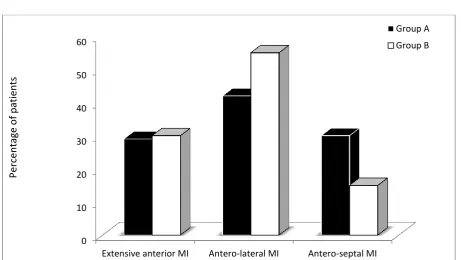
Figure 1. Type of myocardial infarction detected by ECG.
males were 46%, 47% in group A and B respectively, P=0.9), Diabetes was reported in 53% , 65% in group A and B respectively P=0.1, Hypertension was evident in 57 %, 60% in group A and B respectively P=0.7, The percentage of dyslipidemia were 37% , 40% in group A and B respectively P=0.7. Smokers were 43% , 42% in group A and B respectively P=0.9, Positive family history of CAD were 22%, 26% in group A and B respectively P=0.5. We did not report statistical significant differences in baseline characteristics between study groups. Table 1.
Time from onset of symptoms to admission
The mean time was 5+2.9 , 5.3+2.9 hours in group A and B respectively, P = 0.4.
ECG on admission
Extensive anterior STEMI was reported in 28%, 30% in group A and B respectively, P=0.5, antero-lateral STEMI was 42%, 55% in group A and B respectively, P=0.3, The percentage of patients presented with antero-septal STEMI was 30%, 15 % in group A and B respectively, P=0.8). (Figure 1)
Reperfusion Therapy
Fibrinolytic therapy was applied to 54%, 53% of group A and B respectively, P=0.9, while PPCI was performed in 46%, 47% of group A and B respectively, p=0.8. The mean door to needle time was 34+16.5, 30+14.7 minutes in group A and B respectively, P=0.4. The 0
10 20 30 40 50 60
Extensive anterior MI Antero-lateral MI Antero-septal MI
Group A
Group B
P
erce
ntag
e
o
f
p
Figure 2. HR at baseline and follow up
mean door to balloon time was 80.2+38.5 minutes was 87+44.5 , 73.5+31 minutes in group A and B respectively, P =0.2.
Heart rate (HR)
The mean HR on admission was 103.7+10.1, 102.2+8.8 bpm in group A and B respectively, P=0.3. The mean HR after reperfusion was 96.5+8.7, 96.1+7.7 bpm in group A and B respectively, P=0.7. The mean HR after 12 hours was 89.8+7.3, 89+7.4 bpm in group A and B respectively, P=0.4. The mean HR on discharge was 77.6+7, 80.8+7.3 bpm in group A and B respectively, P=0.028*. The mean HR on day 10 was 72.1+8.2, 77.8+9.4) bpm in group A and B respectively, P=0.019*. The mean HR on day 30 was 76.8+20.2 bpm (73.7+18.6, 79.8+21.8 bpm in group A and B respectively, P=0.037. The mean percentage of HR reduction from admission to day 10 was 30.5% (31.6 bpm) and 23.8% (24.4 bpm) for group A and B respectively, and Day 30 by 28.9% (30 bpm) and 21.9% (22.4 bpm) for group A and B respectively, whereas The mean percentage of HR reduction from onset of starting Ivabradine at 12 hours after reperfusion to Day 10 was 19.7% (17.7 bpm) and 12.6% (11.2 bpm) for group A and B respectively, and Day 30 by 17.9% (16.1 bpm) and 10.4% (9.2 bpm) for group A and B respectively (Figure 2).
In-hospital outcome
In-hospital mortality was 1 %, 2% in group A and B respectively, P=0.7, re-infarction rate was 9%, 7% in group A and B respectively, P=0.5, new ischemia rate was 33 %, 37 % in group A and B respectively, P=0.6, overt heart failure rate was 17%, 19% in group A and B respectively, P=0.7.
Echocardiographic data
The mean baseline LVEDV was 118.2+25.5, 119.9+26.7 ml in group A and B respectively, P=0.6, the mean baseline LVESV was.2+18, 66.1+13.5 ml in group A and B respectively, P=0.9, the mean baseline LVEF was 44.4+3.2 , 44.5+3.1 % in group A and B respectively, P=0.8. The mean 30 days LVEDV was 119.1+26.1, 118.2+27.9 ml in group A and B respectively, P=0.8, the mean 30 days LVESV was 59.4+15, 61.1+17 ml in group A and B respectively, P=0.5, the mean 30 days LVEF was 49.4+10.3 , 47.3+11.3 % in group A and B respectively, P=0.2). Within groups analysis did not reveal any significant changes from baseline to 30 days. Regional wall motion abnormalities (RWMA) was found in 96% of patients of Group A and 93% of Group B on baseline echocardiographic examination (P=0.7), and was found to be 76% In Group A and 77% of group B (P= 0.2).
30 days outcome
At 30 days, The percentage of patients with primary end point was 28%, 34% in group A and B respectively, P=0.3, 30 days mortality was 4% , 5% in group A and B respectively, P=0.7, re-infarction was reported in11%, 12% in group A and B respectively, P=0.8, Overt heart failure was 26%, 33% in group A and B respectively, P=0.3, Revascularization was done in 10%, 12% in group A and B respectively, P=0.7
Drugs dosage, tolerance and side effects
The average dose of Ivabradine was 5.25 mg b.i.d , 78 % of group A received 5mg b.i.d, 16 % received 7.5 mg b.i.d and 6% received 2.5 mg b.i.d. 92% of patients tolerated Ivabradine, 7% showed significant 0
20 40 60 80 100 120
Heart rate on admssion
Heart rate after reperfusion
Heart rate after 12
hours
Heart rate on discharge
HR Day 10 Heart rate Day 30
Group A
Group B
Me
bradycardia ( 6% improved by reducing dose and 1 % excluded from the study), 2% showed blurring of vision that was improved later on and 2% showed severe headache that was relieved later on.
DISCUSSION
The present study reported significant reduction in heart rate in Ivabradine group compared to placebo group. However, there was no significant differences in 30 days outcome between both groups. Ivabradine in group A induced reduction in the mean heart rate at 30 days by 16 bpm, while group B showed reduction of the mean heart rate by 9 bpm on 30 days. The BEAUTIFUL trial, designed to evaluate if ivabradine improves cardiovascular outcomes in coronary patients with left ventricular (LV) systolic dysfunction, Ivabradine reduced the mean heart rate by 11 bpm from the baseline heart rate (71.6 bpm) to 61 bpm in a mean dose of 12.36 mg/day at one month in comparison to placebo which reduced the mean heart rate by 3 bpm (Ceconi et al, 20011). The SHIFT trial was to evaluate the effect of pure HR reduction by Ivabradine in addition to guideline based treatment on cardiovascular outcomes, symptoms and quality of life in patients with systolic heart failure, Ivabradine reduced the mean heart rate (79.9 bpm) by 16 bpm versus 5 bpm for placebo at one month and maintained throughout the course of the study ( Ekman et al,2011). VIVIFY trial investigated the safety and tolerability of intravenous Ivabradine in patients with STEMI. Overall, HR at 8 hours was reduced to a greater extent with Ivabradine than with placebo (-22.2 ± 1.3 bpm vs. -8.9 ± 1.8 bpm) with most of the reduction achieved by 4 hours after starting therapy. Similarly, patients on Ivabradine demonstrated greater changes in heart rate than placebo on continuous heart rate monitoring (-19.3 ± 10.9 bpm from baseline to last value over 12 hours vs. -8.4 ± 11.6 bpm; P < 0.001). After the infusion, HR returned to placebo levels by 48 hours, and remained similar in both groups at hospital discharge (Pathak et al, 2012). The SIGNIFY trial compared effect of Ivabradine in chronic stable angina in contrast with placebo, the mean heart rate was reduced to 60.7±9.0 beats per minute with Ivabradine and to 70.6±10.1 beats per minute with placebo at 3 months (Bangalore et al, 2012). In a study where patients with an acute anterior wall MI, who had a heart rate of more than 100/min and systolic pressure < 100mmHg with one or more signs of left ventricular failure and requiring
inotropic support were given Ivabradine, mean heart
rate reduction was 28 beats per minute (bpm) within 24 hours with Ivabradine compared to 8.3 bpm. with
placebo (Canet et al, 2012).
This study showed improvement of LVEF at 30 day from a mean of 44.4 ± 3.2 to 49.4 ± 10.3 % for Ivabradine group, and from a mean of 44.5 ± 3.2 to 47.3 ± 11.3 % for conventional group. In almost all trials involving Ivabradine, there were no significant differences in the changes in LVEF. A trial that have investigated the feasibility, tolerability, and the effects
after 30 days of follow up of Ivabradine versus metoprolol in early phases of anterior STEMI reperfused by PPCI that showed significant increase in LVEF with Ivabradine group (Fasullo et al, 2009). Another recent study to assess the effect of Ivabradine on left ventricular remodeling after reperfused myocardial infarction, there was a significant improvement in LVEF in the Ivabradine group compared with in the control group (Gerbaud et al, 2014)
In this study, the mortality rate at 30 day was 4 % for Ivabradine group and 5% for conventional group. Most of the trials of the Ivabradine did not show significant reduction of mortality rate whether as primary or secondary composite endpoints. The SHIFT trial showed significant reduction of primary and secondary composite endpoints by 18%, but cardiovascular and all-cause deaths were not significantly reduced by Ivabradine. Sudden cardiac death did not seem to be affected by ivabradine (Ekman et al ,2011) A subgroup analysis of the BEAUTIFUL, Ivabradine was associated with a 24% reduction in the primary endpoint cardiovascular mortality (Heusch et al, 2008). In this study, the incidence of re-infarction at 30 days was 11% for Ivabradine group and 12% for conventional group, and need for revascularization at day 30 was 10% for Ivabradine group and 12% for placebo group. In the overall study population in the BEAUTIFUL trial, treatment with Ivabradine did not result in a significant reduction of the primary composite end point admission to hospital for acute MI. However in patients with baseline HR more than 70 bpm, Ivabradine significantly reduced the risk of hospitalization for fatal and non-fatal myocardial infarction by 36% and the risk of coronary revascularization by 30% (Tardif et al, 2005) A subgroup analysis of the BEAUTIFUL trial in patients with limiting angina, Ivabradine was associated with a 42% reduction in hospitalization for MI . In patients with heart rate ≥70 b.p.m., there was a 73% reduction in hospitalization for MI and a 59% reduction in coronary revascularization.11 Non of the other trials showed significant reduction in the incidence or re-infarction or revascularization.
difference in the underlying disease that can alter the outcomes in response to reduction in heart rate, as the patients in BEAUTIFUL study had chronic stable angina and those in the SHIFT study had heart failure compared to the patients in our study who had STEMI. So, the effect of heart rate reduction on the adverse events may be more in patients with chronic stable angina and heart failure compared to those with ACS
CONCLUSION
The study reported that adding Ivabradine to the conventional therapy for patients presented with anterior STEMI that was successfully reperfused did not improve clinical outcome.
STUDY LIMITATIONS
Small sample size. Short follow up.
We didn’t use Ivabradine alone in the study because beta blockers are one of the cornerstone medications used in treatment of patients with STEMI. Lack of randomization.
Lack of placebo
RECOMMENDATIONS
Further studies with larger sample size, longer follow up and in a randomized study design.
Studying the effect of Ivabradine alone without beta blockers on reduction of major adverse cardiac events in patients with ST elevation myocardial infarction.
REFERENCES
Bangalore S, Steg G, Deedwania P, Crowley K, Eagle KA, Goto S et al.: β-Blocker use and clinical outcomes in stable outpatients with and without coronary artery disease. JAMA 2012;308:1340-1349. Canet E, Lerebours G and Vilaine JP : Innovation in
coronary artery disease and heart failure: clinical benefits of pure heart rate reduction with ivabradine. Ann N Y Acad Sci 2011; 1222:90–99.
Ceconi C, Freedman SB, Tardif JC, Hildebrandt P, McDonagh T, Gueret P et al. Beautiful Echo-BNP Investigators. Effect of heart rate reduction by ivabradine on left ventricular remodeling in the echocardiographic substudy of BEAUTIFUL. Int J Cardiol 2011; 14:408–414.
Ekman I, Chassany O, Komajda M, Böhm M, Borer JS, Ford I et al. Heart rate reduction with ivabradine and health related quality of life in patients with chronic heart failure: results from the SHIFT study. Eur Heart J 2011; 32(19):2395–2404.
Fasullo S, Cannizzaro S, Maringhini, Basile I, Cangemi D, Terrazzino G et al.: Comparison of ivabradine versus metoprolol in early phases of reperfused anterior myocardial infarction with impaired left ventricular function: Preliminary findings. J. Card. Fail. 2009, 15, 856–863.
Gerbaud E, Montaudon M, Chasseriaud W, Gilbert S, Cochet H, Pucheu Y, et al.: Effect of ivabradine on left ventricular remodelling after reperfused myocardial infarction. Arch Cardiovasc Dis. 2014 Jan;107(1):33-41.
Heusch G, Skyschally A, Gres P, Martin C, Haude M, Erbel R, et al.: Improvement of regional myocardial blood flow and function and reduction of infarct size with ivabradine: Protection beyond heart rate reduction. Eur. Heart J. 2008, 29, 2265–2275.
Lucats L, Ghaleh B, Colin P,Monnet X, Colin P, Bizé A et al. Heart rate reduction by inhibition of If or by beta-blockade has different effects on postsystolic wall thickening. Br J Pharmacol 2007; 150(3): 335– 341.
Pathak A, Berdeaux A, Mulder A, and Thuillez C : Ivabradine dans la maladie coronaire: Pharmacologie expérimentale et clinique. Thérapie 2010; 65: 483– 489.
Swedberg K, Komajda M, Böhm M, Borer JS, Ford I, Dubost-Brama A, et al. Ivabradine and outcomes in chronic heart failure (SHIFT): a randomised placebo controlled study. Lancet 2010;376,9744:875-85. Tardif JC, Ford I, Tendera M, Bourassa MG and Fox K.:
Efficacy of ivabradine, a new selective If inhibitor, compared with atenolol in patients with chronic stable angina. Eur Heart J. 2005; 26(23):2529–2536.
Thygesen K, Alpert JS, Jaffe AS, Simoons ML, Chaitman BR, White HD. Joint ESC/ACCF/AHA/WHF Task Force for the Redefinition of Myocardial Infarction. Universal definition of myocardial infarction. Eur Heart J 2007; 28:2525-2538.
Tendera M, Talajic M, Robertson M ,Tardif JC, Ferrari R, Ford I et al. Beautiful Investigators. Safety of ivabradine in patients with coronary artery disease and left ventricular systolic dysfunction (from the BEAUTIFUL Holter Substudy). Am J Cardiol 2011; 107(6):805–811.
Accepted 9 November, 2015.
Citation: Salem M, El Sayed M, Abdel Kader M, Abdel Moniem A (2015). Safety and efficacy of Ivabradine in patients with acute ST-segment elevation myocardial infarction (STEMI). International Journal of Cardiology and Cardiovascular Research, 2(1): 007-011.