http://avr.tums.ac.ir
RESEARCH ARTICLE
Evaluation and comparison of auditory processing problems in
temporal lobe epileptic patients and normal subjects with
Persian staggered spondaic word test
Sahar Shahbazi1, Fahimeh Hajiabolhassan*1, Ghassem Mohammadkhani1, Shohreh Jalaie2, Taher Taheri3, Abbas Tafakhori4
1
- Department of Audiology, School of Rehabilitation, Tehran University of Medical Sciences, Tehran, Iran
2- Biostatistics, School of Rehabilitation, Tehran University of Medical Sciences, Tehran, Iran 3-Clinic of Neurology, Shefa Neuroscience Research Center, Khatam-ol-Anbia Hospital, Tehran, Iran 4
- Department of Neurology, School of Medicine, Tehran University of Medical Sciences, Tehran, Iran
Received: 24 Aug 2016, Revised: 19 Sep 2016, Accepted: 26 Sep 2016, Published: 29 Nov 2016
Abstract
Background and Aim: Electrical discharges in temporal lobe epilepsy can cause disorders in auditory pathways. Staggered spondaic word test (SSW) is one of the most common behavi-oral tests to evaluate the central auditory ner-vous system. This study aimed to evaluate and compare auditory processing problems in tem-poral lobe epileptic patients and normal subjects with Persian SSW test.
Methods: This cross-sectional descriptive-analytic study was conducted on 25 patients with left temporal lobe epilepsy aged 18-46 years and 25 controls aged 18-42 years using SSW test. Corrected spondaic word test (C-SSW) was compared between groups.
Results: Significant differences were found in the mean scores of right non-competitive, left non-competitive, right competitive, left compe-titive, right ear, and left ear between groups (p<0.05) and there was poor direct relationship between duration of epilepsy and the total score (r=0.38, p=0.04). There were significant
corre-lations between temporal lobe epilepsy and ear effect, order effect, and reversals (p<0.05).
Conclusion: This study revealed a high preva-lence of central auditory processing disorders in patients with left temporal lobe epilepsy that is increased with increasing duration of disorder of temporal lobe epilepsy.
Keywords: Staggered spondaic word test; audi-tory processing; epilepsy
Introduction
Epilepsy is one of the most common CNS disea-ses which almost one percent of the world population suffer from it [1]. The prevalence in Iran is estimated to be approximately 5% [2] which increases by increasing of age. Also there is the possibility of incidence of epilepsy in all ages, races and social classes [3]. Epilepsy occurs due to abnormal electrical discharges of cerebral neurons and asynchronous neural acti-vity and can cause abnormal spasm on sensori-motor system and deficits in consciousness [4,5]. Various reasons cause epilepsy, such as heart attack, ototoxicity, pre-and post birth infections, trauma, cerebrovascular diseases, developmental disorders of the brain e.g. cerebral palsy, mental retardation and some-times unknown reasons [4,6]. About 40% of
* Corresponding author: Department of Audiology,
patients with epilepsy are resistant to drug trea-tments from which for instance temporal lobe epilepsy is very resistant to drugs and nearly 60-80% of cases encounter this kind of epilepsy [1,7]. Different studies mentioned disorders in speech recognition and cognition in these pati-ents including memory problems, decreasing of binaural function, and reduced speed of infor-mation processing [8,9]. Since temporal lobe epilepsy is caused by structural and functional temporal lobe abnormalities and accurate inter-pretation of hearing information requires normal functioning of hearing pathway, despite normal hearing these patients might suffer from speech perception disorder which indicates impaired central auditory processing disorder (CAPD) in these patients [8]. According to ASHA, central auditory processing disorder is introduced by weakness of one or more than one of hearing skills including sound lateralization and locali-zation, auditory discrimination, recognition patt-ern, temporal aspects of hearing (repetition, res-olution, summation and time masking) and hea-ring function in expose to distorted and com-petition noise [10]. Specialists use electrophysi-ological and behavioral tests to diagnose CAPD, since the latter is available and cost benefit, it is more applicable. One of the most popular tests is staggered spondaic word (SSW) test [11]. Limited studies conducted on central auditory processing in temporal lobe epilepsy indicate that most children with epilepsy suffer from CAPD [12], and the scores of frequency pattern test in 78.6%, temporal pattern in 57.1% and dichotic tests in 20.6% of adults are abnormal [8]. In sound lateralization, duration pattern, dichotic test and non-verbal dichotic shows more deficits in comparing to those without cortical damage [4].
As SSW test is very useful in auditory function evaluation [13] and researches indicate the exis-tence of CAPD in the patients with temporal lobe epilepsy, also considering the limited num-ber of investigations in these patients’ central auditory system and lack of studies on Persian version of SSW test [11], this study aimed to evaluate and compare auditory processing prob-lems in people with temporal lobe epilepsy and
normal subjects using SSW test.
Methods
This comparative-analytic study was conducted on 25 temporal lobe epilepsy (as experimental group) and 25 normal subjects (as control group) aged 18-59 years old, in Audiology sec-tion, Imam Khomeini Hospital, Tehran, Iran. Temporal lobe epilepsy participants were dia-gnosed by a neurologist using Monitoring Video EEG and MRI. Subjects were selected using the convenience sampling method. Subjects in control group were selected among patients’ families and the hospital personnel. All subjects completed the history questionnaire regarding their personal information, hearing status, and general health and they all signed written con-sents. Inclusion criteria were having normal otoscopic findings including hearing better than 25dB in 500, 1000, 2000 and 4000 Hz, with type An tympanogram and normal acoustic reflex (less than 100dB), no history of neuro-logic disorders, ear, brain, or brainstem surgery, tumors, mental problems, ototoxic drugs, and metabolic and cerebral-vessel conditions, being Persian monolingual and right handed deter-mined using Persian version of Edinburg ques-tionnaire. Those with no tendency to continue the test or with fatigue and drowsiness, and weakness of attention and lack of cooperation were excluded from the study. Subjects in control group were matched with the patients in terms of age, sex, and hearing threshold. Temporal lobe epilepsy patients, based on the age of seizure onset, were divided into two groups of early (0-5 years old) and late (10 years old or more) starters, and based on seizure frequency, into two groups of high (weekly) and low (monthly) remission rates. Testing SSW was conducted using clinical audiometer (Orbiter 922, Madsen, Denmark) with CD con-nected to it. Afterwards, patients were instructed on how the test goes on and how they should respond.
http://avr.tums.ac.ir Aud Vest Res (2016);25(4):241-247. (as noncompeting) with the second syllable of
the second spondaic word are separately heard binaurally, whereas, the second syllable of the first spondaic word is presented simultaneously with the first syllable of the second spondaic word (as competing) in both ears (binaurally). The scores of SSW test are interpreted in two quality and quantity methods. A table with 8 columns is used to record the results in which the first four columns are for sections which begin by presenting the first word to the right ear and the next four ones are for those which present the first word to the left ear. The quan-tity method includes C-SSW scores (C; correct), and the response bias is attributed to quality method response bias. The includes reversals (the order of repeating words inverse to what had been heard, > 2 errors is, considered abnor-mal), ear effect (comparing total errors when beginning the item from right ear with when beginning from the left ear, if the difference is more than 5 errors it will be considered abnor-mal), type A pattern (one of the eight cardinal numbers in columns B or F is twice as large as each of the others, and there are at least two or three differences), and type B pattern (one of the eight cardinal numbers in columns C or G is twice as large as each of the others, and there are at least two or three differences) [11].
For analysis, normality of data distribution was
tested by Kolmogrov-Smirnov test. Since nor-mality was not confirmed. For comparing the mean C-SSW scores between two groups Mann-Whitney U test was used. For comparing the quality test results, we used Fisher and Chi square tests, and to evaluate the relationship bet-ween duration of sickness and age with C-SSW scores, Spearman correlation was used.
Results
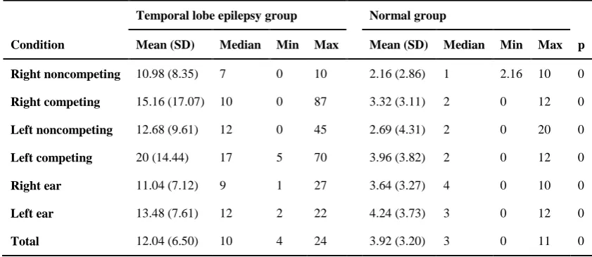
Twenty five (14 females, 11 males) normal subjects and 25 (13 females, 12 males) temporal lobe epilepsy patients aged 18-42 years old, par-ticipated in this study. Mean age of patient group and normal group were 30.52 (SD=8.14) and 32.36 (SD=6.35), respectively. Evaluation of four conditions, ear effect, and total SSW score showed that left competing condition had the highest mean score and the right noncom-peting condition had the lowest.
As indicated in Table 1, we compared the mean scores of conditions, ear and total in both groups. There were significant differences bet-ween mean scores of right noncompeting, right competing, left noncompeting, left competing, right ear, left ear, and total of the temporal lobe epileptic patients and normal subjects (p<0.001). In all of the above-mentioned items, mean scores were higher in patients than in normal subjects.
Table 1. Conditions, ear, and total scores in temporal lobe epileptic patients (n=25) and normal group (n=25)
Temporal lobe epilepsy group Normal group
Condition Mean (SD) Median Min Max Mean (SD) Median Min Max p
Right noncompeting 10.98 (8.35) 7 0 10 2.16 (2.86) 1 2.16 10 0
Right competing 15.16 (17.07) 10 0 87 3.32 (3.11) 2 0 12 0
Left noncompeting 12.68 (9.61) 12 0 45 2.69 (4.31) 2 0 20 0
Left competing 20 (14.44) 17 5 70 3.96 (3.82) 2 0 12 0
Right ear 11.04 (7.12) 9 1 27 3.64 (3.27) 4 0 10 0
Left ear 13.48 (7.61) 12 2 22 4.24 (3.73) 3 0 12 0
We used Mann-Whitney and Chi-square tests to examine the difference in C-SSW quality scores of both genders. No difference was observed (p>0.05). To compare the C-SSW quality and quantity scores in groups of early
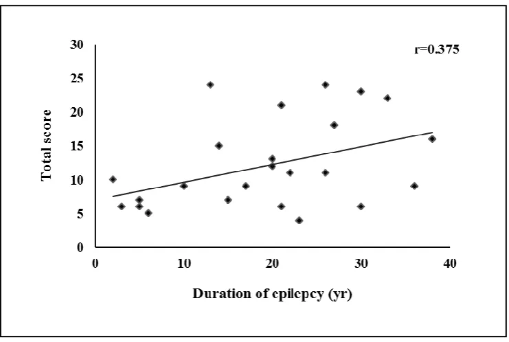
and late age of onset and also to compare these scores in two groups of high and low remission rates, Mann-Whitney and Chi-square tests were used. The results showed with increasing the age of onset, C-SSW quantity scores decrease, however, C-SSW scores had no significant diff-erence in groups of high and low remission rates. The mean of duration of epilepsy was 18 years and 11 months with the minimum duration of 2 years and maximum duration of 38 years. To evaluate the relationship between the duration of temporal lobe epilepsy and C-SSW quantitative responses Spearman correlation test was used (r=0.38, p=0.04), that means there was a poor direct relationship between the duration of epilepsy and the total score. This means with increasing the duration, the total score slightly increases (Fig. 1).
Chi-square test has been used for comparing the quality scores in two groups. It showed
signi-ficant difference in order effect (p=0.048), ear effect (p<0.001) and reversals (p=0.008) which means that mean scores in the patients were more than normal subjects. The results of com-paring A and B patterns indicate that temporal lobe epilepsy has no effect on the occurrence of pattern A or B. (Table 2) (p=1.000).
Discussion
In the present study, patients with temporal lobeepilepsy obtain higher scores in both quality and quantity scores than normal subjects. Tem-poral lobe structural and functional disorders may be one of the reasons for higher score of C-SSW in these patients. Disorders in these pati-ents may be related to integrated functional dis-orders in areas such as brainstem, thalamus, hip-pocampus, corpus callosum or areas of the cor-tex, such as frontal, or temporal lobe [12]. Stru-ctural, histopathological and biochemical disor-ders accompanied with impaired blood flow, decrement of metabolism and loss of neurons and dendrites in epileptic patients can explain the difference in processing between epileptic and normal subjects [12,14].
http://avr.tums.ac.ir Aud Vest Res (2016);25(4):241-247. The results of different studies with SSW test in
epileptic patients show high number of errors in both quality and quantity, and also in dichotic digit and non-verbal dichotic digit tests. Addi-tionally, it showed weak functioning in temporal order, binaural and auditory information proce-ssing accompanied by damaged phonological, semantic, verbal-working memory and CAPD in these patients [4,8,9,12,15]. Studies using tra-ctography showed temporal lobe epilepsy is accompanied by disorder in integrity of fronto-temporal connection, decrement in structural relationship of epileptic hemisphere, and prob-able increase of compensatory connections in the opposite hemisphere [16]. Using electroen-cephalography (EEG), researchers found increa-sed Bandgama activities in areas related to pho-nological network such as Broca, auditory cor-tex, prefrontal corcor-tex, hippocampus and fusi-form gyros [15]. Zhiqiang [17] reported changes in understandding network and decreased func-tional connections in hearing and sensorimotor networks in patients. In Boatman et al. study all patients with benign rolandic epilepsy, despite having normal hearing, showed some disorders in speech recognition in undesirable auditory conditions, such as background noise, acoustic filter and competitive speech [18]. fMRI studies indicated that temporal lobe epilepsy is accom-panied with deficits in organization of cortical networks which are involved in semantic pro-cessing [16].
In present study all of the high scores in con-ditions, ear and total scores, which was related to left competing condition and C-SSW score in patient was higher than normal subjects in left competing condition. This shows functional weakness in the integrity of auditory informa-tion and tolerance fading memory (TFM). Considering qualitative indicators in left tem-poral lobe epilepsy, increase in order effect, ear effect and reversals was observed. It usually seems that ear effect and order effect occur remarkably in perceptive area disorders. Increa-sed reversals shows deficit in organization [13]. The present study is consistent with Ellis et al. that reported wide spread defects in memory including short-term memory [19]. Researchers also reported weak functioning in duties of recent memory including verbal processing dis-order, save or retrieve information, and disorder in primary information processing. They believe that the frequent occurrence of electrical dis-charge may affect memory function during the recording and combination process of new information. This difficulty may relate directly to hippocampus or primary focus areas of the cortex [12]. Some studies reported that destruc-tion of the hippocampus and its surrounding areas following repeating seizures may cause memory defect [20].
In our study there was a poor direct relationship between duration of epilepsy and total score, such that by increasing the duration of temporal Table 2. Correlation between the disorder and ear effect, order
effect, Type A, Type B, and reversals in temporal lobe patient (n=25) and normal group (n=25)
Temporal lobe epilepsy group Normal group
Disorder Yes No Yes No p
Ear effect 13 12 0 25 <0.001*
Order effect 7 18 1 24 0.048*
Reversals 6 19 0 25 0.008
Type A 1 24 0 25 1.000**
Type B 0 25 0 25 1.000**
lobe epilepsy, the total scores slightly increase, which is consistent with some studies. In Ham et al. study, patients with longer duration of epilepsy had lower scores both in dichotic and duration tests. Although the exact mechanism is yet to be known, findings of some studies indi-cate structural and functional decline of tem-poral lobe, and also decrease in hippocampus volume and cerebral glucose metabolism by increasing the duration of epilepsy [8]. Some researchers reported increasing of duration with delayed latency in p300 in epileptic patients [21]. Also it was observed that in the middle temporal lobe epilepsy there is a significant relationship between the duration and decreased functional connection of temporal lobe [17]. In present study there was no correlation between gender and responses of C-SSW which is consistent with other studies [8].
In our research with increasing the age of seizure onset, C-SSW scores decrease, whereas in another study, patients with earlier age of seizure onset achieve significantly lower scores on intelligence tests than patients with later age of onset. This result indicates that age of seizure onset affects cognitive function, especi-ally, in the area of intelligence and memory, as in Lespinet et al. study in which, patients with early onset of seizure, encounter more defect on verbal and non-verbal memory [22]. In this study there was no relationship between seizure frequency and received scores. Some resear-chers reported that recurrent seizure is inversely related to memory, whereas others did not believe it [23].
We encountered some limitations for example some patients used several kind of drugs that could not go without them which could cause some cognitive disorders and yield to abnormal scores. Variables such as the kind of drugs, pro-gnosis, age of onset and seizure frequency may also deface the results. Therefore, it is recom-mended to investigate studies with more partici-pants’ volume.
Conclusion
Our findings indicate that, the patients with left temporal lobe epilepsy in comparing to normal
subjects showed more CAPD with SSW test. There was quality and quantity difference in SSW test between two groups that shows these patients are weaker in auditory processing than normal subjects. Due to importance of auditory processing, and as there was no diagnostic and therapeutic procedures for these patients, we suggest to form teams for their appropriate reha-bilitation.
Acknowledgments
This study is emerged from part of S. Shahbazi’s MSc. thesis in Audiology submitted in Tehran University of Medical Sciences, Tehran, Iran.
REFERENCES
1. Lauritzen F, Eid T, Bergersen LH. Monocarboxylate transporters in temporal lobe epilepsy: roles of lactate and ketogenic diet. Brain Struct Funct. 2015;220(1):1-12.
2. Sayehmiri K, Tavan H, Sayehmiri F, Mohammadi I, V Carson K. Prevalence of epilepsy in Iran: a meta-analysis and systematic review. Iran J Child Neurol. 2014;8(4):9-17.
3. Afzalaghaee M, Dehghani M, Alimi R, Mehdinejad M. Predictors of quality of life in patients with epilepsy. Journal of Knowledge & Health. 2015:10(1):11-8. Persian.
4. Meneguello J, Leonhardt FD, Pereira LD. Auditory processing in patients with temporal lobe epilepsy. Braz J Otorhinolaryngol. 2006;72(4):496-504.
5. Valizadeh L, Barzegar M, Akbarbegloo M, Zamanzadeh
V, Rahiminia E, Ferguson C. The relationship between psychosocial care and attitudes toward illness in adole-scents with epilepsy. Epilepsy Behav. 2013;27(1):267-71.
6. Adams RD, Victor M. Principles of Neurology. 5th ed. New York: McGraw-Hill; 1993.
7. Farias-Serratos F, Kensuke K, Nobuhito S. Temporal lobe epilepsy. Arch Neurocien (Mex). 2014;19(2):88-94.
8. Han MW, Ahn JH, Kang JK, Lee EM, Lee JH, Bae JH,
et al. Central auditory processing impairment in patients with temporal lobe epilepsy. Epilepsy Behav. 2011;20(2):370-4.
9. Ehrlé N, Samson S, Baulac M. Processing of rapid auditory information in epileptic patients with left
temporal lobe damage. Neuropsychologia.
2001;39(5):525-31.
10. Domitz DM, Schow RL. A new CAPD battery--multiple
auditory processing assessment: factor analysis and comparisons with SCAN. Am J Audiol. 2000;9(2):101-11.
11. Hajiabolhassan F, Lotfi Y, Azordegan F. Introducing and evaluating a Farsi - language version of the stag-gered spondaic word test in normal hearing subject. Audiol. 2006;15(1):39-46. Persian.
12. Ortiz KZ, Pereira LD, Borges AC, Vilanova LC.
http://avr.tums.ac.ir Aud Vest Res (2016);25(4):241-247.
Paulo Med J. 2002;120(6):185-8.
13. Fallahzadeh Z, Tahaei SAA, Hajiabolhassan F, Jalaie S, Shahbodoaghi MR, Rouhbakhsh N. Comparing the results of Persian staggered spondaic word test in persistent developmental stutterers and normal subject. Audiol. 2013:22(3):102-11. Persian.
14. Cherlow DG, Dymond AM, Crandall PH, Walter RD, Serafetinides EA. Evoked response and after-discharge thresholds to electrical stimulation in temporal lobe epileptics. Arch Neurol. 1977;34(9):527-31.
15. Boscariol M, Casali RL, Amaral MIR, Lunardi LL, Matas CG, Collela SMF, et al. Language and central temporal auditory processing in childhood epilepsies. Epilepsy Behav. 2012;53:180-3.
16. Jaimes-Bautista AG, Rodríguez-Camacho M, Martínez-Juárez IE, Rodríguez-Agudelo Y. Semantic Processing Impairment in Patients with Temporal Lobe Epilepsy. Epilepsy Res Treat. 2015;2015:746745.
17. Zhang Z, Lu G, Zhong Y, Tan Q, Liao W, Chen Z, et al. Impaired perceptual networks in temporal lobe epilepsy revealed by resting fMRI. J Neurol.
2009;256(10):1705-13.
18. Boatman DF, Trescher WH, Smith C, Ewen J, Los J, Wied HM, et al. Cortical auditory dysfunction in benign rolandic epilepsy. Epilepsia. 2008;49(6):1018-26. 19. Ellis AW, Hillam JC, Cardno A, Kay J. Processing of
words and faces by patients with left and right temporal lobe epilepsy. Behav Neurol. 1991;4(2):121-8.
20. Briellmann RS, Newton MR, Wellard RM, Jackson GD.
Hippocampal sclerosis following brief generalized seizures in adulthood. Neurology. 2001;57(2):315-7.
21. Sunaga Y, Hikima A, Ostuka T, Nagashima K, Kuroume
T. P300 event-related potentials in epileptic children. Clin EEG Neurosci. 1994;25(1):13-7.
22. Lespinet V, Bresson C, N'Kaoua B, Rougier A, Claverie B. Effect of age of onset of temporal lobe epilepsy on the severity and the nature of preoperative memory deficits. Neuropsychologia. 2002;40(9):1591-600. 23. Voltzenlogel V, Vignal JP, Hirsch E, Manning L. The