ISSN: 1735-0344 Tanaffos 2014; 13(1): 26-34
Human Epidermal Growth Factor Receptor 2 and Estrogen
Receptor Status in Respect to Tumor Characteristics in
Non-Metastatic Breast Cancer
Hanifeh Mirtavoos-Mahyari 1, Adnan
Khosravi 1, Zahra Esfahani-Monfared 2
1 Chronic Respiratory Disease Research Center, National
Research Institute of Tuberculosis and Lung Disease (NRITLD), Shahid Beheshti University of Medical Sciences, Tehran, Iran., 2 Mycobacteriology Research
Center, NRITLD ,Shahid Beheshti University of Medical Sciences, Tehran, Iran
.
Received: 19 November 2013
Accepted: 29 December 2013
Correspondence to: Khosravi A
Address: Chronic Respiratory Diseases Research
Center, National Research Institute of
Tuberculosis and Lung Diseases (NRITLD),
Shahid Beheshti University of Medical Sciences,
Tehran, Iran
Email address: adkhosravi@yahoo.com
Background: The expressions of estrogen receptor (ER) and cell surface receptor, Tyrosine Kinase Human Epidermal Growth Factor Receptor 2 (HER 2), have emerged as the most important molecular biomarkers determining the breast cancer prognosis. In this study, interactions between ER and HER2 were assessed to determine if they modulate tumor characteristics.
Materials and Methods: Tissue samples from 120 patients with early stage breast cancer receiving adjuvant chemotherapy were reviewed to evaluate ER and HER2 status quantified by immunohistochemistry and fluorescence in situ hybridization, and the correlation of ER and HER2 with patient characteristics and tumor pathology was studied.
Results: A total of 37(30.8%) and 80(66.6%) out of 120 samples were HER2 (3+ by immunohistochemistry or positive by fluorescent in situ hybridization) and ER positive (by immunohistochemistry), respectively. ER-negative tumors were significantly more likely to be HER-2 positive than were ER-positive tumors (21.25%; odds ratio, 0.270; 95% CI, 0.119 to 0.612; P=0.002). ER positivity was associated with <2 cm tumor size and higher histological grade (P=0.007 and 0.019, respectively). No significant correlation was seen between the co-expression of HER2 and ER and tumor characteristics.
Conclusion: HER2 positive tumors were less common compared to ER positive tumors in early stage breast cancer Iranian patients. Also, higher histological grade among ER negative tumors showed higher aggressiveness of the tumor. Future studies are needed to evaluate the effect of receptor status on prognosis.
Key
words:
Breast cancer, Tumor, Estrogen receptor,
Human Epidermal
Growth Factor Receptor 2 (HER2)
INTRODUCTION
Breast cancer is among the most common cancers affecting females worldwide (1-3). According to a report by the Iranian Ministry of Health and Medical Education in Iran, breast cancer ranks first among the malignancies affecting females (4). Some areas have a higher incidence of breast cancer such as East Africa (5) and the Middle East (including Iran) (6). In Iran, the incidence of breast
malignancies is increasing. Patients are affected at a
younger age and mostly detected at advanced stages (7, 8). In breast cancer, determining the expression status of ER and cell surface receptor tyrosine kinas human
epidermal growth factor receptor (HER2/neu or c-erb-B2) plays a critical role in choosing appropriate therapy (9).
and cancerous breast tissues (10). Several studies have demonstrated this role in proliferation and progression of breast tumors by generating multiple growth-promoting
signals (11-13). Evidence suggests that ER located on or near the cell membrane can activate HER2 (14).
Proliferation of breast tumoral cells and cell migration (15, 16) occur due to HER2 gene amplification and the
relationships between HER2 and lymph node involvement, tumor size and grade have been documented(17). It seems that HER2 over-expression or amplification in tumor cells is associated with a poorer outcome (18).
The crosstalk between the ER and HER2 and the roll of HER2 in ER adjustment and balancing have been well known (19, 20). Some investigators suggest that HER2 activates multiple intracellular signaling pathways leading
to ER regulation. In normal breast tissue, current activation causes estrogenic effect. In addition, ER actively contributes to this pathway by down-regulation (21) of HER2 expression and activation of intracellular pathways
leading to increased HER2 activity. However, in breast cancer, when estrogen concentrations are low, activation of HER 2 may affect ER and increase tumor growth (22).
It was hypothesized that ER may act as a mediator in
regulation of HER2 function. To the best of our knowledge, there are few studies regarding the relationship of ER with HER2 with respect to tumor characteristics in Iran. Thus, the results of this study can
provide basic information on breast cancer in Iranian females, and may help predict patient prognosis.
MATERIALS AND METHODS
This survey was a retrospective single-institute study
on 120 early stage breast cancer female patients referred to
Iranmehr Hospital from August 1997 to January 2011.
Written informed consent was obtained prior to patient
enrollment in accordance with the guidelines of the
medical ethics and scientific committees of Shahid Beheshti
Medical University.
The study protocol was in compliance with the
Declaration of Helsinki. Two anthracyclines-containing
regimens were administered: CAF (n=28) vs. TAC (n=22)
regimens. The administered doses were: 5 -fu 500 mg/m2,
Doxorubicin 50 mg/m2, Cyclophosphamide 500 mg/m2 in
CAF (23) and Docetaxel 75mg/m2, Doxorubicin 50 mg/m2,
Cyclophosphamide 500 mg/m2 for TAC (24), which were
repeated every 3 weeks.
Two-hundred files of breast cancer patients were
reviewed and 120 cases were selected. The inclusion
criterion was early stage breast cancer. The exclusion
criterion was metastatic disease.
To determine the status of hormone receptors and
HER2, immunohistochemical (IHC) methods alone (for ER
and PR), or in combination with fluorescent in situ
hybridization test (FISH) were used.
As recommended by the American Society of Clinical
Oncology/College of American Pathologists (ASCO/CAP)
(25) consensus panel and ESMO guidelines (26), first we
assessed HER2 gene status by IHC. If IHC was 2+, the
tumor block underwent confirmatory FISH test.
HER2 positivity was defined as samples with more
than 10% of cells staining 3+ by IHC or 2+ by IHC along
with FISH confirmation (a ratio of HER-2/neu
gene/chromosome 17 ≥ 2.0). HER2 expression was
determined by HerceptTestTM DAKO test. Breast cancer
was classified according to the World Health Organization
(WHO) classification of breast tumors.
In post-treatment follow-ups, patients underwent
physical examination at least once every 4 months for the
first 3 years, and every 6 months thereafter. Yearly
mammograms, bone scans, and chest X rays were
Statistical analysis
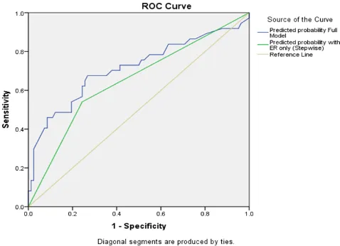
For testing the differences in categorical variables between the two groups, the chi-square test or Fisher’s exact test was used. The difference in quantitative variables between the two groups was compared using the Student's t-test or non-parametric Mann-Whitney test. Estimated probabilities of HER2 positivity by significant factors were obtained from the models. Sensitivity and specificity of these models were derived, along with the receiver operating characteristic (ROC) curves, to assess how good the models were at predicting HER-2 positivity. All analyses were performed using SPSS version 21.
RESULTS
Tumor PathologyOne-hundred twenty patients were studied. Basic demographics of patients and pathological characteristics are shown in Table 1.
The mean age of menarche was 13.8 years. Malignancy was seen in the right breast in 47.6% of patients and the remaining had tumors in their left breast (no one had bilateral disease).
The median tumor size was 1 cm. Invasive ductal carcinoma was found to be the most frequent pathology. Modified radical mastectomy (MRM) and lumpectomy were performed for 88 and 32 patients, respectively. All patients received chemotherapy, and radiotherapy was performed in 56.7% of patients.
Association of HER2 positivity with other prognostic parameters
HER 2 over-expression was seen in 30.8 %( n=37) of the analyzed samples. All patients with over-expression of HER2 had invasive ductal carcinoma. The incidence of lymph node involvement was 51.1% among patients with known HER2 over-expression, vs. 43.2% in group without HER2 over-expression (P=0.237).
Association of ER expression with other prognostic parameters
A significant association was found between ER and tumor size (P=0.007). It means that large tumors were significantly more ER negative. Also, a significant correlation was seen between the histological grade and ER expression (P=0.019). However, given the ER status, no association was found between age, nuclear grade, lymph node involvement and menopausal status (Table 2).
Relationship between HER-2 Status and clinical and pathological variables
ER negative tumors were significantly more likely to be HER2 positive than were ER positive tumors (21.25%; odds
ratio, 0.270; 95% CI, 0.119 to 0.612; P =0.002, Table 1). Thus, we selected the stepwise model including only the ER without the insignificant variables. The ROC curve from the reduced model is shown in Figure 1.
Figure 1. ROC curve from the reduced model.
Association of ER and HER2 with other prognostic parameters
Table 1. Clinicopathological characteristics of patients and association between HER2 and other parameters.
Age All
(n=120)
HER 2 Over expressed
(n=37)
HER 2 non-Over expressed
(n=83) P value
Mean± SD Median IQR <40 40-49 50-59 >60 50.42±11.61 50.0 44.0-58.7 24 (20.0%) 33 (27.5%) 39 (32.5%) 24 (20.0%)
51.27±11.68 50.0 46.50-59.0
7 (18.9%) 10 (27.0%) 13 (35.1%) 7(18.9%) 50.04±11.62 50.0 42-58 17(20.4%) 23(27.71%) 26(31.3%) 17(20.4%) 0.981
Histological grade I
II III
29 (24.1%) 42 (35%) 49 (40.8%)
6(26.2%) 13(35.1%) 18(48.6%) 23(27.7%) 29(34.9%) 31(37.3%) 0.333
Nuclear grade 0
1 2 3
7 (5.8%) 22 (18.3%) 63 (52.5%) 28 (23.3%)
1 (2.7%) 3(8.2%) 24(64.8%) 9(24.3%) 6(7.2%) 19(22.8%) 39(46.9%) 19(22.8%) 0.137 Vascular invasion Absent Present 100(83.3%) 20 (16.7%)
33 (89.2%) 4 (10.8%)
67(80.7%)
16(19.3%) 0. 250
†Lymph node involvement
None
1 to 3 4-9 >9 Unknown
66 (55.4%) 32(26.7%) 17 (14.2%) 4(3.3%) 1(0.8%)
18 (48.6%) 11 (29.7%) 5 (13.5%) 3(8.1%) 0 48(57.8%) 21(25.3%) 12(14.4%) 1(1.2%) 1(1.2%) 0.237 ER + - 80 (66.7%) 40(33.3%) 17 (45.9%) 20 (54.1%) 63(75.9%) 20(24.1%) 0.002*
‡Tumor size
<2 2-5 >5
27 (22.5%) 83 (69.1%) 10 (8.3%)
6 (16.2%) 27 (73.0%) 4 (10.8%) 21(25.3%) 56(67.4%) 6(7.2%) 0.486 Pathology
Invasive ductal carcinoma Lobular carcinomas Others 111(92.5%) 6(5%) 3(2.5%) 37(86.04%) 0 0 74(89.1%) 6(7.2%) 3(3.6%) 0.144 Menopausal status Yes No 70(58.3%) 50(41.6%) 23(62.1%) 14(37.8%) 47(56.2%) 36(43.3%) 0.57
Abbreviations: HER-2, human epidermal growth factor receptor 2; ER, estrogen receptor.
† No. of nodes involved: 0, node negative, 1 to 3:1 to 3 positive nodes, 4 to 9: 4 to 9 positive nodes; >9: >9 positive nodes.
Table 2. Association of ER expression with other prognostic parameters.
Variables ER positive (n=80) ER negative (n=40) P value Age
<40
40-49
50-59
>60
12(15%) 26(32.5%) 26(32.5%) 16(20%)
12(30%) 7(17.5%) 13(32.5%) 8(20%)
0.15
Histological grade
I
II
III
25(31.25%) 28(35%) 27(33.75%)
4(10%) 14(35%) 22(55%)
0.019*
Nuclear grade
0
1
2
3
Unknown
6(7.5%) 16(20%) 42 (52.2%)
16(20%) 0
1(2.5%) 6(15%) 21(52.5%)
12(30%) 0
0.449
Vascular invasion Absent
Present
65(81.25%) 15(18.75%)
35(87.5%) 5(12.5%)
0.386
†Lymph node involvement None
1 to 3
4-9
>9
Unknown
40(50%) 22(27.5%) 13(16.3%) 4(6.25%)
0
26(65%) 10(25%) 4(10%)
0 0
0.27
‡Tumor size
<2
2-5
>5
24(30%) 52(65%) 4(5%)
3(7.5%) 31(77.5%)
6(15%)
0.007*
Pathology
Invasive ductal carcinoma Lobular carcinomas Others
74(92.5%) 4(5%) 2(2.5%)
37(92.5%) 2(5%) 1(2.5%)
0.885
Menopausal status Yes
No
44(55%) 36(45%)
26(65%) 14(35%)
0.29
Abbreviations: ER, estrogen receptor.
† No. of nodes involved: 0, node negative, 1 to 3: 1 to 3 positive nodes,4 to 9: 4 to 9 positive nodes; >9: >9 positive nodes .
Table 3. Association of ER and HER2 with other prognostic parameters
ER Positive (n=80) ER negative (n=40)
HER2 non-over
expressed(n=63)
HER2 over expressed(n=17)
P-value HER2 non-over expressed(n=20) HER2 over expressed(n=20) P value Age <40 40-49 50-59 >60 8(12.6%) 21(33.3%) 19(30.1%) 15(23.8%) 4(23.5%) 5(29.4%) 7(41.1%) 1(5.8%) 0.288 9(45%) 2(10%) 7(35%) 2(10%) 3(15%) 5(25%) 6(30%) 6(30%) 0.95
Histological grade
I II III 21(33.3%) 22(34.9%) 20(31.7%) 4(23.5%) 6(35.2%) 7(41.1%)
0.682 2(10%) 7(35%) 11(55%) 2(10%) 7(35%) 11(55%) 0.999
Nuclear grade
0 1 2 3 5(7.9%) 14(22.2%) 32(50.7%) 12(19.04%) 1 2 10(4.1%) 4(1.6%) 0.776 1(5%) 5(25%) 7(35%) 7(35%) 0 1(5%) 4(70%)
5(25%)
0.096 Vascular invasion Absent Present 50(79.3%) 13(20.6%) 15(88.2%) 2(11.7%)
0.406 17(85%) 3(15%)
18(90%) 2(10%)
0.633
†Lymph Node involvement None
1 to 3
4-9 >9 Unknown 33(52.3%) 17(26.9%) 11(17.4%) 1(1.5%) 1(1.5%) 7(41.1%) 5(29.4%) 2(11.7%) 3(17.6%) 0 0.058 15(75%) 4(20%) 1(5%) 0 0 11(55%) 6(30%) 3(15%) 0 0 0.365
‡Tumor size
<2 2-5 >5 19(30.1%) 42(66.1%) 2(3.1%) 5(29.4%) 10(58.8%) 2(11.7%)
0.349 2(10%)
14(70%) 4(20%) 1(5%) 17(85%) 2(10%) 0.597
Abbreviations: HER-2, human epidermal growth factor receptor 2; ER, estrogen receptor.
† No. of nodes involved: 0, node negative, 1 to 3:1 to 3 positive nodes,4 to 9: 4 to 9 positive nodes; >9: >9 positive nodes,‡ <=2cm: tumors less than 2 cm in size; 2-5cm: tumors between 2 and 4.99 cm in maximum diameter; >5 cm, tumors >5 cm in maximum diameter.
DISCUSSION
Determination of factors, which may affect tumor characteristics and clinical behavior, can provide basic, important information on cancer. HER2 positive tumors were found to be less common (30.8%) compared to ER positive tumors (66.6%) and were inversely associated with ER positivity status (Table 1). Likewise, a significant
(Table 2). Also, a significant correlation was seen between ER negative tumors and high histological grade. In early stage breast cancer patients, data suggests that HER 2 status has a strong correlation with hormone receptors, especially ER.
countries such as Lebanon a higher percentage was reported. HER2 overexpression in this study was in accordance with the data from Egypt and another study in Iran (29, 30).
We also confirmed that over-expression of HER2 was infrequent in invasive lobular cancers. However, our sample size was not large enough to exclude these cases from HER2 screening.
In several studies, nearly 50% of patients with HER2 amplification were also ER positive which is similar to the results of the present current study (31). Also, the data of our study were similar to those of other studies in that HER2 over expression in breast cancer was associated with ER-negative status (32,33). Amplification of HER2 oncogene is related to increased proliferation and cell migration (16,17).
Moreover, the expression ratio of HER2 and ER varies between different geographical regions. ER expression was seen in 66.6% of our patients, which was similar to a study by Bartlett et al, (20) and higher than the result of Moradi-Marjaneh et al (30). An insignificant correlation was found between younger age, larger size and higher nuclear and histological grade and ER negativity, which indicates worse prognosis. This result is similar to that of a report by Walker et al (34).
In relation to breast cancer biology, many parameters are known, but tumors expressing ER have a relatively favorable prognosis. Results of the current study showed that ER negative tumors had significantly higher histological grade than ER positive ones (Table 2); which may reflect higher aggressiveness of the tumor.
Some investigators have shown that only 10% of ER positive breast tumors at the time of diagnosis show HER2 over-expression but this rate was higher in our study (about 20); which may be due to the higher frequency of HER2 over expression (35).
It is assumed that the impact of HER2 on balancing ER, is applied via different, separate pathways such as RAS/MAPK or AKT/PI3Kinase (36, 37).ER may therefore modify the effect of HER2 expression on breast tumor
pathology presumably via ER/HER2–mediated crosstalk. A number of potential pathways, which mediate this effect, are known and additional research may provide insight into the potential of this interaction to function as a therapeutic target. Considerations relative to ER and tumor differentiation provide a possible explanation for the dichotomy of response to adjuvant chemotherapy observed in pre- and postmenopausal women. We acknowledge the limitations of this study. First, this was strictly a single-institute investigation. Second, tumor grading, as well as tests for ER, PR and HER2, were performed by different laboratories without central supervision. Third, more than half the patients lacked information about tumor grading and vascular invasion, with the latter constituting the bulk of missing data.
This study was undertaken in early breast cancer patients and it would be useful to study this relationship more widely in other stages. Despite these shortcomings, our study is of value because 1) it highlights the importance of the ER and HER2 relationship and crosstalk between them; 2) it emphasizes the higher percentage of HER2 in our patients comparing to some countries as an important risk factor. Further research regarding the contribution of each of the tumor markers is underway with survival analyses adjusting for multiple risk factors.
Finally, the crosstalk between HER2 and ER status may help adopt multi-targeted strategies in the hope of improving patient outcome.
Acknowledgement
The authors wish to thank all the patients who participated in this study. Also, the authors appreciate the help of Dr. Naser Etamdi Moghadam.
REFERENCES
1. Siegel R, Ward E, Brawley O, Jemal A. Cancer statistics, 2011:
the impact of eliminating socioeconomic and racial disparities on premature cancer deaths. CA Cancer J Clin 2011; 61 (4):
2. Assi HA, Khoury KE, Dbouk H, Khalil LE, Mouhieddine TH,
El Saghir NS. Epidemiology and prognosis of breast cancer in
young women. J Thorac Dis 2013; 5 (Suppl 1): S2- 8.
3. Zaha DC, Lazăr E, Lăzureanu C. Clinicopathologic features
and five years survival analysis in molecular subtypes of
breast cancer. Rom J Morphol Embryol 2010; 51 (1): 85- 9.
4. Goya M. Iranian Annual Cancer Registration Report
2005/2006. Ministry of Health and Medical Education, Health
Deputy, Center for Disease Control and Prevention (In
Persian). Tehran, Iran; 2007
5. Kraft P, Haiman CA. GWAS identifies a common breast cancer
risk allele among BRCA1 carriers. Nat Genet 2010; 42 (10): 819-
20.
6. Runnak MA, Hazha MA, Hemin HA, Wasan AA, Rekawt RM,
Michael HD. A population-based study of Kurdish breast
cancer in northern Iraq: hormone receptor and HER2 status. A
comparison with Arabic women and United States SEER data.
BMC Womens Health 2012; 12: 16.
7. Montazeri A, Vahdaninia M, Harirchi I, Harirchi AM, Sajadian
A, Khaleghi F, et al. Breast cancer in Iran: need for greater
women awareness of warning signs and effective screening
methods. Asia Pac Fam Med 2008; 7 (1): 6.
8. Mousavi SM, Montazeri A, Mohagheghi MA, Jarrahi AM,
Harirchi I, Najafi M, et al. Breast cancer in Iran: an
epidemiological review. Breast J 2007; 13 (4): 383- 91.
9. Zubeda S, Kaipa PR, Shaik NA, Mohiuddin MK, Vaidya S,
Pavani B, et al. Her-2/neu status: a neglected marker of
prognostication and management of breast cancer patients in
India. Asian Pac J Cancer Prev 2013; 14 (4): 2231- 5.
10. Haslam SZ, Counterman LJ, Nummy KA. Effects of epidermal
growth factor, estrogen, and progestin on DNA synthesis in
mammary cells in vivo are determined by the developmental
state of the gland. J Cell Physiol 1993; 155 (1): 72- 8.
11. Yager JD, Davidson NE. Estrogen carcinogenesis in breast
cancer. N Engl J Med 2006; 354 (3): 270- 82.
12. Benson CS, Babu SD, Radhakrishna S, Selvamurugan N,
Sankar BR. Grade Dependent Expression of Growth Factor
Receptors and Signaling Molecules in Breast Cancer. Journal
13. Shou J, Massarweh S, Osborne CK, Wakeling AE, Ali S, Weiss H, et al. Mechanisms of tamoxifen resistance: increased
estrogen receptor-HER2/neu cross-talk in ER/HER2-positive
breast cancer. J Natl Cancer Inst 2004; 96 (12): 926- 35. 14. Razandi M, Pedram A, Park ST, Levin ER. Proximal events in
signaling by plasma membrane estrogen receptors. J Biol
Chem 2003; 278 (4): 2701- 12.
15. Somerville JE, Clarke LA, Biggart JD. c-erbB-2 overexpression
and histological type of in situ and invasive breast carcinoma.
J Clin Pathol 1992; 45 (1): 16- 20.
16. Mylonas I, Makovitzky J, Jeschke U, Briese V, Friese K, Gerber
B. Expression of Her2/neu, steroid receptors (ER and PR),
Ki67 and p53 in invasive mammary ductal carcinoma associated with ductal carcinoma In Situ (DCIS) Versus
invasive breast cancer alone. Anticancer Res 2005; 25 (3A):
1719- 23.
17. Tovey SM, Witton CJ, Bartlett JM, Stanton PD, Reeves JR,
Cooke TG. Outcome and human epidermal growth factor
receptor (HER) 1-4 status in invasive breast carcinomas with proliferation indices evaluated by bromodeoxyuridine
labelling. Breast Cancer Res 2004; 6 (3): R246- 51.
18. Petrelli F, Barni S. Role of HER2-neu as a prognostic factor for survival and relapse in pT1a-bN0M0 breast cancer: a
systematic review of the literature with a pooled-analysis. Med
Oncol 2012; 29 (4): 2586- 93.
19. Osborne CK, Shou J, Massarweh S, Schiff R. Crosstalk between
estrogen receptor and growth factor receptor pathways as a
cause for endocrine therapy resistance in breast cancer. Clin Cancer Res 2005; 11 (2 Pt 2): 865s- 70s.
20. Bartlett JM, Ellis IO, Dowsett M, Mallon EA, Cameron DA,
Johnston S, et al. Human epidermal growth factor receptor 2 status correlates with lymph node involvement in patients
with estrogen receptor (ER) negative, but with grade in those
with ER-positive early-stage breast cancer suitable for cytotoxic chemotherapy. J Clin Oncol 2007; 25 (28): 4423- 30.
21. Antoniotti S, Taverna D, Maggiora P, Sapei ML, Hynes NE, De
Bortoli M. Oestrogen and epidermal growth factor
down-regulate erbB-2 oncogene protein expression in breast cancer
22. Qui WS, Yue L, Ding AP, Sun J, Yao Y, Shen Z, et al. Co-expression of ER-beta and HER2 associated with poorer
prognosis in primary breast cancer. Clin Invest Med 2009; 32
(3): E250- 60.
23. Martin M, Villar A, Sole-Calvo A, Gonzalez R, Massuti B,
Lizon J, et al. Doxorubicin in combination with fluorouracil
and cyclophosphamide (i.v. FAC regimen, day 1, 21) versus
methotrexate in combination with fluorouracil and
cyclophosphamide (i.v. CMF regimen, day 1, 21) as adjuvant
chemotherapy for operable breast cancer: a study by the
GEICAM group. Ann Oncol 2003; 14 (6): 833- 42.
24. Martín M, Seguí MA, Antón A, Ruiz A, Ramos M, Adrover E,
et al. Adjuvant docetaxel for high-risk, node-negative breast
cancer. N Engl J Med 2010; 363 (23): 2200- 10.
25. Wolff AC, Hammond ME, Schwartz JN, Hagerty KL, Allred
DC, Cote RJ, et al. American Society of Clinical
Oncology/College of American Pathologists guideline
recommendations for human epidermal growth factor
receptor 2 testing in breast cancer. Arch Pathol Lab Med 2007;
131 (1): 18- 43.
26. Aebi S, Davidson T, Gruber G, Castiglione M; ESMO
Guidelines Working Group. Primary breast cancer: ESMO
Clinical Practice Guidelines for diagnosis, treatment and
follow-up. Ann Oncol 2010; 21 Suppl 5: v9- 14.
27. Ross JS, Slodkowska EA, Symmans WF, Pusztai L, Ravdin PM,
Hortobagyi GN. The HER-2 receptor and breast cancer: ten
years of targeted anti-HER-2 therapy and personalized
medicine. Oncologist 2009; 14 (4): 320- 68.
28. Fountzilas G, Valavanis C, Kotoula V, Eleftheraki AG,
Kalogeras KT, Tzaida O, et al. HER2 and TOP2A in high-risk
early breast cancer patients treated with adjuvant
epirubicin-based dose-dense sequential chemotherapy. J Transl Med
2012; 10: 10.
29. el-A Helal T, Khalifa A, Kamel AS. Immunohistochemical
expression of p53 and c-erbB2 proteins in breast cancer in
Egypt. Anticancer Res 2000; 20 (3B): 2145- 50.
30. Moradi-Marjaneh M, Homaei-Shandiz F, Shamsian SAA,
Mashhadi IEZ, Hedayati-Moghadam MR. Correlation of
HER2/neu over expression, p53 protein accumulation and
steroid receptor status with tumor characteristics: An Iranian
study of breast cancer patients. Iranian Journal of Public
Health 2008; 37 (3): 19- 28.
31. Vaz-Luis I, Winer EP, Lin NU. Human epidermal growth
factor receptor-2-positive breast cancer: does estrogen receptor
status define two distinct subtypes? Ann Oncol 2013; 24 (2):
283- 91.
32. Choi Y, Pinto M. Estrogen receptor beta in breast cancer:
associations between ERbeta, hormonal receptors, and other
prognostic biomarkers. Appl Immunohistochem Mol Morphol
2005; 13 (1): 19- 24.
33. Moriki T, Takahashi T, Ueta S, Mitani M, Ichien M. Hormone
receptor status and HER2/neu overexpression determined by
automated immunostainer on routinely fixed cytologic
specimens from breast carcinoma: correlation with histologic
sections determinations and diagnostic pitfalls. Diagn
Cytopathol 2004; 30 (4): 251- 6.
34. Walker RA. Immunohistochemical markers as predictive tools
for breast cancer. J Clin Pathol 2008; 61 (6): 689- 96.
35. Fu X, Osborne CK, Schiff R. Biology and therapeutic potential
of PI3K signaling in ER+/HER2-negative breast cancer. Breast
2013; 22 Suppl 2: S12- 8.
36. Gutierrez MC, Detre S, Johnston S, Mohsin SK, Shou J, Allred
DC, et al. Molecular changes in tamoxifen-resistant breast
cancer: relationship between estrogen receptor, HER-2, and p38 mitogen-activated protein kinase. J Clin Oncol 2005; 23
(11): 2469- 76.
37. Ignatoski KM, Maehama T, Markwart SM, Dixon JE, Livant
DL, Ethier SP. ERBB-2 overexpression confers PI 3'
kinase-dependent invasion capacity on human mammary epithelial