ABSTRACTS
Free Communications
FC1_01
No place for laparotomy—ovarian cyst in pregnancy Fawzia Sanaullah, Ashwini K Trehan
Dewsbury and District Hospital, UK
Laparoscopic surgery during pregnancy has been reported to be safe. Advantages of laparoscopic approach in pregnancy include•panoramic magnified view,•reduced intraoperative uterine manipulation and reduced risk of miscarriage,•reduced postoperative pain and need for opiates,• early mobility and reduced risk of DVT and•Less risk of poor scar healing. This video presentation of three cases will demonstrate laparoscopic approach to ovarian cyst impacted in the pouch of Douglas and massive ovarian cyst (30 cm). All three cases are examples of safe laparoscopic approach to adnexal cyst. It demonstrates that a benign cyst of any size and at any location can be done laparoscopically even in the second trimester of pregnancy.Case 1: 20 weeks pregnancy with 11 cm adnexal cyst at ultrasound. At laparoscopy, the cyst was impacted in the pouch of Douglas behind the uterus. Laparoscopic aspiration of the left ovarian cyst (800 ml) and cystectomy was performed.Case 2: 18 weeks pregnancy with 15 x 14 cm cyst at ultrasound. Laparoscopic aspiration of ovarian cyst of 1500 ml clear fluid followed by Left salpingo-oophorectomy.
Case 3: 16 weeks with 30 × 32 cm large simple cyst originating from the left adnexa at ultrasound. 8150 ml of clear fluid was aspirated. At laparoscopy, an excision of the left large parovarian cyst and salping-ectomy was done. All three were discharged home the following day. The histology was benign. All three delivered at term with good outcome. References:
1. Nezhat F, Nezhat C, Silfen SL, et al. Laparoscopic ovarian cystectomy during pregnancy. J Laparoendosc Surg 1991; 1:161–164 2. Al-Fozan H, Tulandi T. Safety and risks of laparoscopy in pregnancy. Curr Opin Obstet Gynecol 2002; 14(4):375–9.
3. Yuen PM, Chang AM. Laparoscopic management of adnexal mass during pregnancy. Acta Obstet Gynecol Scand 1997; 76(2):173–6. 4. Mathevet P, Nessah K, Dargent D, Mellier G. Laparoscopic management of adnexal masses in pregnancy: a case series. Eur J Obstet Gynecol Reprod Biol 2003; 108(2):217–22.
FC1_02
Laparoscopic treatment of interstitial (cornual) pregnancy Metin Capar
Selcuk University, Turkey
Objectives:Interstitial (cornual) pregnancy is the least common type of ectopic pregnancy.the incidence of interstitial ectopic is 1 in 2500– 5000 livbe births and it accounts for 2–6% of all ectopic pregnancies’
risk factors predisposing to an interstitial ectopic pregnancy are the same as those for tubal ectopics and include previous ectopic pregnancy, assisted reproduction treatment and sexually transmitted infections and previous ipsiateral salpingectomy.
Design and methods:32 years old patient with history of laparoscopic adhesolysis for pelvic adhesions presented with lower abdominal pain in pelvic examination she had a slightly enlarged uterus, Hgb 10 gm/dl, serum B-HCG 2680, ultrasonography suspected a left cornual ectopic pregnancy, with positive fetal heart pulsation. arrangements were made for emergency laparoscopy and possible laparotoy, laparoscopy con-firmed left cornual ectopic pregnancy, with single trochar entry the ectopic pregnany was recognized. First the pregnancy material was aspirated then a cut was made in cornual region by the help of endograsper the ectopic pregnancy was extirpated and sutured. Conclusion: Interstitial (cornual) pregnancy can be treated by laparoscopic surgery.
FC1_03
Translation and validation of the German“ICIQ
Vaginal Symptoms Questionnaire”(The ICIQ-VS German): An observational study
Carolin Banerjee1, Marc Banerjee2, Martin Hellmich3, Günter Karl Noé1 1
Hospital Dormagen, Teaching Hospital Univ. Cologne
2
Cologne Merheim, Univ. Witten-Herdecke
3
Instit. Medical Statistics, Univ. Cologne, Germany
Introduction:In 2006 the International Continence Society developed a validated vaginal questionnaire contenting the sexual aspect as well as pain, pressure and quality of life items. The ICIQ-VS has not been translated yet into German. We here report about the translation and validation process of the ICIQ-VS (German).
Design:Observational Study
Setting: Gynaecological and internal Department of a German teaching hospital.
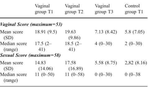
Main outcome measures:Consistency, reliability, sensitivity, validity Results:In our pre-test analysis all items were well interpreted and filled in by all ten patients. In the main study mean age was 64 years (35 to 87 years) in the vaginal group and 56 years (range 25–86 years) in the control group. No changes from the original format were observed after translation and cultural adaptation. Internal consistency was high (standardized Cronbach alpha coefficient range 0.71–0.78). The test retest reliability (stability over time) was measured by weighted Kappa index and was stable for both groups (0.71–0.88). Sensitivity to change was excellent (Wilcoxon sign rank test, p<0.01–p<0.001). The impact of vaginal symptoms of quality of life was worse in the vaginal than in the control group. Construct validity revealed statistical significant differ-ences between groups (Tab.1). Response rate was 96 % in the vaginal group and 98 % in the control group. We had low rates of missing data (vaginal group 3.6 %, control group 3.6 %). Thus the content validity was excellent. We cannot state concurrent validity as no comparable questionnaire concerning vaginal symptoms was available in German. Conclusion: The ICIQ-VS has been translated and evaluated successfully into German. To our knowledge this is the first evaluated vaginal questionnaire which is available in German. During the last years the broad use of the English version has proven its relevance for clinical research. We expect the same to this German version.
Vaginal group T1
Vaginal group T2
Vaginal group T3
Control group T1
Vaginal Score (maximum=53) Mean score
(SD)
18.91 (9.5) 19.63 (9.86)
7.13 (8.42) 5.8 (7.05)
Median score (range)
17.5 (2– 41)
18.5 (2– 41)
4 (0–30) 2 (0–30)
Sexual Score (maximum=58) Mean score
(SD)
14.83 (14.06)
17.58 (16.89)
5.58 (8.75) 2,82 (8.16)
Median score (range)
11 (0–50) 11 (0–58) 0 (0–30) 0 (0–38
Table 1:Verification of the scoring system: Statistical results comparing the mean vaginal and sexual symptoms score for vaginal group at time one, two and three (T1-T3) and control group at time one
FC1_04
Evaluation of SprayShield in a Center: Randomized Controlled Study in Women Undergoing Myomectomy
G. Tchartchian, B. Dittert, R.L. De Wilde
Pius Clinic, Oldenburg, Germany
Evaluation of SprayShieldTM in a Single Center, Randomized Controlled Study in Women Undergoing Laparoscopic Myomectomy Tchartchian G; Dittert B, De Wilde RL Dept.Ob/Gyn, Pius Clinic, Oldenburg, Germany. Adhesions are a major cause of chronic, recurrent pelvic pain and infertility in a significant percentage of operative patients. Adhesions also have major financial implication as well; indeed, in the United Kingdom alone, adhesion-formation related readmissions following lower abdominal surgery over a ten year period cost over 500 million pounds . Though many different materials have been evaluated for their potential ability to prevent adhesion formation to date, the need for comprehensible, site-specific
protection remains high. The SprayShieldTM Adhesion Barrier has been developed as a product to be sprayed onto adhesiogenic tissues. It is a site specific adhesion barrier that solidifies within seconds of being applied onto tissues and is intended to protect tissues from adhesion formation while the tissues heal. The SprayShieldTM Adhesion Barrier System includes a polymer kit and an air-assisted sprayer applicator. The SprayShieldTM Polymer kit contains two solutions, a polyethylene glycol (PEG) ester trylisine amine solution and a borate buffer solution referred to as the blue and the clear precursors, respectively. When sprayed, the blue and clear precursors react in-situ in an electrophilic-nucleophilic reaction, causing the PEG molecules to cross-link and form a hydrogel within seconds with no heat evolved and no external energy source required (i.e., light source). Within 7 days of application, the hydrogel will hydrolyze and the individual components (PEG and Trylisine) are readily absorbed into the circulatory system and excreted via renal filtration . The SprayShieldTM Adhesion Barrier System has been shown to prevent adhesions in a laparoscopic porcine model of gynecological surgery versus control (good surgical technique). To further evaluate the ability of SprayShieldTM to reduce adhesions following gynecolog-ical surgery, a single center, fifteen subject, single blinded, random-ized controlled study was undertaken comparing SprayShieldTM versus good surgical technique alone (control group) in subjects undergoing laparoscopic myomectomy. Briefly, the study was con-ducted in women age 18 and older, who were scheduled to undergo laparoscopic myomectomy, met all study inclusion and exclusion criteria and signed an informed consent. Subjects were randomly assigned in a 2:1 ratio to the SprayShieldTM group or to a control group. In subjects randomized to the SprayShieldTM group, SprayShieldTM was applied to all myomectomy suture lines, any other areas surgically treated or at sites the surgeon deemed to be prone to adhesion formation. Subjects assigned to the control group received no anti-adhesion treatment, of any kind, only good surgical technique. Subjects were then asked to return 8–12 weeks post002Dmyomectomy for a second look laparoscopic (SLL) procedure to evaluate adhesion formation at 21 anatomical sites. Both the myomectomy procedure and the SLL were recorded. The videos were then edited to remove the SprayShieldTM application from treated subjects and all videos were assigned a unique blind code to blind the reviewer to treatment. The videos were then sent to an independent gynecological surgeon to obtain a stan-dardized, unbiased evaluation of adhesion formation. All adhesions were evaluated for Incidence (Number of adhesions from the uterus to other anatomical structures), Severity (1=filmy, avascular; 2=vascular and/or dense; 3=cohesive) and Extent (1=covering 51% of total area). The area of the uterus, in millimeters, covered by adhesions was also assessed.
References:
–
Practice Committee of the American Society for Reproductive Medicine in Collaboration with the Society of Reproductive Surgeons. Pathogenesis, consequences, and control of peritoneal adhesions in gynecologic surgery. Fertil Steril 2006;86(Suppl 4): S1–S5.–
Wilson MS, Menzies D, Knight A, Crowe AM. Demonstrating the clinical and cost-effectiveness of adhesion reduction strate-gies. Colorectol Dis. 2002;4:355–360.–
Campbell PK, Bennett SL. Technology of a next generation abdominopelvic adhesion barrier. Abstract. 2008.FC1_05
CEGPA–a new peruvian-german teaching center for gynecologic endoscopy in Lima - Peru
H. Krentel, Th. Römer
Bethesda Hospital Wuppertal, Germany
CEGPA—a new peruvian-german teaching center for gynecologic endoscopy in Lima - Peru Following the educational concept of the german association of gynecologic endoscopy (AGE) in March 2009 we realized the first workshops for gynecologic laparoscopy in the new Centro de Endoscopia Ginecologica Peruano Aleman–CEGPA in Lima / Peru. Live surgery has been demonstrated followed by a program of scientific presentations and a practical part using various laparotrainers. The CEGPA has been presented during the first german-peruvian congress of gynecology and obstetrics in Lima in October 2008 which had been organized in cooperation with the AGE and the Peruvian Society of Obstetrics and Gynecology (SPOG) and the Peruvian Society of Ultrasound in Gynecology and Obstetrics (SPUOG). The CEGPA is a new teching center realizing a constant cooperation between german and peruvian gynecologic endoscopists which is supported by the Peruvian University Cayetano Heredia and the departement of gynecology and obstetrics of the University in Tübingen—Germany. In the first workshops 21 peruvian ginecologists from five different peruvian cities recieved the certificates of the AGE after successfull participation. The cooperation offers the exchange of new scientific, social and cultural aspects in the treatment of females by gynecologic endoscopy. The CEGPA is a new link between Europe and South America in the teaching and the use of modern endoscopic techniques.
FC1_06
Two simple and cheap systems for recording surgery digitally M. Granata, I. Korkontzelos, N. Gioulekas, I. Tsimpanakos, S. Charalampos, A. Magos
Royal Free Hospital, London, UK
Introduction:The visual recording of surgical procedures either as still images or movies is becoming standard practice across many specialities. Analogue recording using film or video is gradually being replaced by digital technology for reasons of quality and convenience. Commercial systems are available but are expensive and have limited capabilities and capacity. We have been using a digital system based around a standard personal computer since 2003 to record all our surgery. Methods: We use one of two systems. One system consists of a standard laptop running Windows® operating system linked to a large and fast (7200 rpm) external hard drive which is used to save the video file. The video signal from the theatre camera is processed by an external analogue/digital converter (Ikasu USB video converter); we compress the video using the DivX codec to approximately 1 Gb per hour of video. The second system is based around an Archos AV-500 mobile digital video recorder which also uses the DivX codex to compress the video signal; we use the Archos to make edited recordings which are then transferred to a CD and given to the patient after her surgery.
Findings:Very few patients refused to have their surgery recorded, and we have recorded over 1500 procedures since 2003. All record-ings were of excellent quality and could easily be searched and
reviewed if required (e.g. in cases of operative complications). They proved an excellent medium for teaching trainees, and could easily and conveniently edited if required using standard software (we used the freeware program, VirtualDub). The recordings could also be added to PowerPoint presentations for teaching or demonstration purposes after suitable conversion to an appropriate format.
Conclusions: A digital recording system built around a standard personal computer or a mobile digital video recorder is relatively cheap, versatile and has a huge capacity to record surgical procedures. It is a convenient and useful way to keep a permanent record of surgery.
FC1_07
Training laparoscopic gynaecologic surgery on live animals Pedro González Ramos, Pedro Royo Manero, Cristina Pastor Oliver, Nadia Nassar Melic, Daniel Ordóñez Pérez
Hospital Clínico Universitari "Lozano Blesa", Zaragoza, Spain
A laparoscopic surgery teaching program has already started at the Gynaecology Department Of The `Lozano Blesa` University Hospital. Zaragoza. Spain. This program has been approved by the Ethics Committee for Animal Research Of The Zaragoza University and will use rats, rabbits and sows. This project aims to follow a “Step By Step”process, with progressive surgery complexity from the rat and rabbit models to more difficult surgery in sows. We worked in wistar rats using them in two different ways:—Microsurgery as first level, because like laparoscopic surgery, working with operating micro-scope, surgeons can´t see their hands only the movement of the mnstruments and also work with high vision, then microsurgery improves spacial orientation - inside a pelvitrainer and looking at mobile compact unit telepack, we will be able to use dissecting and grasping forceps, scissors required in laparoscopic surgery. This animal is cheaper than a sow and it is very useful to practise suturing thecniques. Following level would be in new zealands white rabbits: we use this model to teach—abdominal acces: by using a veress needle. Open acces—electrosurgery thecniques—how to perform total hysterectomy using vaginal extraction of uterus, tubes and ovaries (notes)—vaginal closed by laparoscopic approach using suturing techniques interrupted or continuous with intra-corporeal or extra-corporeal knots. Finally we work with sows to perform oncologic laparoscopic surgery, it would be the highest level (master) describing anatomic - surgical landmarks that must be followed to complete this kind of surgery, includindg pelvic lymphadenectomy, creating paravesical, pararectal spaces and to be prepared for casual injuries that could be happen during laparoscopic surgery.
FC1_08
Colpotomy for transvaginal NOTES: should we be afraid? J. Nassif, S. Perretta, B. Dallemagne, M. Vix, J. Marescaux, I. Miranda, A. Wattiez
IRCAD / EITS, Strasbourg, France
Endoscopic Surgery (NOTES) is a new era of minimal invasive surgery. For the moment, transvaginal approach is mostly used since other routes (ie, transgastric, transrectal and transvaginal) require incision closure techniques to be defined. Recently, a study using a questionnaire assessed the acceptance of gynaecologists to this route. They would not advise their patients to use this route. Infection and dyspareunia are the most feared complications reported in this study. We present the results of colpotomy in NOTES transvaginal procedures.
Methods:The study enrols 17 patients operated by NOTES trans-vaginal procedures. Ten patients had cholecystectomy and seven sleeve gastrectomy. Eventual peri operative complications such as rectal injury, fever and pain are reported. Follow up is done at one and six months. Colpotomy is assessed by vaginal exam and direct visualization. Patients fill a questionnaire about sexual function, dyspareunia and infection.
Results: Ongoing. Primary results do not confirm the fear of gynaecologists.
Conclusion:It seems that the fears from complications related to the colpotomy in NOTES are not supported by clinical data. Even though, patient’s acceptance should also be assessed.
FC1_09
First clinical experience of Argon neutral plasma energy in gynaecological surgery
Thumuluru Kavitha Madhuri, Dimitri Papatheodorou, Anil Tailor, Christopher JG Sutton, Simon Butler-Manuel
Department of GynaeOncology Royal Surrey County Hospital, Guildford, UK
Introduction: We present the first series of in-vivo use of neutral argon plasma energy in gynaecological surgery. PlasmaJet™was used in 65 cases in a tertiary referral centre for gynaecological oncology and minimal access surgery.
PlasmaJet™is a new device designed to produce a fine jet of argon plasma by heating argon gas. Originally developed for jet engines in the Russian space exploration programme, it utilizes neutral plasma energy to desiccate and vaporize soft and hard tissues.
Methods: A prospective, observational study in women with a wide spectrum of benign and malignant conditions was undertaken between June and May 2009. Patients were offered information on the use of PlasmaJet™ during their procedure. PlasmaJet™ Version-2 was used in 47 cases and Version-3 in 18 cases. Efficacy and safety data was collected on 57 laparoscopies, 7 laparotomies and 1 groin node dissection. Data related to use of PlasmaJet™ including procedure performed, power settings used and tissue effect were recorded. Effectiveness of PlasmaJet™ was measured in terms of precision, ease of use, coagulation and cutting effects, safety and complications.
Results:The PlasmaJet™device was easy to use and set up with a disposable hand piece and no moving parts. The length of the jet produced is approximately 2cms with no risk of overshoot to distant organs. Depth of penetration appears very superficial with minimal lateral heat spread; no neighbouring tissue damage was observed. Neutral plasma energy removes the risk of arcing.
Conclusion: shows promising results as a cutting and coagulating instrument for both laparoscopic and open gynaecological surgery.
FC1_10
Advantages of successful laparoscopic approach vs conversion laparotomy and total laparotomy
E. Martínez, A. Zapico, P. Garbayo, J.A. Solano, F. Cardinali, B. Ochoa, R. Garcia
Universitary Hospital Príncipe de Asturias. School of Medicine. Alcalá University. Madrid, Spain
Objective: To value the advantages of laparoscopic approach in endometrium cancer vs conversion laparotomic approach and total laparotomic access.
Design and methods:A retrospective study between 1996 and 2008, over 201 consecutive patients with endometrium cancer. Three groups were defined LPS (successfull laparoscopic approach) NLM (Conver-sion into laparotomy needed) and TLM (total laparotomy). We have studied different items such as epidemiological data, diagnosis procedures, surgical access, operating time, complications rate, hospital stay, transfusion rate, and pathological findings. Statistical analysis was done using SPSS 15.O.
Results: No differences were observed in age or BMI ( body mass index). Endometrial risk factors were seen in 103(51,2%). In 123 patients (89,13%) successfull LPS approach was feasible while in the remaining 15 cases (10,86%) NLM and TLM was chosed in 63 cases like initial approach. Lymphadenectomy was possible in 118 (95,93%) of LPS group vs NLM 10(66,66%) and in TLM 41(65,07%). Operating time time was longer for NLM 160,71±10,7 (85–270) and LPS 154,07 ± 3,85(50–285) vs TLM 142,06± 5,99 (45–270) minutes. Hospital stay was shorter for LPS 5,13+0,67(2–22) and TLM 7,76±0,68(3–33) vs NLM 13,20±4,68(3–65) days (p<0.05). Haemoglobin balance was better p<0.001 for LPS 2,89±0,13(0,4– 8,8) vs NLM 3,78±0,4(0,4–7,8) gr/dl and TLM 3,16±0,2(0,4–7,8 gr/ dl. Global transfusion rate was higher in NLM 46,7% (p<0,01). No differences in nodes collected were found. Survival rate were similar. Conclusions:When laparoscopic approach is feasible the results are better. Lymphadenectomy were more sucessfully performed in LPS group with less haemoglobin balance, operating time and fewer transfusion rate. Hospital stay was also shorter in LPS approach. When TLM is permorfed from initial access the surgical results are better than in LPS conversion. Avoid NLM should help to improve operative morbidity.
FC1_11
Hysteroscopy after uterine artery embolization (UAE)
or laparoscopic uterine artery occlusion (LUAO) for leiomyomas Michal Mara1, Petr Horak1, Kristyna Kubinova1, Jana Maskova2, Pavel Dundr3, David Kuzel1
1
Department of Obstetrics and Gynecology, 1st Medical Faculty of Charles University. Prague, Czech Republic
2
Department of Radiology, Royal Infirmary Hospital, Aberdeen, Scotland
3
Department of Pathology, 1st Medical Faculty of Charles University, Prague, Czech Republic
Design:Prospective comparative study.
Methods:Women with reproductive plans and symptomatic intramu-ral uterine fibroid/s larger than 4 cm treated with UAE or LUAO were assigned for hysteroscopy and biopsy from 3–6 months after primary procedure. Only patients without simultaneous or subsequent myo-mectomy were included.
Results:111 women entered the study (mean age: 35 years, mean dominant fibroid size before treatment: 51 mm, mean number of fibroids: 2; no significant differences between groups); 74 with previous UAE and 37 with LUAO. Completely normal hysteroscopic finding was assessed in 30 patients after UAE (40.5 %) and in 36 after LUAO (97.3 %) = the difference was significant (p=0.0001, chi-square test). Intrauterine necrotic changes were present and verified by histology in 32 patients (43.2 %) after UAE, in compare with only 1 patient (2.7 %) after LUAO (p =0.0001, chi-square test). Vital, functional endometrium was histologically proved in 64 women after UAE (86.5 %), and in 35 (94.6 %) after LUAO (non-significant difference, Fisher’s test).
Conclusion: Our results show that uterine fibroid embolization in patients with intramural leiomyomas could significantly impair uterine cavity, which could subsequently worsen their fertility prognosis. Except one patient (from 37) no damage of endometrium and uterine cavity was observed after LUAO.
FC1_12
Single port laparoscopy in gynaecology, what can we perform: a series of 35 cases
Stefanos Chandakas
Iaso Hospital, Athens, Greece
Introduction:Minimally invasive surgery has influenced the techni-ques used in gynaecology, with an overall minimisation of complica-tions and increased patient satisfaction.
Study objective:To demonstrate the safety and feasibility of Single Port Laparoscopic (SPL) Surgery in Gynaecology
Methods and procedures: Retrospective, descriptive, non random-ized study, in the setting of Iaso Hospital and Attikon University Hospital, Athens, Greece. 35 patients were selected who underwent SPL Surgery between October 2008 and February 2009.
Indications included 55% Salpingo-oophorectomy, 26% Diagnostic Laparoscopy and treatment of Stage 1/2 Endometriosis, 19% cases for cystectomy, 1 case of Total Hysterectomy.
Results:Duration of operation and of hospital stay, safety (morbidity and mortality), and patient satisfaction were assessed Estimated blood loss was 35 ml (range 10–230 ml) Intraoperative complications: 0% vascular injuries and 0% nerve or ureter injuries. Early postoperative morbidity included no major complications, 0.1% bladder infection and dysfunction and 0.3% of incision infection. All patients were discharged to home the same day with an average length of stay for these patients of 8 hours.
Conclusions:Single port Laparoscopic Surgery seems to be a safe alternative to traditional Laparoscopy for the procedures performed in this study. Surgical time, safety and feasibility is similar, were as the cosmetic result and the post operative pain levels seem to be better accepted by the female patient. Further studies need to be performed and new instrumentation is necessary in order to perform more complicated cases.
FC1_13
The efficacy of a polyethylene glycol adhesion barrier in the prevention of post-operative adhesions in patients undergoing gynecologic laparoscopy
N. Kok, E. Bakkum, R. ten Broek
OLVG Hospital, Amsterdam, The Netherlands
Background: Postoperative adhesions are the most frequent complication of peritoneal surgery, causing small bowel obstruc-tion, female infertility and chronic pelvic pain. This study assessed the efficacy of a sprayable polyethylene glycol barrier in reducing adhesion formation.
Methods: 16 patients aged 20–38 years undergoing laparoscopic gynecologic surgery were randomly assigned to receive either the adhesion barrier or no adhesion prevention. Incidence and severity of adhesions were scored systematically on 8 sites in the pelvis. After 9– 60 days second look laparoscopy was performed to reassess adhesions. Success was defined as a reduction in the number of sites covered with adhesions or preservation of an adhesion free abdomen. Results:10 patients were randomized to treatment and 6 patients to control group. One patients in the treatment group was excluded because no complete adhesiolysis could be achieved during initial laparoscopy. Patients who received the polyethylene glycol adhesion barrier had significantly more often successful prevention (100% vs. 50% P=0.04) and a significant greater reduction in the number of sites covered with adhesions (−2.4 ± 2.0 vs. 0.8 ± 2.3, P=0.01). Further, patients in the treated group had a significant greater reduction of LABS score at second look laparoscopy (−2.6 vs.−0.06, P=0.03). Conclusion: Polyethylene glycol significantly reduces postoperative adhesion formation. Further studies are necessary to confirm benefi-cial effects on small bowel obstruction and infertility.
FC1_14
Uterine artery embolization versus hysterectomy in the treatment of symptomatic uterine fibroids: Five-years’outcomes
from the randomized EMMY trial
Sanne M. van der Kooij1,2,4, Wouter J.K. Hehenkamp1, Nicole A. Volkers2, Erwin Birnie3, Willem M. Ankum1, Jim A. Reekers2
1
Academic Medical Centre, Amsterdam, Department of Gynaecology
2Academic Medical Centre, Amsterdam, Department of Radiology 3
Erasmus Medical Centre, Rotterdam, Institute of Health Policy and Management
4
Sint Lucas Andreas Ziekenhuis, Amsterdam, Department of Gynaecology
assessed by validated questionnaires and patients’ satisfaction after five years.
Results: 177 patients were randomly assigned UAE (n =88) or hysterectomy (n=89) and followed for 5 years (median of 59 months). Five years after treatment 23/81 (28.4%) UAE patients had undergone a hysterectomy because of insufficient improvement of complaints (24.7% after successful UAE). Menorrhagia was controlled in 75.9% of the UAE patients that still had their uterus. HRQOL measures improved significantly and remained stable in both groups from 6 months on. UAE had a positive effect both on urinary and defecation function. Hysterectomy patients and UAE patients were equally satisfied after five years of follow up.
Conclusions:UAE avoided hysterectomy in 3 out of 4 cases after 5 years of follow-up. Based on re-interventions, menorrhagia and HRQOL results in both groups, UAE is a good alternative to hysterectomy and patients should be counselled on the possibility of UAE.
FC1_15
Natural Orifice Transluminal Endoscopic Surgery (NOTES): retroperitoneoscopic approach for sentinel
limph-node-preliminary report in the porcine model (with video) N. Bourdel1,2, R. Botchorisvili1,2, L. Poincloux1,3, J. Niro1,2, W. Kondo1, B. Rabischong1,2, MA Bruhat1,2, G. Mage1,2, M. Canis1,2 1
CHU Clermont-Ferrand. Department of Obstetrics and Gynecology, Polyclinique Clermont-Ferrand, France
2
CICE (Centre International de Chirurgie Endoscopique), Faculty of medicine, Clermont-Ferrand, France
3
CHU Clermont-Ferrand. Department of Gastroenterology, Hôtel Dieu, Clermont-Ferrand, France
Introduction:NOTES is a new concept of surgery and it seems to obey one of the basic tenets of modern day surgery which is decreased invasiveness. The search for the sentinel lymph node (SLN) by this surgical approach after injection of a blue dye can allow improved staging of uterine cancers which may ultimately change the manage-ment of early stage uterine cancer.
Objective:To evaluate the feasibility of accessing the SLN from the uterine cervix by a transvaginal retroperitoneoscopic approach in the porcine model (NOTES technique).
Material and methods:Female pigs weighing between 12 and 18 kg were submitted to general anaesthesia using Stresnil and Propofol according to their weight, were intubated and ventilated. 0.2 ml of patent blue was injected into the paracervical region at the 4 and 8 o'clock position under laparoscopic guidance. A 10-mm vaginal incision in the right side was performed to allow the introduction of a double-channel flexible endoscope (Videogastroscope Karl Storz®, Tuttlingen, Germany). The retroperitoneal space was created using carbon-dioxide dissection and the endoscope. Exposure of pelvic and lumbar-aortic lymph nodes was obtained and SLN coloured in blue were excised. Then a conventional laparoscopy followed by laparot-omy were performed to evaluate the number of residual SLN and the presence of any complications. All procedures were recorded. Results:11 pigs were operated. Mean operative time was 56 (±15) minutes. Mean number of SLN retrieved was 2 (±1.41). All but one SLN were identified and retrieved by NOTES procedure. In two pigs the SLN could not be extracted by NOTES due to violation of the peritoneum. In these cases, 2 SLN retrieved by laparotomy had been
accurately identified by NOTES. No major complication was observed. During laparoscopic surgery performed immediately after NOTES procedure, the peritoneum returned to its original anatomic place in most cases.
Conclusion:Transvaginal retroperitoneal NOTES can allow a simple and satisfactory exposure of pelvic and lumbar-aortic nodes. Identification and resection of SLN by this approach is feasible and promising.
FC1_16
Results of a comparative randomized study in adhesion prevention: second-look evaluation shows significant results of PREVADH adhesion barrier
Michel Canis (CHU de Clermont Ferrand, Poclinique de l’ Hôtel-Dieu)—Jean Louis Benifla (AP-HP-Hôpital Armand Trousseau)— RenaudDe Tayrac / Grégory Tiropon (CHU Carémeau- Nîmes)—Emile Darai (AP-HP- Hôpital Tenon- Paris)—Patrick Madelenat (AP-HP-Hôpital Bichat)—Jean Leveque (CHU- Hôpital Sud- Rennes)—Hervé Fernandez(AP-HP- Hôpital Antoine Béclère- Clamart)—Pierre Panel (Centre Hospitalier de Versailles-Hôpital Mignot-Le Chesnay)—Philippe Descamps (CHU-Angers)—Alain Audebert (Polyclinique du Tondu-Bordeaux)—Nicolas Castaing (Centre Hospitaliers des quatre villes-Sèvres)
Polyclinique de l’Hôtel Dieu Boulevard Léon Malfreyt, Clermont Ferrand, France
Introduction: Post-surgical adhesions are a universal phenomenon whatever the surgical discipline is. 60 to 90% of the patients having undergone a major gynaecological surgical operation develop adhesions1. After a myomectomy, rates from 55 to 94% were reported in the literature according to the route 2–8. The aim of this comparative multicenter randomized study was to assess the efficiency and long-term outcomes of a continuous and hydrophilic resorbable film (PREVADH™ -Sofradim-Covidien France) in prevention of post-myomectomy adhesions. Material:From May 2006 to June 2008, sixty patients aged 34 years ±5 years, undergoing myomectomy by open surgery, were randomly allocated to receive either PREVADH™ film or Ringer® Lactate solution, directly applied to the uterine scars. Only patients with intramural and subserosal myomas larger than 6 cm were included in this study. The incidence, severity and extent of postoperative adhesions to the uterine scars, the presence of adhesions to the adnexa and in the abdomino-pelvic cavity were first assessed by the investigators during a laparoscopic second-look performed between 10–20 weeks. A second reviewing of the data was performed by two independent surgeons via videos.
No significant differences were found in terms of AFS and mAFS scores between the two groups, even if the Prevadh™film seems to have a favorable impact on adhesions formation.
Although the surgeons tend to underevaluate the presence of adhesions, the reviewers’ assessment concludes to the same results in terms of incidence (67% in the Prevadh group versus 100% in the control group (p=0.004), severity (78% of adhesions to the uterine scars are considered as severe in the control group versus 54% in the Prevadh group (p=0.04) and extent of adhesions to the uterine scars, and in terms of AFS and mAFS scores (no significant differences). The reviewers also concluded that there are significantly morede novo
adhesions on the treated sites in the Ringer group than in the Prevadh group (54% in the Prevadh group versus 68% in the control group p=0.05).
No serious adverse event related to PREVADH™or control group and no adhesion-related complication were reported.
Conclusion: Myomectomy operations frequently result in pelvic adhesions which may impair fertility. In this prospective comparative randomized study, it has been clearly demonstrated that the Prevadh™ film prevents adhesions formation to the uterine scars.
Bibliography:
1. Monk BJ. et al. Adhesions after extensive gynecologic surgery: clinical significance, etiology, and prevention. Am J Obstet Gynecol. 1994; 170(5):1396–403.
2. Diamond MP et al. Reduction of adhesions after uterine myomectomy by Seprafilm membrane (HAL-F): a blinded, prospective, randomized, multicenter, clinical study. Fertil Steril 1996; 66: 904–910
3. Carta G. et al. Postoperative adhesion prevention in gynecologic surgery with hyaluronic acid. Clin Exp Obtet Gynecol 2004; 31(1): 39–41 4. Pellicano M. et al. Effectiveness of Autocrosslinked hyaluronic acid
after laparoscopic myomectomy in infertile patients: a prospective, randomized, controlled study. Fertil Steril 2003; 80(2):441–444 5. Mettler L. et al. A randomized, prospective, controlled, multicenter
clinical trial of a sprayable site-specific adhesion barrier system in patients undergoing myomectomy. Fertil Steril 2004; 82(2):398–40. 6. Tulandi T. et al. Adhesion formation and reproductive outcome after myomectomy and second look laparoscopy. Obstet Gynecol 1993; 82:761–7
7. Sawada T. et al. Postoperative adhesion prevention with an oxidized regenerated cellulose adhesion barrier in infertile women. J Reprod Med 2000; 45(5): 387–9
8. Mais V. et al. Prevention of de-novo adhesion formation after laparoscopic myomectomy: a randomized trial to evaluate the effectiveness of an oxidized regenerated cellulose absorbable barrier. Hum Reprod 1995; 10(12): 3133–5
FC1_17
Recent possibilities of suturing-free techniques in gynaecological operations
J. Adam, T.J. Bielik, M.Šima
Department of Gynaecology and Obstetrics, Faculty Hospital Presov, Slovakia
Department of Gynaecology and Obstetrics, Faculty Hospital Banska Bystrica, Slovakia
Introduction:The operative techniques in gynaecological surgery are strongly changing in last decades. There are new special
surgical materials, new more sofisticated surgery hardware, high definition cameras, electrosurgery, new instruments, surgery tools etc.
There is a new revolution in laparoscopic surgery last 30 years. Thanks to modern miniinvasive operative tools, instruments, graspers, scissors, morcellators, endobags, trocars, endo-suturing materials, electrosurgical instruments, laparoscopic approach becomes simply, safe, cost-effective method. Laparoscopic with vaginal approach are more preferable in simply hysterectomy recently. There are more advantages of laparoscopic approach, medically and econom-ically more effective, short surgery-time, short time of hospital-isation and time of reconvalescency, less painful, better cosmetic effect.
Background:One of the modern electrosugery technique is using of LigaSure. LigasSure is unique vessel sealing technology that provides a unique combination of pressure and energy to create vessel fusion. LigaSure permanently fuses vessels up to and including 7 mm in diameter and tissue bundles without dissection or isolation. An optimized combination of pressure and energy creates the seal by melting the collagen and elastin in the vessel walls and reforming it into a permanent, plastic-like seal. It does not rely on a proximal thrombus.
Besides it reduces thermal spread: it minimizes thermal spread to approximately 2 mm for most LigaSure instruments.
Between october 2007 and may 2009 we operated on 162 patients, 68 by laparoscopic, 94 by laparotomic approach, using LigaSure. Endoscopy, 45 laparoscopic assisted vaginal hysterectomy / LAVH, 23 adnexal operations. Open surgery: 54 abdominal hysterectomy, 27 vaginal hysterectomy, 13 adnexal operations.
Results:In surgery using LigaSure didn’t occur severe complications, blood-loss was minimal / occured in few very first operations/, handling of instrument was easy, ergonomic. In LAVH operations after laparoscopic step followed vaginal step, in which after exstirpation of uterus vagina was closured with only one suture (Caprosyn, Vicryl). The average operating time was 55–118 minutes, blood-loss from 150–350 ml and hospitalisation time from 3 to 5 days.
Conclusion: LigaSure technique was excellent in both laparoscopic and laparotomic approach. Using of LigaSure is absolutely safe, it enables doing surgery without classic suturing, LigaSure technique preserves fysiologic anatomy and minimalizes tissue damage. This technique spare operation time and also special surgical materials. Using of LigaSure technique is very perspective in gynaecological surgery.
FC1_18
A Comparison of Veress needle versus direct trocar entry technique in laparoscopy
B. Thirumagal, N. Berugoda, S. De Silva
Dudley Group of Hospitals NHS Trust, Russells Hall Hospital, Dudley, West Midlands, UK
Objective: To determine the adherence of operators to the Middlesbrough consensus document and compare the complica-tions between Veress needle and direct trocar entry.
sets of notes for Veress entry were analysed. Main outcomes looked at were the adherence to the consensus and the complication rates for both techniques.
Results:No major injuries or failed laparoscopies were identified in both groups.
3 minor injuries were reported in Veress group. 12% of Veress group and 10% of direct trocar group led to laparotomy for various reasons but they were not due to injuries. Suboptimal documentation was a prominent feature. In this study none of the cases were adhere to the Middlesbrough consensus in regard to pressure reached, alternative entry site or ascertain entry.
Conclusion:Direct trocar entry technique is not widely used in UK. But various case control studies were published from different countries did not show any major hazards with the technique. However they did not analyse the experience of the operator or the case selections. Based on our study, the direct trocar entry is a safe technique for abdominal entry and to achieve pneumoperitoneum.
FC1_19
Conservative laparoscopic treatment of adnexal torsion M. Marín Palazón, E. Cazorla Amorós, R.M. Díaz, C. Pace
Department of Obstetrics and Gynecology, Hospital de Torrevieja, Alicante, Spain
Objective:Adnexal torsion (AT) is a common surgical gynecologic emergency thath should be suspected in any patient presenting with abdominal pain, mainly in the childbearing age group. Ultrasound findings (pelvic mass) and Doppler flow studies may help to achieve diagnosis in addition to the clinical symptoms. Definitive diagnosis would be with laparoscopy or laparotomy examination. We present the first two cases of AT treated at the Hospital de Torrevieja, since its opening in October 2006.
Material and methods: Case report 1 A 15-year-old patient was admitted to the hospital after the onset of right lower abdominal pain in the last 24 hours. There was tenderness on deep abdominal palpation due to a mass depending on right adnexa. A transvaginal ultrasound scan revealed a biloculated cyst of dense content that appeared to depend on the right adnexa. Color and spectral Doppler findings were absence of vascularisation both peripher-ally and inside the mass. It measured 90x50 mm. An emergency laparoscopy was performed. The examination of the pelvis showed a right AT due to an ovarian haemorragic cyst and a para-ovarian cyst that was removed. After detorsion and cystectomy of the para-ovarian cyst, both ovary and tube were of trophic aspect.Case report 2A 29-year-old woman, gravida 2, para 2, presented with lower and right abdominal pain for 6 days, associated with nausea, diarrea, pirexia and abdominal distension in the last day. There was abdominal guarding and ultrasound scan showed a cyst at right adnexa with peripheral vascularisation that was placed upon the uterus. Its size was 47x45 mm. Right AT due to a cyst was found. We achieved laparoscopic detorsion and cystectomy.
Conclusion: AT is considered a surgical emergency to preserve reproductive function. The gold standard in its management is laparoscopy with a conservative approach.
FC1_20
Vaginal myomectomy in surgical treatment of uterine myomas A. Shaparnev, B. Tsivyan, S. Vardanyan
Department of Gynecology, State City hospital of Sestroretsk, St. Petersburg, Russia
Objective:To evaluate the safety and efficacy of vaginal approach in surgical treatment of uterine myomas.
Materials and methods: Retrospective case study of twenty-four patients operated with vaginal approach from April 2004 to March 2009 by the authors. Age of patients was from 24 to 48 years, uterus size from 7 to 12 weeks, diameter of myomas—from 1 to 8 cm, number of myomas—from 1 to 4.
Results: Mean +/− SD operating time, blood loss, and length of hospital stay were 68+/−19 minutes, 230+/−44 ml, and 3.10+/ −0.75 days, respectively. Conversion to laparotomy was performed in one case (4,2%), laparoscopic assistance was used in four cases (16,7%).
Conclusion:The results of this study show the feasibility of vaginal myomectomy, using traditional and cheap surgical instruments and thus avoiding the trauma of laparatomy, minimal operative blood loss, reduced operating time and postoperative recovery. In our opinion, vaginal myomectomy could be useful for treatment of selected cases with fundal or posterior wall uterine myomas.
FC1_21
A 15 years experience with the open entry technique M.E.K. Mie, C. Dettori
Department of Obstetrics and Gynaecology, Ospedale del Mugello, Borgo San Lorenzo (Florence), Italy
Objective: Open laparoscopy eliminates blind manoeuvres with needles and sharp trocars that are requested in traditional entry technique, so it is unlikely that rare but serious complications as trauma to great vessels or injury to bowel happen. Hasson in 1974 reported other advantages of the open technique.
Materials and methods: We started our activity in gynaecological operative laparoscopy in 1993 with the traditional blind entry technique. Such method was used in our first 70 operative laparoscopies, and we had no major complications.
From 1996, after adequate training period, we changed the entry method by using the Hasson's technique. After a brief period we standardized the technique which is now a little modified in comparison with original Hasson's report:
-Step 2: Expose the fascia by blunt dissection with S-shaped retractor with synchronic medium-lateral movements.
-Step 3: Lift the fascia with 2 Kocher clamp, the first cranial, the second caudal, seizing again the fascia for a better and deeper hold. -Step 4: Incise transversely the fascia lifted by the two Kocher clamps at the maxima elevation.
-Step 5: Enter the peritoneum with the curved Klemmer (or dissector) clamp, in closed position, applying a sharp but controlled movement. -Step 6: Spread the Klemmer and substitute it with the two retractors. Step 7: Confirm peritoneal entry. Suture of the fascial edges (cranial and caudal).
Step 8: Insert Hasson Trocar.
Step 9: Fix sutures and insufflate abdomen.
Step 10: At the end of the operation, close the fascial incision using the sutures in place, following the original technique of Hasson. Results:In our Hospital we perform about 80 laparoscopies a year, from infertility to oncology indications, and we don’t remember unsuccessful cases of open entry. In cases of very obese patients a few longer incision may be necessary. In cases of difficulties by entering the peritoneum or identifying the deep fascia, it may be necessary to repeat the steps from the beginning and to confirm the anatomy. Sometimes for the entry of peritoneum it can be necessary to grasp it and to incise with scissors. During one entry a damage happened to the little bowel that adhered to the peritoneum under the umbelicus: This exceptional case has been solved enlarging the incision of the umbilicus and applying stitches to the organ, continuing afterwards the laparoscopic surgery.
From then on we try to use as a routine an ultrasound examination, conceived by A. Fedi (unpublished data), to avoid similar complications.
Conclusion:It is an original diagnostic approach preliminary to the surgery that identifies adhesions under the umbelicus. Using an ultrasound examination we explore the umbelical region in real time inviting the patient to do deep respiratory movements. In this way we can demonstrate the anatomy of the parietal peritoneum under the umbilical region: the free movement of the viscerus underneath the peritoneal membrane suggests the absence of adhesions in this region. The negativity of the test means reassurance. In case of doubts we estimate if it is better to use the technique of open entry with great attention or to use alternative techniques.
FC1_22
Initial experience with a synthetic adhesion barrier SprayShield™ on fertility patients and pelvic pain patients-small prospective study including second look procedures
C. Van de Mosselaer, M. Francx, B. De Vree, B. Tas, B.J. van Herendael
ZNA, Antwerp, Belgium
Methods:Twelve prospective patients were included.
Materials and method: Three distinct groups. Group one: laparo-scopic myomectomy and tubal reanastomosis patients with no initial gynaecological adhesions and with a second look (SLL). (n=3 Study,
n=1 Control). Group Two: endometriosis patients with SLL (n=2 Study) and Group Three: laparoscopic adhesiolysis patients for pelvic pain (n=6).
Results: Group One: An average of 13.3 ml’s of SprayShield was applied in 2.7 min. Ne Novo adhesions were not present during SLL SprayShield™. De Novo adhesions at the surgical site requiring an additional 15 min theatre time at myoma control SLL. One myoma patient in the SprayShield Study excluded because of pregnancies after the initial laparoscopy. Group Two: Endometriosis stage III (n= 2) 10 ml’s in 3.3 min on average of nine adhesion sites. SLL adhesion area reduced 96% in n=1 and severe adhesions reforming as mild. 43% reduction in patient 2 were two severe adhesions were not lysed at first procedure. Group Three: 10 ml’s of product on average five sites in 2.6 min. Average time to lyse adhesions 55 min.
Conclusion: SprayShield™ may be applied accurately within a few minutes to multiple site specific ashesiogenic areas usually with only one vial 10 ml. Initial SprayShield™adhesion results based on SLL appear promising in both preventing De Novo adhesions and in preventing lysed adhesions from reforming.
FC1_23
Prevention and treatment pelvic adhesions R. Pabuccu
Ufuk University, Department of Obstetrics and Gynecology-Ankara-TURKEY
In gynecology, adhesions can be differentiated on the basis of location as being intraabdominal or intrauterine. Postoperative adhesions may, in addition to infertility, cause chronic pelvic pain and intestinal obstruction. The pelvic adhesions that cause subsequent serious sequelae, including small bowel obstruction, infertility, chronic pelvic pain, and difficulty in postoperative treatment, including complexity during subsequent surgical procedures. Adhesions may affect fertility adversely by distorting adnexal anatomy and interfering with gamete and embryo transport. Adherence to microsurgical principles, mini-mally invasive surgery, and some peritoneal instillates may help to reduce postoperative adhesions. Microsurgical methods should applied to particularly in infertile patients.
When performing conservative surgery, the gynecologist must avoid using clamps, heavy suspension sutures to obtain visibility; and unnecessary and potentially harmful ancillary procedures. Use of atraumatic technique, irrigation with balanced salt solution such as Ringer’s lactate warmed at 35°C, complete excision of the pathologic tissues, meticulous hemostasis, and the precise alignment and approximation of tissue planes are microsurgical principles to be applied irrespective of the access route, laparotomy or laparoscopy. References:
1. Gomel V. Tubal reanastomosis by microsurgery. Fertil Steril 1977; 28: 59–65.
2. Tulandi T. Introduction-prevention of adhesion formation (the journey continues). Hum Reprod Update 2001;7:545–546. 3. Al-Musawi D, Thompson JN. Adhesion prevention (state of the
4. Liakakos T, Thomakos N, Fine PM, et al. Peritoneal adhesions: etiology, pathophysiology, and clinical significance. Dig Surg 2001;18:260–73.
5. Nappi C, Di Spiezio Sardo A, Ereco E, Guida M, Bettocchi S and Bifulco G. Prevention of adhesions in gynaecological endoscopy. Hum Reprod Update 2007;13:379–394.
6. Gomel V. Tubal reanastomosis by microsurgery. Fertil Steril 1977; 28: 59–65.
7. Gomel V. Microsurgery in Female Infertility. Boston, USA: Little, Brown and Co. 1983.
8. Milingos S, Kallipolitis G, Loutradis D, et al. Adhesions: laparoscopic surgery versus laparotomy. Ann NY Acad Sci 2000;900:272–85.
9. Johns A. Evidence-based prevention of postoperative adhesions. Hum Reprod Update 2001;7:577–9.
10. Lower AM, Hawthorn RJS, Ellis H, O’Brien F, Buchan S, Crowe AM. The impact of adhesions on hospital readmissions over ten years after 8849 open gynaecological operations (an assessment from the Surgical and Clinical Adhesions Research Study) .Br J
Obstet Gynecol 2000;107:855–862.
FC1_24
Adhesion formation following laparoscopic myomectomy K Myrillas, GA Pistofidis, N Bardis, P Balinakos, M Filippidis
Athens, Greece
Objective:To investigate factors that affect the occurrence and extent of adhesion formation following laparoscopic myomectomy. Materials and methods: Prospective study including 63 women (aged 26 to 46 years) who underwent laparoscopic myomectomy between 2003 and 2005.. All of them had second look laparoscopy within 2–6 months of the first operation to assess the presence, severity (filmy or dense) and extent of adhesions. The factors analysed included the fibroid size, number of fibroids removed, myoma position, duration of surgery, and blood loss. All cases were perforformed by the same surgeon.The statistical analysis was performed using the SigmaStat 2.03 statistical software.
Results: Thirtythree (52.5%) women developed post operative adhesions.The median number of fibroids removed per patient was 2
(min: 1–max: 4). In total 111 fibroids were removed in all patients with a median size of 4 cm(min: 2 cm–max: 8 cm). Neither the size nor the number of the fibroids removed per patient were significantly related to either the presence or severity of adhesions at second look laparoscopy. From the fibroids removed 43 (38.7%) were anterior, 57 (51.4%) posterior and 11 (9.9%) fundal. Following removal of posterior fibroids adhesions were formed in 59.6% (dense in 36.8% and filmy in 22.8%) compared with 54.6% (dense in 27.3% and filmy in 27.3%) after removal of fundal fibroids and 48.8% (dense in 25.6% and filmy in 23.2%) after removal of anterior fibroids. These differences were not statistically significant. The adhesion formation was significantly affected by both the duration (p= 0.028) and blood loss during the first procedure (p = 0.019). However, these factors did not affect the severity of adhesions. Finally the organs involved and the topography of adhesions during the second look laparoscopy were not significantly related to their severity, with exception of omental involvement where more dense adhesions were noted (p = 0.004).
Conclusions: Although laparoscopic surgery is considered less adhesiogenic, over 50% of patients developed post operative adhesions. The most significant factors in our study were duration and blood loss. In our study posterior myomectomies seemed to cause more adhesions but the difference was not significant.
FC1_25
Transvaginal hydrolaparoscopy: investigation of infertile women in a portuguese tertiary referral centre
A. Quintas, G. Fornelos, M. Barbosa, S. Maia, A. Barbosa
Centro Hospitalar Vila Nova de Gaia, Espinho/EPE, Porto, Portugal
Objective:Transvaginal hydrolaparoscopy (TVHL) is a powerful tool in infertility evaluation.
Materials and methods: Clinical files of 104 infertile women in whom TVHL was attempted were reviewed. The indications, clinical findings, therapeutic acts and complications were ana-lysed. When appropriate, pre and post surgery hormonal levels were registered.
Results: From August 2001to June 2009, TVHL was attempted in 104 infertile women and no overt pelvic pathology. Access to the Douglas pouch was not possible in 1, 9% of patients. Complete visualization of the pelvic organs was accomplished in 94, 1% of patients. The most common clinical findings were OPC stigmata (20,1%) tubo-ovarian adhesions (15,7%) and mild endometriosis (7,8%). Adhesiolysis was performed in 2 patients. Hysteroscopy was performed in all women and dye test for tubal patency in 96,1%. There was unilateral tubal occlusion in 16,3% and bilateral in 6,1% of women. Ovarian drilling was performed in all patients with PCOS, with normalization of FSH/LH in most of the women. Spontaneous pregnancy was reported in 14,7% of women. In 6,9% of women, elective operative laparoscopy for endometriosis and/or adhesions was recommended.
Conclusions: TVHL has already found its place amongst other methods of infertility investigation. The combination of different minimally invasive techniques allows a unique and most complete exploration of the reproductive tract while performing some thera-peutic acts.
FC1_26
Laparoscopic myomectomies for large fibroids Waseem Kamran, Thumuluru Kavitha Madhuri, Dimitri Papatheodorou, Simon A. Butler-Manuel
Department of Gynaecology, Royal Surrey County Hospital, Guildford, United Kingdom
Objective: A retrospective study to assess safety and efficacy of performing laparoscopic myomectomy using Harmonic ACE® irre-spective of size, number and location of uterine fibroids.
Myomectomies were undertaken using Harmonic ACE®. Selected cases with large fibroids (8/30) were combined with pre-operative uterine artery embolisation (UAE). Two patients had previous UAE with sub-optimal results. Fibroid tissue retrieval was performed with GYNECARE X-TRACT® morcellator. Laparoscopic repair of uterine defect was carried out with sutures in 50% of cases. Results:
Fibroid Measures
Range Mean Median 95% CI
No of fibroids removed
1–10 2.87 2 1.98– 3.75
Weight 18–1540 gm 267.8 gm 115 gm 135.3– 400.3 Size (largest
dimension)
3–20 cm 7.9 cm 6 cm 5.9–9.8
Blood loss 50–1000 ml 146.7 ml 50 51.8– 241.5 Operating Time 17–200 min 112.9 min 105 min 93–132
None of the 30 patients suffered organ injury or received blood transfusion. One patient developed wound infection requiring anti-biotics. Following an overnight stay all patients were discharged within 24hours. Histology confirmed benign leiomyomas in all cases. Conclusions: Laparoscopic myomectomy is a safe procedure in experienced hands for removal of large fibroids.
FC1_27
Parasitic disseminated leiomyomatosis: an unusual complication following laparoscopic myomectomy. Report of a case
G. Cucinella, R. Granese, G. Calagna, S. Gueli Alletti, M. Giunta, A. Perino
Università di Palermo, Italy
Parasitic leiomyomatosis following laparoscopic myomectomy is a very rare condition, characterized by one or more myomas which may have an iatrogenic pathogenesis correlated with the use of an electric tissue morcellator. It is believed that myomatous fragments, if not properly removed, may occasionally lead to peritoneal tumor implants, with further development of one or more leiomyomas in ectopic areas. Very few cases have been described in literature. A case of parasitic, iatrogenic leiomyomatosis, recently diagnosed and treated at the Operating Unit of Obstetrics, Gynecology of the Polyclinic of Palermo, Italy, is here presented. A 41-year-old woman who had undergone laparoscopic myomectomy in 2001, and laparotomic hysterectomy in 2004, was referred to our unit in June 2008 for a control scan. Two oval formations were found in the pelvic area, which were then examined by nuclear magnetic resonance (RMN); this showed 3 unhomogeneous neoformations, a few centimeters in diameter presumed to be leiomyomas. The patient then underwent laparoscopic surgery and the 3 intraperitoneal masses were removed. Histologic analysis identified the excised formations as leiomyomas, with no atypic factors or necroses.
Keywords: laparoscopic myomectomy, parasitic disseminated leiomyomatosis
FC1_28
Retrospective review of 55 cases of laparoscopic sacrocolpopexy: Evaluating the demographics, the operating time, the intra-operative complications and the Hospital stay
E. Manzo, K. El-Farra, M. Al Samarrai
Princess Alexandra Hospital, Harlow, Essex, UK
Objectives:This is a retrospective review to evaluate the demograph-ics, as well as the intra and post-operative performance of 55 cases of laparoscopic sacrocolpopexy.
Materials and methods:55 cases of laparoscopic sacrocolpopexy. Results: Demographic evaluation showed that the mean age was 53 years (SD+/−11.3, age range from 44 to 81, median was 61). The mean body mass index was of 29.4 (SD +/- 4.8; range 20 to 42, median was 30). 20 (36.3%) patients had previously had vaginal hysterectomy, 4 cases (7.27%) had had laparoscopic hysterectomy and 18 (3.27%) cases had had total abdominal hysterectomy (not recorded in 13 cases). The mean number of years from hysterectomy was 9.5 years (range: 1 to 35 years). Pelvic floor repair had been performed once in 22 (40%) cases and twice in 3 (5.45%) cases. The mean operating time was 62 minutes (SD of +/−22 minutes) and a median time of 55 minutes. This was inclusive of pelvic floor repair and TVT-O in 32 and 8 cases respectively. The mean intra-operative blood loss was 175mls (SD of +/−86mls, median was 200mls range 45 to 300mls). No intra-operative complications were reported. The mean hospital stay was 2.1 days for LSCP alone and 2.5 days (SD of +/−1 day) with PFR +/−TVO-O (median was 3, range 1 to 5 days).
Conclusions: Laparoscopic sacrocolpopexy is a safe and quick procedure for vaginal vault prolapse.
FC1_29
Training the fellows to the use of PI value: learning curve and pitfalls
A. Fagotti, F. Fanfani, L. Mereu, G. Vizzielli, V. Gallotta, C. Rossitto, M.L. Gagliardi, B. Costantini, F. Romano, G. Scambia
Department of Obstetrics and Gynecologic, Catholic University, Rome, Italy
Objective:We previously demonstrated the efficacy of a laparoscopic score to predict optimal cytoreduction in primary advanced ovarian cancer (AOC) patients. The aim of this study was to evaluate the performances of the laparoscopic model when utilized by a fellow in GYO with respect to a senior assistant.
Materials and methods:All patients with suspicious AOC presenting at the Division of Gynecologic Oncology of the University of the Sacred Heart of Rome and Campobasso underwent a staging laparoscopy (S-LPS) by both a fellow and an expert laparoscopist, sequentially. The surgical team consisted of 3 fellows in GYO and 3 senior assistants. They investigated the presence of omental cake, peritoneal and diaphragmatic extensive carcinosis, mesenteric retrac-tion, bowel and stomach infiltraretrac-tion, liver superficial metastasis, to calculate a PIV in each patient. Operative time, complications, PIV, surgical outcome were registered and compared for each procedure. Moreover, discrepancies were pointed out and discussed.
additional trocars were always utilized. PIV concordance between the surgeons was obtained in 11 cases (73.3%); in the remaining 4 discrepant cases, subsequent laparotomy confirmed that the correct evaluation was carried out by the expert surgeon. In particular, major critical points of disagreement were bowel infiltration and diaphrag-matic carcinosis. No complications were registered at the end of the procedure. At laparotomy, optimal residual tumour (RT<1 cm) was obtained in 9 of 15 cases (60%).
Conclusions:In this study, the performance of the laparoscopic model achieved by the fellow in GYO corresponds to the score obtained by the senior in a high percentage of cases (73.3%). This result suggests that the knowledge of the natural history of the disease and experience in ultra-radical surgical procedures are the bases to correctly identify by laparoscopy the AOC patients optimally resectable at primary surgery.
FC1_30
Sonohysterography with Constant Infusion Pressure (SHG-CP) as the method of choice in the assessment of free margin of myometrium over the myoma qualified to hysteroscopic myomectomy
A. Ludwin, I. Ludwin, P. Basta, A. Basta
Department of Gynecology and Oncology of Jagiellonian University, Krakow, Poland
Objective: To compare the three methods of assessment of free myometrial margin over myoma with the evaluation during hystero-scopic myomectomy under control of transrectal intraoperative ultrasonography (TRUS).
Materials and methods:58 women with submucous and intramural myomas in which hysteroscopic myomectomy under control of TRUS was performed. In all cases the free margin of myometrium over the myoma was measured using USG TV, SHG and SHG-CP (120 mmHg) and this results were compared with intraoperative assessment by TRUS performed during hysteroscopic myomectomy. Statistical analysis: comparison of correlation R and determination R2 indexes. Results:The median free margin of myometrium over the myoma in assessing by USG TV was 8.91 mm, by SHG 6.9 mm, and by SHG-CP 6.7 mm, however in intraoperative assessment by TRUS was 6.84 mm. The highest correlation with intraoperative result has SHG-CP(R=0.99), high has SHG (R=0.95), and the lowest USG TV (0.67). Conclusions:SHG-CP gives the results of free myometrial margin evaluation most similar to intraoperative anatomy and prevents qualification to operation the myomas that in reality during action of intrauterine pressure are in contact with perimetrium.
FC1_31
Effects of training the non-dominant upper extremity on laparoscopic performance: a randomized controlled trial Th.E. Nieboer, V. Sari, K.B. Kluivers, M.J.N. Weinans, M.E. Vierhout, D.F. Stegeman
Radboud University Nijmegen Medical Centre, The Netherlands
Objective: Handedness, or hand dominance, is one of the most frequently occurring functional asymmetries. In laparoscopy, where
tasks are often more complex than in open surgery, surgeons will execute these complex tasks with less effort by the dominant side. This may lead to a relative overuse of muscles on the dominant side. We hypothesized that training the non-dominant arm would improve skills on that side. As a consequence, a more equal distribution of tasks and decreased work load of the dominant extremity may occur. Materials and methods: Subject were recruited from surgical departments of the Radboud University Nijmegen Medical Centre. At baseline, all participants performed 3 tasks on a Virtual Reality (VR) simulator (LapSim). After randomization, subjects in the intervention group were assigned training tasks (everyday activities as hand writing, cutting paper en painting pictures). For purposes of this study, a diary was developed in which subjects in the training group had to record or perform the tasks. Both groups were instructed not to practice on box trainers or VR simulators during the study period. Results: Twenty-six participants were included, 13 in both groups. One subject in the control group was lost to follow-up. At baseline, there were no differences between groups on all tested parameters. Compliance to training tasks was good. After 3 weeks training, subjects in both groups showed similar improvement of skills on the non-dominant side. On the dominant side, however, subjects in the training group showed significant better improvement of skills on 4 out of 8 parameters.
Conclusions:Specific training of the non-dominant upper extremity leads to improvement of skills on the dominant side. In literature, this phenomenon is known as intermanual transfer of skill learning. To improve laparoscopic skills, bimanual training is recommended.
FC1_32
Which way of myomectomy helps on infertility? Béla G. Molnár, Klára Áron
Department of Obstetricain and Gynecology, University of Szeged, Szeged, Hungary
Objective:There is a long-standing debate on the possible connection between uterine fibroids and infertility. The focus of our research was on the connection between the size and position of the myomas and the chosen methods of myomectomy, and on the possible causal connection between uterine fibroids and infertility.
Materials and method: This retrospective study is based on the statistical analysis of a nonrandomized sample of 458 patients underwent myomectomy during a two year period having succeeding two year follow up. 114 of the 458 patient (25%) had their fibroid removed by endoscopic (hysteroscopy, laparoscopy or both) proce-dure, the rest of the patients (75%) had myomectomy by laparotomy. 55 of the 458 patient suffered from infertility. 31 of them developed pregnancy—13 spontaneously, 18 after infertility treatment (IVF-ET or AIH)—resulting 23 childbirth at term.
Conclusions:Endoscopic myomectomy was much more effective in resulting pregnancy than open surgery.
FC1_33
Virtual reality technology in laparoscopic surgical education Fatih Sendag, Ali Akdemir, Onur Bilgin, Kemal Oztekin
Department of Obstetrics and Gynecology, Ege University School of Medicine, Izmir, Turkey
Objective:The purpose of this study was to determine the effectivity of the newely usable computer based virtual reality (VR) surgical simulators and traditional box trainers and also was to compare the VR simulators with the traditional box trainers and determine whether one has advantages over the other.
Methods: Twentyfour novice residents which has no previous laparoscopic surgical experience, randomised into following three groups. LapSim, box trainer and control. After one week didactical laparoscopic surgery and laparoscopic tubal ligation lessons, first group had trainig on the LapSim® and second group on the box trainer for 3 weeks for 60 minutes weekly, and the control group did not had any training. Consequently both groups performed a laparoscopic bilateral tubal ligation under the supervison of the expert laparosco-pist. All operations was validated for the global rating scales and operation time.
Results: LapSim group scored significantly higher than the box trainer group on all 5 surgical evaluating tools: respect for tissue, confidence of movement, flow of operation, knowledge of specific procedure, operation time (for all parameters p<0,004). Box trainer group performed significantly better than control group on all surgical evaluating tolls except knowledge of specific procudere and operation time (for all parameters p < 0,004). Also LapSim group scored significantly higher than the control group on all 7 surgical evaluating tools (for all parameters p<0,003).
Conclusions:This study demostrated that novice residents trained on a computer based virtual reality simulators performed better live laparoscopic bilateral tubal ligation procedure as compared with those trained with a traditional box trainer. But further trials are needed to determine the adequate time periods for exercises for both trainer methods.
Keywords:Laparoscopy Education, Virtual Reality, LapSim, Simulation, Box Trainer
FC1_34
Ultrasound guided high intensity focused ultrasounds (US-gFUS) therapy for uterine fibroids. A 70 patients series. Evaluation of the results and comparison with other treatments Jordi Isern, Antoni Pessarrodona, Jordi Rodriguez, Elena Vallejo
Hospital Universitari Mutua de Terrassa, Spain
Introduction:In the last years, the treatment of the uterine fibroids has experimented an important change. New technologies allows non invasive, or less invasive, approaches. HIFU is an emerging technology for thermal ablation of solid tumors, truly non-invasive, that is developing quickly. It has been used in Western countries to treat uterine fibroids guided with MRI since 2000.
Objectives: We present the preliminary data of our series of uterine fibroids treated with USgFUS, and we evaluate safety issues of the procedure and effectivity. We compare HIFU results with other non-invasive methods, as arterial embolization.
Methods:We included the first 70 patients treated with the HAIFU JC System. Each patient was diagnosed by US and contrast enhanced MRI and all of them were closely followed-up, with a specially designed questionnaire regarding immediate postoperative pain, discomfort and recovery, about the possible discomfort in the first week after treatment, and one month after, this time together with a new UFS-QOL questionnaire, contrast-enhanced MRI and blood analysis.
Results:There were no clinically relevant complications during or immediately after treatment. All treatments were performed at high power with an average of 370 W. The postoperative pain was EVA= 0 ( at 4 hours) and all patients recovered to their normal life 24 to 48 h after the treatment. The treated area covered more than 80% of the fibroid in most cases .The data shows a significant improvement in the UFS-QOL scores. We found less complications and lower pain scores compared with the data of other treatments at use.
Conclusions:We didn't find any clinically significant complications related to the procedure neither immediately nor in the follow-up. Patients recovered very soon after the procedure. The results at one year are similar to the published data with other devices, but we think a better pre-treatment valorization of the imaging data, the clinics and the type of fibroids will improve the results. Also, the increasing range of therapeutic options requires to refine the selection of the cases to offer the best treatment.
FC1_35
Disclosing the secret. Fertility after myomectomy A. Hackethal, A. Westermann, F. Oehmke, D. Brueggmann, K. Muenstedt, H-R Tinneberg
Dept. Gynecology and Obstetrics, University of Giessen, Germany
Introduction: Today it remains unclear, how far uterine myomas influence the fertility and whether myomectomy might enhance the fertility outcome. Once a myomectomy has been indicated, the surgical approach becomes subject of discussion.
Patients and methods: In this retrospective, multicenter study, data from 159 patients after laparoscopic, laparotomic or converted surgery for infertility and uterine myomas were evaluated. Patient characteristics, surgical data (i.e. surgical approach, blood loss, number of suture layers, complications) were evaluated from patient files. To assess the fertility outcome after myomectomy, a questionnaire comprising follow-up data was send to the patients.